Considerations when devising a protocol for
pre-eclampsia
Geraldine O’Sullivan
Department of Anaesthetics, St Thomas’ Hospital, London
Correspondence to: Geraldine O’Sullivan, Department of Anaesthetics, St Thomas’ Hospital, London SE1 7EH. E-mail: [email protected]
Hypertension is the most frequent medical complication of pregnancy. Pre-eclampsia is one of the main causes of maternal and fetal morbid-ity and mortalmorbid-ity. Hypertension is the most common first sign of pre-eclampsia. Pre-eclampsia is also associated with fetal growth restriction, low birth weight, preterm delivery, small for gestational age infants and respiratory distress syndrome.
Key words: hypertension, complication of pregnancy, pre-eclampsia
INTRODUCTION
HYPERTENSION is the most frequent medical complication of pregnancy, occurring in up to 10% of pregnancies. PRE-ECLAMPSIA is one of the main causes of maternal and fetal morbidity
and mortality. Hypertension is the most common first sign of pre-eclampsia. Pre-eclampsia is also associated with fetal growth restriction, low birth weight, preterm delivery, small for gestational age (SGA) infants and respiratory distress syn- drome (1).
Table 1. Definitions
Hypertension A diastolic blood pressure of 90 mmHg or more
New hypertension Hypertension at or after 20 weeks gestation in a woman with a diastolic blood pressure of less than 90 mmHg before 20 weeks
Pre-existing hypertension A diastolic blood pressure pre-pregnancy or at booking (before 20 weeks) of 90 mmHg or more
New proteinuria The presence of proteinuria as shown by 1+ (0.3 g/l) or more on proteinuria dipstick testing, a protein / creatinine ratio of 30 mg/mmol or more on a random sample or a urine protein excretion of 300 mg or more per 24 hours Significant proteinuria Urine protein excretion ≥300 mg per 24 hr
Pre-eclampsia New hypertension and significant proteinuria at or after 20 weeks of pregnancy, confirmed if it resolves after delivery
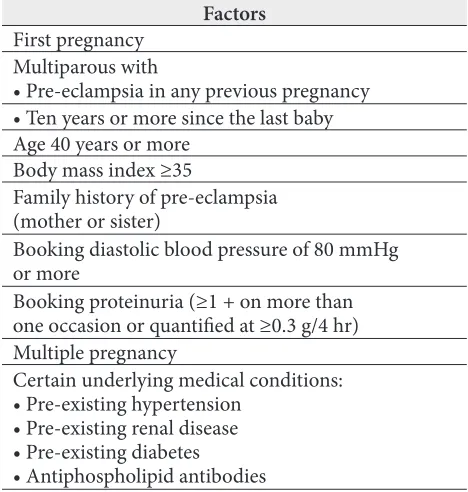
Table 2.Factors that increase the likelihood of pre-ec-lampsia developing in any given pregnancy
Factors First pregnancy
Multiparous with
• Pre-eclampsia in any previous pregnancy • Ten years or more since the last baby Age 40 years or more
Body mass index ≥35
Family history of pre-eclampsia (mother or sister)
Booking diastolic blood pressure of 80 mmHg or more
Booking proteinuria (≥1 + on more than one occasion or quantified at ≥0.3 g/4 hr) Multiple pregnancy
Certain underlying medical conditions: • Pre-existing hypertension
• Pre-existing renal disease • Pre-existing diabetes • Antiphospholipid antibodies
Blood tests relating to pre-eclampsia
• Blood tests relating to pre-eclampsia are plate -let count, serum creatinine, transaminases (AST or ALT as per local availability).
• Laboratory test results should be available
within no more than 24 hours of the woman attend-ing and the same day where practically possible, with a mechanism to review the tests and talk to the woman concerned, also within 24 hours.
• Use pregnancy-specific ranges for platelets,
serum urate, transaminases and creatinine and a gestational age specific range for serum urate, as shown in Table 3.
Laboratory tests for proteinuria
• Test to exclude or confirm significant proteinu -ria in women with 1+ dipstick proteinu-ria. A labo-ratory urinary protein creatinine (PCR) ratio from a random sample of less than 30 mg/mmol excludes
Table 3. Liver function tests: gestation specific 95% reference ranges (2.5th centile – 97.5th centile) in normal population (2)
Non
pregnancy Trimester 1 Trimester 2 Trimester 3
AST, iu/L 7–40 10–28 11–29 11–30
ALT, iu/L 0–40 6–32 6–32 6–32
• Platelet count <150 × 109/L. • Creatinine ≥90 micromol/L.
significant proteinuria (A PCR > 30 mg/mmol does not reliably confirm or quantify proteinuria). A 24 hour urine collection of ≥300 mg/24 hr both confirms and quantifies proteinuria.
• For women with new hypertension between
90–99 mmHg and 1+ proteinuria, the decision to admit can be deferred until the results of the PCR are known. This is appropriate only when there are no maternal symptoms or clinical suspicion of fetal compromise. Arrange an umbilical artery Doppler and blood tests while waiting for the result of the PCR.
Umbilical artery Doppler
• Umbilical artery Doppler is the best test for
predicting an at-risk fetus in women with pre-eclampsia. Abnormal umbilical artery Doppler thresholds include:
• Umbilical artery PI > 2SD;
• Absent or reverse end diastolic flow.
Antenatal inpatient management
The rationale for admitting women with a diag-nosis of pre-eclampsia is the increased risks of pla-cental abruption and eclampsia. These are likely to be managed more expeditiously if the woman is an inpatient.
A TPR chart of blood pressure (at least twice a day), urinalysis (daily) should be kept.
Any symptom relating or possibly relating to pre-eclampsia (headache, abdominal pain, breath-lessness) should prompt a full set of observations (temperature, pulse, respiratory rate, blood pres-sure, oxygen saturations if breathless) and medical review.
Blood tests relating to pre-eclampsia should be performed at least twice weekly and recorded on
the flow chart in the woman’s handheld notes.
Assessment of a woman with pre-eclampsia
Symptoms
• Visual disturbance; • Headaches;
• Nausea;
• Epigastric pain / right upper quadrant (RUQ) pain; • Decreased fetal movements.
Signs
• Raised blood pressure; • Oedema (facial in particular); • Clonus (>1 beat is significant); • Liver edge or epigastric tenderness.
If BP ≥140/90 mmHg, check every four hours with a manual sphygmomanometer.
Initial investigations
• Urinary protein (Dipstix, start 24 hr collection),
protein / reatinine ratio.
• Midstream specimen of urine (MSU) to exclude
infection (if leucocytes and / or nitrite+).
• Urine output must be accurately monitored and
recorded.
• Full blood count (FBC) and platelets.
• Clotting screen is only required if platelets <100 × 109/l or liver function test (LFT) is
abnor-mal.
• Urea and electrolytes (U & Es) (creatinine should be <90 µmol/l).
• LFTs (serum aspartate amino transferase (AST)
and alanine amino transferase (ALT) should be
<30 iu/l).
• Glucose if ALT >150, acute fatty liver of pregnan -cy (AFLP) or diabetic.
• Blood film if HELLP suspected (haemolysis,
ele-vated liver enzymes and low platelets – associated with higher maternal mortality).
• Group and save (G & S) and antibody screen.
Fetus
Clinical
• Assessment of gestation.
• Fundal height, presentation and estimated fetal
weight (EFW).
• Fetal movements. Investigations
• Cardiotocograph (CTG).
• Ultrasound scan for presentation, EFW and
li-quor volume.
• Umbilical artery Doppler studies (and uterine).
• Uterine Doppler studies (outcome worse if high
respiratory infection / notches).
If BP is controlled and proteinuria is 0.3–0.5 g, the woman may be allowed home. However, she will need to be reviewed three times per week with weekly 24-hour urine collection. She should be ad-mitted if the BP becomes out of control or proteinu-ria is >0.5 g/24 hr. The rationale for this strategy is the low incidence of complications if proteinuria is
<0.5 g/24 hr.
Managing women with isolated new-onset pro-teinuria in pregnancy
Women with new-onset isolated proteinuria in pregnancy are usually regarded as falling some-where within the pre-eclampsia spectrum. They may be regarded as a group that could go on to develop full-blown pre-eclampsia and kept under surveillance in the same way as women who have isolated pregnancy-induced hypertension. In the BEST study 10% of women with isolated proteinu-ria went on to eclampsia within one week. The comparable figure for women with isolated hyper-tension was 22% (3).
Management
These women should have the following monitor-ing:
• Day assessment unit twice per week for BP check,
review of symptoms and PET bloods. Admission if BP ≥140/90 mmHg, symptoms or abnormal PET bloods.
• Repeat 24-hour protein collection every week.
Admit if protein rises to ≥3 g/24 hours.
• Fetal growth scan every 4 weeks. • Consider renal investigations.
• MSU on the first referral and review within
48 hours by an obstetric registrar.
Follow-up
It is extremely important that these women con-tinue to have a regular review by their obstetrician.
MANAGEMENT OF HYPERTENSION
Pre-existing hypertension
β-blockers and diuretics should be discontinued once pregnancy is confirmed. If treatment is
re-quired to keep the blood pressure <150/100 mmHg
pres-sure in the first trimester), then treatment with methyl dopa should be commenced. If the blood pressure is satisfactory without treatment (which is often the case in women with well-controlled hy-pertension on monotherapy pre-pregnancy), the woman should be counselled that antihypertensive medication may be required at 26–30 weeks ges-tation. A plan should be made for monitoring of the blood pressure at least 3–4 weekly throughout pregnancy.
Women receiving calcium antagonists (nifedi-pine or amlodi(nifedi-pine) or labetalol may continue these drugs in pregnancy.
Women receiving ACE inhibitors and angio-tensin receptor blockers should ideally discontinue these drugs prior to pregnancy.
Pregnancy induced hypertension (PIH)
Management aims to control the blood pressure and undertake surveillance.
The aim of treatment of hypertension is to avoid severe hypertension (SBP >160 mmHg) and pro-long gestation. Women without pre-eclampsia treatment of hypertension also avoid admission to hospital.
The antihypertensive agents used are the same regardless of the underlying cause of hypertension.
Pre-eclampsia
Methyldopa is the first-line treatment. Nifedipine is the second-line drug. Labetolol is a useful second- or third-line drug where there are no contra-indica-tions to beta blockade. Labetalol or hydralazine are used for severe hypertension once the decision has been taken to deliver. Labetalol should be avoided in women with asthma.
Oral antihypertensive therapy
Drugs for the treatment of hypertension in preg-nancy are given in Table 4.
INTRAPARTUM MANAGEMENT
The decision to deliver should be made by the con-sultant obstetrician. Remember that initial stabi-lisation of the maternal condition leads to a safer delivery by whatever route.
DO NOT RUSH into delivery. If <34 weeks, try
to stabilise and gain time for betamethasone to act. If >34 weeks, usually labour will be induced. Assess
the state of the cervix, maternal condition and fetal wellbeing. Indications for operative vaginal delive-ry are not altered by hypertension. Inform neona-tologists at an early stage.
Pre-eclampsia may progress intrapartum and a close watch should be kept on the blood pressure and symptoms remembering that PET is a multi-system disease, where end-organs (e. g. cardio-vas-cular, renal, central nervous, hepatic, coagulation and placenta) may be affected to a greater or lesser extent.
Intravenous access is required for all women with pre-eclampsia during labour and delivery. Oral an-tihypertensive treatment should continue through labour. Stabilisation of BP with oral treatment be-fore induction or caesearan section (CS) makes hy-pertension in labour, delivery and the postpartum period easier to manage, and may avoid the need for parenteral therapy.
Analgesia during labour
Severe PET is not a contra-indication to epidural analgesia providing clotting is normal and platelets
>75 × 109/l. All women with proteinuric
hyperten-sion should have platelets and clotting checked on admission, otherwise delay in implementing epi-dural analgesia may occur. The anaesthetist need not wait for the results of a clotting screen unless
the platelet count is <100 × 109/l. If the platelet
count is between 75–100, the epidural should only be sited if the clotting screen is normal.
Remember that vasoconstriction is a part of the pathophysiology of PET, and a very careful
atten-tion to fluid balance is mandatory.
Third stage:
Oxytocin 5IU should be given intramuscularly. NO syntometrine or ergometrine.
Anticonvulsant therapy in eclampsia
If seizure: Recovery position & administer
oxygen.
Ensure adequate IV access.
Initial treatment (loading dose)
(prophylaxis or treatment)
4 g (diluted to 20 ml in 5% dextrose or 0.9% sodium chloride) 10% MgSO4 infused IV over 5–10 min.
Maintenance dose 5 g MgSO4 in 50 ml running at 10 ml per hour (1 g/hr). This volume needs to be deducted from hourly maintenance fluid.
Contraindications Cardiac disease, acute renal failure (must be discussed with senior obstetrician, usually same loading dose plus reduced maintenance dose).
Myaesthenia gravis
Use diazemuls, 10 mg, then 2.5 mg/hr OR rectal diazepam 10 mg/2.5 ml.
Duration of infusion While patient on HBC, i. e. until 24–48 hrs after delivery or 24 hrs after last seizure.
Monitoring Clinical Level of consciousness: hourly. Respiratory rate should be >10/min. Patellar reflex:
After completion of loading dose. Hourly whilst on maintenance dose (Use arm reflexes if epidural). ECG / pulse
oximetry Electrocardiogram (ECG) mandatory during and for 1 hr after loading dose.Pulse oximetry whilst on Mg. Mg levels Only if oliguric, creatinine >150, urea >10 or recurrent seizures.
2 ml in plain (clotted) tube.
Mark URGENT and ask lab to phone the result. Therapeutic range: 2–4 mmol/l (4.0–8.0 mg/dl).
Presence of patellar reflex, oxygen saturation and Mg levels MUST be recorded on the PET chart.
Dose
alterations Oliguria (≤80 ml over 4 hr)OR urea >10 mmol/l, cr 6 hourly >150 mmol/lMeasure Mg levels ALT > 250 IV/l Measure Mg levels 6 hourly Mg level > 4 mmol/l Decrease maintenance dose to 0.5 g/hr Table 4. Drugs for the treatment of hypertension in pregnancy
Preferred agents Dose Comments
Methyldopa 250 mg bd – 1 g tds Possible drug of choice; safety after first trimester well documented, including 7-year follow-up of offspring. May cause lethargy and dizziness or rarely depression. Labetalol 200 mg bd – 500 mg tds May be associated with fetal growth restriction
and neonatal bradycardia. Nifedipine 10 mg SR bd – 20 mg SR
bd Possible interference with labour; may interactsynergistically with magnesium sulphate. May cause headache, flushing, swollen lower legs.
Doxasocin 1 mg od – 8 mg bd Third-line therapy.
Contraindicated ACE inhibitors
and ARB (angio-tensin receptor
antagonists)
Leads to fetal loss in animals; human first trimester use associated with fetal abnormalities; use in the second and third trimester associated with fetopathy, oligohydramnios, growth restriction,
Toxicity includes • Loss of patellar reflex • Nausea
• Feeling of warmth • Flushing
• Weakness • Somnolence • Double vision • Slurred speech
5 mmol/l
• Muscle paralysis
• Respiratory arrest 6–7.5 mmol/l
• Cardiac arrest >12 mmol/l
Management Loss of patellar reflex or respiratory
rate <10/min
1. Stop maintenance infusion.
2. Send Mg level to laboratory URGENTLY. 3. Withhold further Mg until patellar reflexes return or blood Mg level known. Restart at 1 g/hr and check levels at 1 hr.
Oxygen saturation persistently <95%
(on air)
1. Commence O2.
2. Stop maintenance infusion and send Mg level.
3. Inform anaesthetist. Cardio-respiratory
arrest 1. “Crash call”.2. Woman should be in left lateral tilt position and institute cardiopulmonary resuscitation (CPR).
3.Stop maintenance infusion.
4. Administer 10 ml 10% calcium gluconate IV (antidote) slowly.
5. Intubate immediately and manage with assisted ventilation until resumption of spontaneous respirations.
6. Send Mg level to lab URGENTLY.
Recurrent seizures after starting Mg
1. Treat recurrent seizure with a further bolus of magnesium 2 g over 5 minutes.
2. If possible, take blood for Mg prior to addi-tional bolus.
3. If further seizures occur despite the above, consider:
a) Diazemuls 10 mg IV bolus and then an
infu-sion (2.5 mg/hr);
b) Thiopentone infusion (on intensive care
unit);
c) Paralysis and ventilation.
If recurrent seizures, inform the anaesthetist and the intensive care unit and consider giving another anticonvulsant.
Eclampsia does not usually occur without pre-monitory symptoms (e. g. severe headache, visual disturbance, and epigastric pain). Symptoms should always be taken seriously. 44% of eclamptic fits oc-cur after delivery (3). Thus, post-natal vigilance is
essential, although the disease will resolve sponta-neously in all but a few cases.
Blood Pressure
• A BP of greater than 160/110 requires treat -ment (see below).
Do not forget that acute reduction in BP to
</90 may precipitate fetal distress and therefore
continuous CTG is necessary in the acute situation when fast acting drugs are used. It is important that BP is not allowed to fall precipitately (below 140/90) to avoid the risk of underperfusion of the patient’s vital organs, i. e. brain, kidneys and placenta.
Oliguria is common, especially post delivery and especially in the context of prolonged oxytocin use. Urine output may incorrectly appear to be reduced if the fetal head is well down in the pelvis, so use a large-bore urinary catheter. As long as urine out-put >20 ml/hr, await resolution of PET. In case of
-atinine. Remember the risk of pulmonary oedema
and avoid the temptation to give intravenous fluids
to treat oliguria unless there is evidence of blood loss or hypovolaemia.
Until a diuresis occurs, blood should be taken regularly (initially 4–6 hourly) to monitor electro-lytes, creatinine, FBC, clotting and LFTs. Diuresis usually signals resolution unless there is underlying disease, and gradual reduction in antihypertensives and anticonvulsants can begin.
ANAESTHETIC PROTOCOL FOR PET
Labour
Epidural analgesia is the preferred method of an-algesia.
Contraindications: Platelet count <75 × 109/l
(beware of a rapidly falling platelet count).
Elevated activated partial thrombin time (APTT) or
thrombin time (TT).
Symptoms or signs of abnormal bleeding or clotting.
Maternal refusal.
FLUID PRE-LOAD: Hartmann’s solution (250– 500 over one hour).
Then continue with Hartmann’s solution at 85 ml/hr.
Caesarean section
If ranitidine has not been given within the last 12 hr, give 50 mg IV.
Both epidural and spinal anaesthesia are suit-able for CS.
Epidural anaesthesia usually provides very stable cardiovascular parameters.
Spinal anaesthesia can be employed as a part of a combined spinal epidural technique (CSE), the intrathecal dose of heavy bupivacaine 0.5% can be reduced (1.7–2.0 ml). Intrathecal diamorphine
250–300 µg or fentanyl 15–25 µg should be given
with bupivacaine. In greater than 90% of mothers this solution will provide adequate anaesthesia for CS. In rare cases when it does not, small increments (3 ml of ropivacaine 0.75%) can be given through the epidural catheter.
However, if the platelets are falling, a single shot spinal anaesthesia may be more appropri-ate. A 25–27 g spinal needle will cause less trauma than the larger epidural needle. 2–2.2 ml of heavy
bupivacaine with fentanyl or diamorphine should be used. Spinal anaesthesia is not associated with a catastrophic fall in blood pressure. Vasopressors should be used as required.
FLUID PRE-LOAD: Elective CS
If no CVP, preload = 500 ml Hartmann’s solu-tion.
CVP ≥ 2 mmHg – 500 ml Colloid.
CVP > 2 mmHg – 500 ml Hartmann’s (over 15 min) prior to block.
Maintain 85 ml/hr and replace blood loss. Phenylephrine / ephedrine as required to main-tain pressure.
Emergency LSCS
If no CVP, extend epidural block without
addi-tional fluid.
No functioning block: As above. Functioning block:
CVP ≤ 2 mmHg – 500ml Hartmann’s.
CVP > 2 mmHg – Extend block without
addi-tional fluid.
General anaesthesia
In addition to the rapid sequence induction, alfen-tanil 1–2 mg should be given prior to intubation to obtund the hypertensive response. The induction dose of thiopentone should be 5–6 mg/kg. Con-sider using intra-arterial pressure monitoring. Mg-SO4 (2 g) may be necessary prior to surgery. If the mother has received MgSO4,the degree of muscle relaxation must be carefully monitored. Labetolol may still be required at intubation and extubation.
No non-steroidal anti-inflammatory drugs
(NSAIDS) (e. g. diclofenac) for at least 48 hr post-partum due to potential adverse effects on the renal and platelet function.
Received 24 July 2012 Accepted 1 August 2012
References
2. Girling JC, Dow E, Smith JH. Liver function tests in pre-eclampsia: importance of comparison with a reference range derived for normal pregnancy. Br J Obstet Gynaecol. 1997; 104(2): 246–50.
3. Douglas KA, Redman CW. Eclampsia in the Unit-ed Kingdom. BMJ. 1994; 309: 1395–1400.
4. CEMACH Saving Mothers Lives. 7th Report of Confidential Enquiries into Maternal and Child Health 2006–2008. London: CEMACH, RCOG; 2010.
5. Davey DA, MacGillivray I. The classification and definition of the hypertensive disorders of preg-nancy. Am J Obstet Gynecol. 1988; 158(4): 892–8. 6. Eclampsia Trial Collaboration Group. Which an-ticonvulsant for women with eclampsia? Evidence from the Collaborative Eclampsia Trial. Lancet. 1995; 345: 1455–63.
7. Helewa ME, Burrows RF, Smith J, Williams K, Brain P, Rabkin SW. Report of the Canadian Hy-pertension Society Consensus Conference: 1. Defi-nitions, evaluation and classification of hyperten-sive disorders of pregnancy. CMAJ. 1997; 157(6): 715–25.
8. Management of severe pre-eclampsia / eclampsia. Green Top Guideline 10 (A). London: RCOG; 2006.
Geraldine O’Sullivan
PASVARSTYMAI RENGIANT PREEKLAMPSIJOS PROTOKOLĄ
Santrauka
Preeklampsija, kurios pirmas dažniausias požymis yra hi-pertenzija, yra viena pagrindinių motinos ir vaisiaus ser-gamumo ir mirtingumo priežasčių. Hipertenzija – daž-niausia medicininė nėštumo komplikacija, pasireiškianti net iki 10 % visų nėštumų. Preeklampsija taip pat sieja-ma su sulėtėjusiu vaisiaus augimu, sieja-mažu gimimo svo-riu, priešlaikiniu gimdymu, mažais pagal gestacinį am-žių naujagimiais ir respiracinio distreso sindromu (The Magpie Collaborative Group, 2002).