Modeling of Tactile Detection of an Artery in a Soft Tissue
by Finite Element Analysis
Ali Abouei Mehrizii, Siamak Najarian ii ٭and Majid Moiniiii
i A. Abouei Mehrizi is with the Faculty of Biomedical Engineering, Amirkabir University of Technology, Tehran, Iran (email: [email protected]).
ii * Corresponding Author, S. Najarian is with the Faculty of Biomedical Engineering, Amirkabir University of Technology, Tehran, Iran (email: [email protected]).
iii M. Moini is with Sina Trauma and Surgery Research Center, Tehran University of Medical Sciences, Tehran, Iran (email: [email protected]).
ABSTRACT
Nowadays, one of the main problems encountered in minimally invasive surgery and telesurgery is the detection of arteries in tissue. In this study, for the first time, tactile detection of an artery in tissue and distinguishing it from the tumor has been modeled by finite element method. In this modeling, three 2D models of tissue have been created: tissue, tissue including a tumor, and tissue including an artery. After solving three models with similar boundary conditions and loadings, first, the 2D tactile mappings and stress graphs for upper nodes of models, which have the role of transferring tactile data, have been explored. Comparing these results, if stress values of nodes are equal and constant, tissue is without tumor or artery. In addition, it was concluded that if stress graph includes a peak, the tissue has a tumor or an artery and that the stress graph of tissue including artery is time-dependent in comparison with the tissue including the tumor.
KEYWORDS
Soft tissue, tumor, artery, tactile detection, finite element method (FEM).
1. INTRODUCTION
Minimally invasive surgery (MIS) is a kind of surgery in which two or three incisions with nearly 1 cm diameter or so are being created on external surface of the body. Then, tiny long surgery tools are entered through these incisions into the body and the surgeon does the operation by these tools. Because of the numerous advantages of MIS, comparing with traditional open surgery, the researchers are putting a great deal of effort on optimizing the design and capabilities of its related tools. Some of these advantages are: reducing damage to healthy tissue, decreasing trauma, patients heal more quickly, reducing recovery times, etc. MIS suffers from one major drawback: it decreases the sensory perception of the surgeon and the surgeon might accidentally cut or incur damage to some of the tissues. Hence, the design and manufacture of surgical tools equipped with artificial tactile sensing is a new research topic [1].
One of the main difficulties which surgeons encountered in this area is inability in detecting the arteries embedded in tissues [2]. Therefore, in
laparoscopic surgeries like removing the gall bladder out of body, the location of cutting is burnt for bloodshed prevention or some grippers are placed in each side of the cutting location.
Although this subject is new, many studies have been performed about detection of arteries in tissues and their stenosis. Nearly all of these studies have used imaging and ultrasound techniques. Beside their advantages, they also have some limitations including: a) vessels deep in the body are harder to see than superficial vessels, b) smaller vessels are more difficult to image and evaluate than larger vessels, and c) calcification that occurs as a result of atherosclerosis may obstruct the ultrasound beam [3]. Therefore, a practical method which could eliminate these limitations during the surgery process sounds very necessary. Artificial tactile sensing is a new technique for detecting of arteries in soft tissues by palpation [4], [5].
artery and sensing hard nodules in a mock breast [10]. It is noticeable that in this study, numerical solutions were not performed. In another study, a long 10 mm diameter probe was constructed with an array of tactile sensor set in the end of it [11]. This sensor was pressed against the tissue of interest and pressure distribution was read out as electrical signal across contact area. Then this information was processed and the presence of an artery in tissue was concluded. In this study, just like the pervious works, numerical solutions were not employed. The last study on artery detection is related to construction of a tactile sensor that can track a vessel with various curves in artificial tissue with silicon type by a programmable robot [12]. Similar to pervious works, in this study numerical solutions were not performed again.
In this paper, for the first time, we have investigated the detection of an artery and a tumor in a tissue and separation of these two categories from each other by using finite element method. Furthermore, we have modeled a 25% stenotic artery in a tissue and investigated the possibility of distinction of a healthy artery from a stenotic artery and presented a criterion in this regard.
2. NUMERICAL SOLUTION
In every application of tactile sensing method, the physical contact between tactile sensor and object or tissue is in special importance [13]. In this physical contact, according to the design of sensor, a parameter of touch was used as a criterion for measuring or an operative for stimulating a sensor. This criterion can be force, pressure (stress), displacement (strain), temperature, humidity, roughness, stiffness, and softness that is appeared on the surface of the touched object in which tactile data was transferred between it and the sensor. Otherwise, they cannot be suitable criteria. In the present study, for modeling the palpation process, displacement -5 mm was applied on top side of model through an array of tactile sensors (see Fig. 1). The stress parameter was selected as the criterion of detection in this study. For completing the simulation of palpation, the displacement of the bottom side of model in y-direction was considered zero.
A. Definition of Problems
New minimally invasive surgical techniques prevent surgeon from directly touching and palpating internal tissue. Therefore, surgeon cannot be aware of the condition of internal tissue. For example, hard lumps in soft organs are detected by probing the tissue with the fingers and arteries are localized during dissection by feeling for a time varying pressure.
In this paper, we scrutinized on the presence of artery in soft biological tissue and distinction of a tumor to pattern out palpation by finite element method.
B. Modeling, Simplifying, and Assumptions
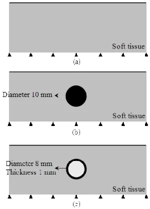
According to physical standard for modeling of soft tissues, we consider three phantoms of soft tissue as three 2D models as shown in Fig. 2. All of three models are 80×30 mm in size.
The tissue and the artery components were assumed elastic and isotropic material with a modulus of elasticity of 15 [7] and 300 [14] kPa, respectively. The modulus of elasticity of tumor has been assumed 20 times larger than tissue [7]. The Poisson’s ratio of the tissue, artery, and tumor were considered 0.49, 0.49, and 0.3, respectively [15].
Figure 1: (a) Schematic representation of the contact of sensor and soft tissue and (b) The finite element model and the boundary conditions.
Figure 2: (a) Soft tissue model, (b) Tissue including tumor, and (c) Tissue including artery.
healthy pulse, which is approximately 0.85 second. This effect has been applied as pressure loading with a maximum and a minimum of inside pressure of artery to be 120 and 80 mmHg, respectively [16]. This loading has been divided into two steps: systolic and diastolic phases which loading time for each phase is 0.425 second. In systolic step pressure increases from 80 to 120 mmHg as linear in during first 0.425 second and in diastolic step it decreases from 120 to 80 mmHg as linear in during next 0.425 second.
C. Finite Element Model and Boundary Conditions
This model has been solved by finite element method through ABAQUS software (release 6.6.1). The length of each model has been considered 80 mm so that the left and the right side of the tissues be far from the artery and the tumor and do not effect on the stress distribution of the artery, the tumor and their environments. Bottom side of the tissue in each model has been fixed in y-direction that is the direction of loading on top side of the model. With prevention of rigid body motion, each model was solved for duration of 0.85 second.
For having continuous strain in touch site of the artery and the tumor with tissue, they were glued to tissue by gluing same nodes together. The model has been meshed by a 4-node bilinear plane stress quadrilateral element.
To gain accurate solution and reduce the duration of solving, the element size of the tumor, the arteries, and the environments that have severe gradients of stress were considered finer than other parts of the model. In Fig. 3, a sample of the mesh of model c was presented.
Figure 3: (a) Soft tissue model, (b) Tissue including tumor, and (c) Tissue including artery with geometrical dimension of each model.
D. The Method of Results Extraction
The aim of this modeling is to explore the effect of an artery and a tumor in the tissue and comparison of them together for finding a criterion for separating them. Therefore, Von Mises stress graph for all of nodes created in top side of four models, shown in Fig. 2, and 2D tactile image of the models were explored.
3. NUMERICAL RESULTS
For every model, outputs in two different situations were derived: 1) stress distribution on top side of tissue model that is the site of touch between tissue and sensor. This was named 2D tactile image and 2) Von Mises stress
graph on all of top nodes. According to 2D tactile imaging and stress graph, the results are as follows:
A. Appearance of the Symptoms of Existence of an Artery or a Tumor on Top Side of Tissue Model
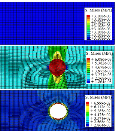
Fig. 4 shows 2D tactile images corresponded by applying -5 mm displacement on top line of three models. From the 2D tactile images a, b, and c, it is understood that applied pressure on the tissue which has the tumor or the artery, caused a non-uniform stress distribution in comparison with the tissue itself. This result supports the suitability of the artificial tactile sensing method in artery or tumor detection.
Figure 4: 2D tactile images a) the tissue itself, b) tissue including a tumor, c) tissue including an artery (tactile image c is related to time 0.425 second).
B. Appearance of a Peak in Stress Graph
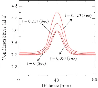
As shown in Figs. 5, 6, and 8, Von Mises stress graphs were derived for the nodes on top side of models b and c, in comparison to model a, include a peak. The peak for these two graphs is a symptom for existence of a tumor or an artery in tissue. Also, the center of the peaks is exactly under the tumor or the artery. Therefore, we can determine the exact location of the artery or the tumor through the stress graph. By comparing Fig. 6 and 7, it is clear that the derived result for model b has a good agreement with other studies [7].
Since pressure inside of artery increases as linear in systolic step from 80 to 120 mmHg and decreases as linear in diastolic step from 120 to 80 mmHg, we derive a graph similar to graphs shown in Fig. 9 for diastolic step.
1. DISCUSSION
In this study, the palpation of physician was modeled and simulated by finite element method. This modeling has been performed for three 2D models: tissue itself, tissue including a tumor, and tissue including an artery.
Figure 5: Von Mises stress graph for nodes on top side of model a (this graph is same during 0.85 second).
Figure 6: Von Mises stress graph for nodes on top side of model b (this graph is same during 0.85 second).
Figure 7: Von Mises stress graph for nodes on top side of the model of tissue including a tumor [7], [8].
By comparing 2D tactile images a, b, and c in Fig. 4, we can understand, with applying the same loading on top side of each model shown in Fig. 2, if 2D tactile image was uniform there is only the tissue itself, if 2D tactile image was not uniform, the tissue includes other material with different mechanical properties of soft tissue like tumor or artery, if 2D tactile image was not uniform but remained constant versus time, the tissue includes tumor, if 2D tactile image was neither uniform nor constant, the tissue includes artery. Therefore, the cases which can be explored from these tactile images include: the existence of a hard embedded object in soft tissue, detection of pulsatile effect, and determination of the location of embedded object approximately.
Figure 8: Von Mises stress graph for nodes on top side of model c at 0.425 second.
Figure 9: Von Mises stress graph for nodes on top side of model c at 0.425 second.
constant, the tissue includes tumor, and 4) if the value of the stress peak be time-dependent, the tissue includes artery. Therefore, the cases which can be explored from these stress graphs include: the existence of a hard embedded object in soft tissue, distinguishing between the artery and the tumor inside the soft tissue, and determination of the exact location of the artery and the tissue inside the soft tissue.
2. CONCLUSION
In this study, by creating three tactile models and exploring 2D tactile image and stress graphs for each one and comparing them together, we could detect existence
of a tumor or an artery in tissue. According to corresponded results, we were able to separate the tissue itself from tissue including a tumor, and tissue including an artery. This distinction is useful for surgeons during minimally invasive surgery. The major point of this study is that we can detect the existence of an artery in the soft tissue through artificial tactile sensing and can differentiate it from the tumor.
3. ACKNOWLEDGMENT
The authors gratefully acknowledge the Center of Excellence in Biomedical Engineering of Iran, for their help in conducting this project.
4. REFERENCES
[1] M. E. H. Eltaib and J. R. Hewit, "Tactile sensing technology for minimal access surgery-a review", Mechatronics, vol. 13, p.p. 1163-1177, 2003.
[2] W. J. Peine, "Remote palpation instruments for minimally invasive surgery," Ph.D. dissertation, Div. of Eng. and Appl. Sci., Harvard University, 1998.
[3] J. Dargahi and S. Najarian, "Human tactile perception as a standard for artificial tactile sensing- a review”, Inter. J. of Med. Rob. Comp. Ass. Surg., vol. 1, pp. 23-35, 2004.
[4] J. Dargahi and S. Najarian, "Advances in tactile sensors design, manufacturing, and its impact on robotics application - review", Indus. Rob.: An Inter. J., vol. 32/3, pp. 268-281, 2005.
[5] J. Dargahi and S. Najarian, "Human tactile perception as a standard for artificial tactile sensing- a review", Inter. J. of Med. Rob. Comp. Ass. Surg., vol. 1, pp. 23-35, 2004.
[6] J. Dargahi and S. Najarian, "A Supported membrane type sensor for medical tactile mapping", Sensor Review, vol. 24, pp. 284-297, 2004.
[7] S. M. Hosseini, S. Najarian, S. Motaghinasab, and J. Dargahi, "Detection of tumors using computational tactile sensing approach", Inter. J. of Med. Rob. Comp. Ass. Surg., vol. 2, no. 4, p.p. 333-340, 2006.
[8] S. M. Hosseini, S. Najarian, S. Motaghinasab, "Analysis of effects of tumors in tissue using of artificial tactile modeling", Amirkabir Journal, to be published.
[9] S. M. Hosseini, S. Najarian, S. Motaghinasab, and S. Torabi, "Experimental and numerical verification of artificial tactile sensing approach for predicting tumor existence in virtual soft tissue", Proc. of the 15th Annual-International Conf. of Mechanical Engineering, 2007.
[10] P. Dario, M. Bergamasco, "An advanced robot system for automated diagnostic tasks through palpation" IEEE Trans. on Biomed. Eng., vol. 35, no. 2, p.p. 118-26, 1988.
[11] W. J. Peine, S. Son, and R. D. Howe, "A palpation system for artery localization in laparoscopic surgery", First Inter. Symp. on Medical Robotics and Computer-Assisted Surgery, Pittsburgh, 1994.
[12] R. A. Beasley and R. D. Howe, "Tactile tracking of arteries in robotic surgery", Proc. of IEEE, International Conf. on Robotics & Automation, Washington, DC, 2002.
[13] M. H. Lee and H. R. Nicholls, "Review article tactile sensing for mechatronics-a state of the art survey", Mechatronics, vol. 9, pp. 1-31, 1999.
[14] D. J. Mozersky, D. Sumnfer, D.E. Hokanson, and D.E. Strandness, "Transcutaneous measurement of the elastic properties of the human femoral artery", Circulation AHA Journal, v. XLVI, 1972. [15] A. E. Kerdoke, S. M. Cotin, M. P. Ottensmeyer, A. M. Galea, R. D.
Howe, and S. L. Dawson, "Truth cube: establishing physical standard for soft tissue simulation", Med. Imag. Anal., vol. 7, pp. 283-291, 2003.
[16] K.B. Chandran, "Cardiovascular Biomechanics", New York