Katarzyna Skórka
1 C, D, F, Nupur Bhattacharya
3, B, F, Paulina Własiuk
1, B, F,
Małgorzata Kowal
2, A, B, F, Daniel Mertens
3, 4, A, B, E, F, Anna Dmoszyńska
2, E, F,
Krzysztof Giannopoulos
1, 2, A–C, E, FThalidomide Regulation of NF-κB Proteins
Limits Tregs Activity
in Chronic Lymphocytic Leukemia
1 Department of Experimental Hematooncology, Medical University of Lublin, Poland 2 Department of Hematooncology and BMT Unit, Medical University of Lublin, Poland 3 Department of Internal Medicine III, University of Ulm, Germany
4 “Mechanisms of Leukemogenesis” Cooperation Unit, German Cancer Research Center DKFZ,
Heidelberg, Germany
A – research concept and design; B – collection and/or assembly of data; C – data analysis and interpretation;
D – writing the article; E – critical revision of the article; F – final approval of article; G – other
Abstract
Background. Thalidomide may represent a novel therapeutic strategy in the treatment of chronic lymphocytic leukemia (CLL). Since the activation of nuclear factor kappa B (NF-κB) causes not only malignant transformation and tumor progression, but also allows tumor cells to evade immune surveillance, NF-κB signaling components might constitute a potential target for future therapy in CLL.
Objectives. The current study is an attempt to characterize proteins regulated by thalidomide. Thalidomide’s influ-ence on NF-κB proteins and on regulatory T cells (Treg) in CLL was investigated.
Material and Methods. A total of 15 patients with CLL were treated with a combined thalidomide/fludarabine regimen. Peripheral blood mononuclear cells were separated by Ficoll density gradient centrifugation. To evaluate glucocorticoid-induced tumour-necrosis-factor-receptor-related protein (GITR) expression in regulatory T cells, cells incubated with anti-CD3, ani-CD4 and anti-CD25 were permeabilized and then stained with anti-FOXP3 and analyzed using flow cytometry. Human TNF enzyme-linked immunosorbent assay (ELISA) was used to determine the tumor necrosis factor (TNF) levels in the serum. To evaluate NF-κB activity, chemiluminescent oligonucleotide-based ELISA was performed.
Results. Itwas found that thalidomide regulates NF-κB activity differentially, and the activity of certain NF-κB components correlated with TNF levels and T regulatory cell (CD4+CD25highGITR+).
Conclusions. These results might indicate that thalidomide not only regulates TNF but also directly interferes with NF-κB components (Adv Clin Exp Med 2014, 23, 1, 25–32).
Key words: nucelar factor kappa B (NF-κB), thalidomide, chronic lymphocytic leukemia (CLL), regulatory T cells (Treg), tumor necrosis factor (TNF).
Adv Clin Exp Med 2014, 23, 1, 25–32 ISSN 1899–5276
ORIGINAL PAPERS
© Copyright by Wroclaw Medical University
Immunomodulatory drugs including thalid-omide [1] represent a novel and promising ther-apeutic option in chronic lymphocytic leukemia (CLL). It has been shown that thalidomide mod-ulates the tumor microenvironment by inhibiting angiogenesis, as well as modulating the immune
system by co-stimulating immune effector cells [1– 3]. Moreover thalidomide therapy in CLL reduces the numbers of regulatory T cells (Tregs). In CLL patients the numbers of regulatory T cells (Treg) are increased, which constitutes a crucial mecha-nism of immunosuppression [4].
Furthermore, it is known that thalidomide blocks nuclear factor kappa B (NF-κB) activation by suppressing the activity of the inhibitor of NF-κB INF-κB kinase (IKK) [5]. NF-NF-κB is a family of struc-turally related transcription factors that play a ma-jor role in the inflammation and immune responses. Moreover, NF-κB inhibits apoptosis, induces pro-liferation and angiogenesis, suggesting that NF-κB has a pivotal role in oncogenesis and tumor pro-gression [6, 7]. In many solid tumors and hema-tological malignancies, including CLL, NF-κB di-mers are located in the cell nucleus, where they are constitutively active [8]. This constitutive activa-tion of NF-κB causes not only malignant transfor-mation and tumor progression, but also enables the tumor cells to evade immune surveillance [7].
The mechanisms involved in the process of NF-κB activation in leukemic cells are mainly as-sociated with microenvironmental stimuli includ-ing cells and cytokines [9, 10]. Functional inter-actions between CD40 (located on CLL cells) and CD154 (located on lymphocytes, T helper lympho-cytes and sometimes, to some extent, on leukemic cells), stimulation by cytokines including IL4 (in-terleukin 4), BAFF (B cell activating factor), a pro-liferation-inducing ligand (APRIL) and vascular endothelial growth factor (VEGF) could be re-sponsible for NF-κB activation in CLL [9, 11, 12]. Tthe prognostic importance of the RelA compo-nent of NF-κB in CLL has been noted [13]. Hence it has been suggested that NF-κB could be a poten-tial target for future CLL therapy.
Since immunomodulatory drugs could be an effective therapeutic option for CLL patients and the mechanism of thalidomide’s action is not fully understood, the present study aimed to define tha-lidomide’s regulation of NF-κB proteins and its in-fluence on Tregs in CLL.
Material and Methods
The Patients
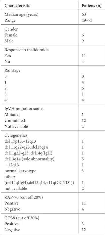
The current study involved 15 patients with CLL (median age 63, range 49–73) who were treated in a clinical study with thalidomide and fludarabi-ne [14]. The patients were treated at the Department of Hematooncology at the Medical University of Lu-blin, Poland. Informed consent was obtained from all patients and the study was approved by the local eth-ics committee. The clinical characteristeth-ics of the pa-tients are shown in Table 1. The papa-tients’ molecular characteristics, including genomic aberrations, were detected using fluorescence in situ hybridization and the IgVH mutational status, which was determined by sequencing as previously described [15].
Study Protocol
Thalidomide (100 mg p.o. per day) was started at day 0. Every 28 days fludarabine was given for 5 subsequent days (25 mg/m2 i.v. per day), start-ing at day 7, for up to 6 cycles. In order to prevent thrombosis, acetylsalicylic acid (100 mg) was ad-ministered. The patients’ clinical response was as-sessed after six cycles of thalidomide/fludarabine therapy according to International Workshop on CLL (IWCLL) response criteria [16].
Table 1. Clinical and molecular characteristics of chronic lymphocytic leukemia patients
Characteristic Patiens (n)
Median age (years)
Range 6349–73
Gender Female
Male 69
Response to thalidomide Yes
No 114
Rai stage 0 1 2 3 4 0 4 6 1 4 IgVH mutation status
Mutated Unmutated Not available 1 12 2 Cytogenetics del 17p13,+12q13 del 11q22-q23, del13q14 del11g22-q23, del14q(IgH) del13q14 (sole abnormality) +12q13 normal karyotype other: (del14q(IgH),del13q14,+11q(CCND1)) not available 1 1 1 5 1 3 1 2 ZAP-70 (cut off 20%)
Positive
Negative 114 CD38 (cut off 30%)
Positive
Negative 312
Peripheral Blood
Mononuclear Cells
Peripheral blood mononuclear cells (PBMC) were separated by Ficoll (Biochrom AG, Berlin, Germany) density gradient centrifugation. After isolation, the cells were stored frozen in liquid ni-trogen until the time of the analyses.
Flow Cytometry
The PBMC were thawed. To evaluate gluco-corticoid-induced TNF receptor (GITR) expres-sion in Tregs, the GITR were stained after incuba-tion of 1*106 cells with antibodies according to the manufacturer’s protocols. To characterize Tregs, the cells were incubated with anti-CD3, ani-CD4 and anti-CD25 permeabilized, then stained with anti-FOXP3 (eBiosciences, San Diego, CA, USA) and analyzed as previously described [4]. The dif-ference in the percentage of Treg GITR+ was cal-culated as a change in the frequency of Treg GITR+ after thalidomide therapy.
ELISA
Serum was separated and stored at –80˚C. To determine the level of tumor necrosis factor (TNF) in the serum, human TNF-alpha Quantikine HS enzyme-linked immunosorbent assay (ELISA, R&D Systems, Minneapolis, MN, USA) was used in accordance with the manufacturers protocols. The difference in TNF levels was calculated as se-rum TNF level after thalidomide therapy/sese-rum TNF level before therapy.
Chemiluminescent
Oligonucleotide-based ELISA
To evaluate the activity of different NF-κB components, chemiluminescent oligonucleotide-based ELISA (co-ELISA) was performed. After
coating single-stranded oligonucleotides, bind-ing NF-κB and detectbind-ing NF-κB-oligo complexes on 96-well plates, chemiluminescent and colori-metric detection were performed as previously de-scribed [17]. The changes in NF-κB proteins were calculated as the average activity of certain pro-teins after thalidomide therapy/average activity of certain proteins before thalidomide therapy.
Statistical Analysis
All results are presented as median values with the range. To assess correlations among the vari-ables Spearman’s rank correlation test was used. All the tests reported were two-sided, and results were considered significant if the P-value was 0.05 or less.
Results
Thalidomide Regulates NF-κB
Proteins Activity Differentially
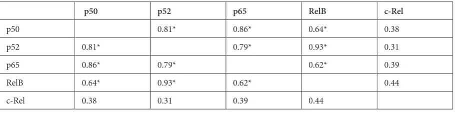
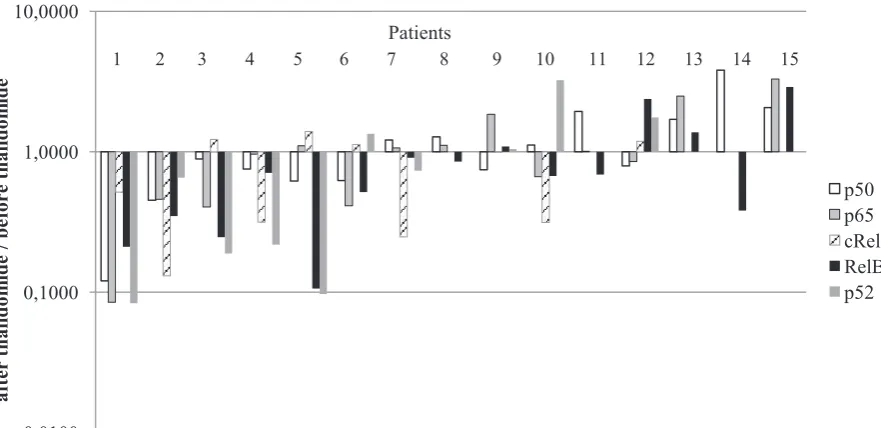
To characterize thalidomide regulation of NF-κB proteins, the activity of all DNA-binding NF-κB proteins before and after therapy was de-termined by co-ELISA. NF-κB proteins were reg-ulated differentially after thalidomide therapy: p50 was downregulated in 8 patients and upregulated in 7 patients; p52 was downregulated in 6 patients and upregulated in 4 patients; p65 was downregu-lated in 7 patients and upregudownregu-lated in 7 patients; RelB was downregulated in 11 patients and upreg-ulated in 4 patients; c-Rel was downregupreg-ulated in 6 patients and upregulated in 4 patients (Fig. 1). Be-fore the therapy the average activity of certain NF-κB subunits, like p50, p52, RelA, RelB, were corre-lated with each other, with the exception of c-Rel (Table 2). Correlations were observed between the average activity of p50 and p52 (R = 0.82, p < 0.05), between p50 and p65 (R = 0.87, p < 0.05), between
Table 2. Correlations between average activity of certain components of NF-κB before thalidomide therapy
p50 p52 p65 RelB c-Rel
p50 0.81* 0.86* 0.64* 0.38
p52 0.81* 0.79* 0.93* 0.31
p65 0.86* 0.79* 0.62* 0.39
RelB 0.64* 0.93* 0.62* 0.44
c-Rel 0.38 0.31 0.39 0.44
p50 and RelB (R = 0.65, p < 0.05), between p52 and p65 (R = 0.80, p < 0.05), between p52 and RelB (R = 0.93, p < 0.05) and between p65 and RelB (R = 0.62, p < 0.05).
Changes in NF-κB Activity
do not Differ in Clinical
and Molecular Subgroups
of CLL Patients
No differences in the activity of NF-κB pro-teins were observed in different clinical and molec-ular subgroups. The activity of p52 was higher in patients who responded to the therapy, but the dif-ference did not reach statistical significance (Ta-ble 3). Interestingly, both c-Rel and p52 showed the largest differences in patients with and with-out a flare reaction (Table 4), in patients with and without mutated IGHV (Table 5) and in patients
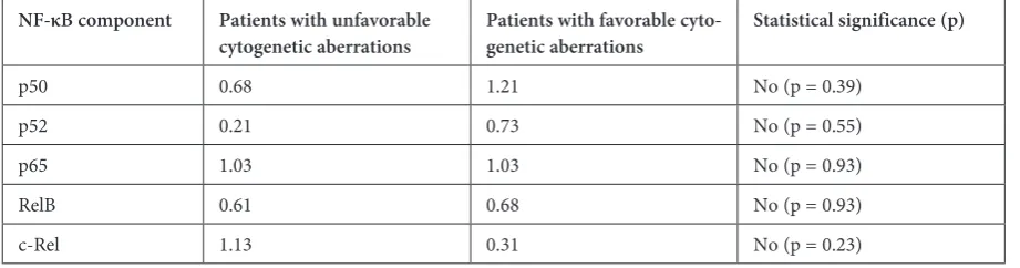
with unfavorable (+12, 11q-, 17p-, complex kary-otype) and favorable (13q-, normal karykary-otype) cy-togenetic aberrations (Table 6), although statistical significance was not reached here either.
Changes in NF-κB Activity
did not Correlate with ZAP-70
and CD38 Expression
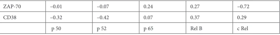
To characterize the association of NF-κB pro-teins with prognostic markers, their activity was correlated with ZAP-70 as well as with CD38 ex-pression. Changes in p50 did not correlate with ZAP-70 (R = –0.01, p > 0.05) and CD38 expression R = –0.32, p > 0.05). Changes in p52 did not corre-late with ZAP-70 (R = –0.07, p > 0.05) and CD38 expression (R = 0.05, p > 0.05). Changes in p65 did not correlate with ZAP-70 (R = 0.24, p > 0.05) and CD38 expression (R = 0.07, p > 0.05). Changes
Table 3. Median of changes in the activity of NF-κB components in responders and non-responders after thalidomide therapy
NF-κB component Responders to thalidomide
therapy Non-responders to thali-domide therapy Statistical significance (p)
p50 1.16 0.75 No (p = 0.55)
p52 0.88 0.15 No (p = 0.13)
p65 1.01 1.10 No (p = 0.45)
RelB 0.68 0.70 No (p = 1.00)
c-Rel 0.99 0.85 No (p = 0.40)
Fig. 1. Thalidomide’s differentialregulation of NF-κB activity in 15 CLL patients. Ratios were calculated as the avarage activity of particular NF-κB proteins after thalidomide therapy in reference to the avarage activity of particular NF-κB proteins before thalidomide therapy, assessed by chemiluminescent oligonucleotide-based ELISA (co-ELISA)
0,0100 0,1000 1,0000 10,0000
af
te
r
th
al
id
om
ide
/ b
ef
or
e
th
al
id
om
id
e
in RelB did not correlate with ZAP-70 (R = 0.27, p > 0.05) and CD38 expression (R = 0.37, p > 0.05). Changes in c-Rel did not correlate with ZAP-70 (R = –0.72, p > 0.05) and CD38 expression (R = 0.29, p > 0.05) (Table 7).
Changes in c-Rel Activity
Correlated with Lymphocyte
Doubling Time (LDT)
Interestingly, changes in c-Rel showed a sig-nificant inverse correlation with lymphocyte dou-bling time (LDT), (R = –0.83, p < 0.05). However,
changes in p50 (R = 0.04, p > 0.05), in p52 (R = 0.08, p > 0.05), in p65 (R = –0.10, p > 0.05) and in RelB (R = –0.27, p > 0.05) did not correlate with LDT.
The Effect of Thalidomide
Modulation of NF-κB Proteins
on TNF Regulation of
T
Cells
Correlations in the changes in the average ac-tivity of NF-κB subunits with the levels of TNF were investigated, as well as the expression of the specif-ic TNF receptor GITR, whspecif-ich characterizes Treg. There was a strong correlation between changes
Table 4. Median of changes in the activity of NF-κB components in CLL patients with or without flare reaction
NF-κB component Patients with flare reaction Patients with no flare
reaction Statistical significance (p)
p50 0.86 0.88 No (p = 0.51)
p52 0.37 0.88 No (p = 0.45)
p65 0.83 1.01 No (p = 0.74)
RelB 0.51 0.85 No (p = 0.21)
c-Rel 0.41 0.06 No (p = 0.59)
Table 5. Median of changes in the activity of NF-κB components in CLL patients with mutated IgVH and unmutated IgVH
NF-κB component Patients with mutated
IgVH Patients with unmutatedIgVH Statistical significance (p)
p50 1.11 1.04 No (p = 1)
p52 3.22 0.65 No (p = 1)
p65 0.66 1.06 No (p = 1)
RelB 0.67 0.69 No (p = 1)
c-Rel 0.31 0.99 No (p = 1)
IgVH – Immunoglobulin heavy chain variable region.
Table 6. Median of changes in the activity of NF-κB components in CLL patients with unfavorable and favorable cytogenetic aberrations
NF-κB component Patients with unfavorable
cytogenetic aberrations Patients with favorable cyto-genetic aberrations Statistical significance (p)
p50 0.68 1.21 No (p = 0.39)
p52 0.21 0.73 No (p = 0.55)
p65 1.03 1.03 No (p = 0.93)
RelB 0.61 0.68 No (p = 0.93)
c-Rel 1.13 0.31 No (p = 0.23)
in NF-κB (p50) and differences in TNF concen-tration (before and after treatment) (R = 0.69, p < 0.05), and a strong correlation between chang-es in NF-κB (p52) and percentagchang-es of Treg GITR+ (R = 0.97, p < 0.05).
Discussion
The NF-κB family includes five proteins that can be divided into two groups according to dif-ferences in their structure, function and posttran-scriptional modification. One group includes the RelA, RelB and c-Rel proteins, while the second group comprises NF-κB1 proteins (p50/p105) and NF-κB2 proteins (p52/p100) [18]. These compo-nents can form homo- or heterodimers, which are active forms that bind to their target site on DNA. The most common dimer present in the hu-man organism is dimer p50/RelA [19]. Based on the structure of the dimers, NF-κB regulates the expression of different genes caused by their di-verse affinities to specific gene promoter binding sites. The regulation of gene transcription is also dependent on the time of the dimers’ diffusion into the nucleus [19]. Moreover, the type of gene whose expression is regulated depends on the microenvi-ronment and the cell type, since the NF-κB ing pathway is integrated with many other signal-ing pathways [20].
According to recent data, the level of NF-κB activity in CLL cells is highly diverse, and the con-stitutive activity of NF-κB in unstimulated CLL cells is always higher than the activity in normal B cells. In CLL cells, prevailing NF-κB components include p50, RelA and c-Rel proteins [9, 13]. More-over, it has been shown that NF-kB activity is in-creased in CLL patients and correlates with the sur-vival of CLL cells in vitro [13]. Interestingly, CLL cells with higher NF-κB activity undergo apopto-sis upon NF-κB inhibition more easily, which may suggest that blocking NF-κB could be a therapeu-tic option for CLL. CLL cells are even more sen-sitive to pharmacological inhibitors of NF-κB sig-naling pathway than normal lymphocytes [19].
Keifer et al. [5] demonstrated that thalidomide can block NF-κB DNA binding through a mecha-nism that involves the suppression of IKK activity.
The current study showed that thalidomide regu-lates the activity of NF-κB differentially.
Thalidomide is a drug with pleiotropic activi-ties, but it is known to have an anti-TNF and an-ti-Treg effect [14]. It has been shown that Treg cells, whose numbers are increased in CLL pa-tients and correlate with serum levels of TNF, con-stitute a crucial mechanism of immunosupression in CLL patients [4]. Hence the present study in-vestigated correlations between changes in the av-erage activity of NF-κB subunits before and after thalidomide therapy and changes in immunolog-ical parameters connected with T cells, including the frequency of TregGITR+ and TNF serum level. The results showed a strong correlation between changes in NF-κB (p52) and differences in Treg-GITR+, and a strong correlation between changes in NF-κB (p50) and differences in TNF. Why this correlation is limited only to certain components of NF-κB remains unclear.
This study represents the first report of a new possible mechanism of thalidomide action associ-ated with reducing regulatory T cells expressing GITR, which is a TNF receptor, through the mod-ulation of NF-κB proteins. It is possible to specu-late that along with the regulation of TNF levels, thalidomide might modulate the activity of the p50 and p52 components of NF-κB and subsequent-ly reduce Treg GITR+. It has been demonstrated that GITR is a member of the TNFR superfamily, and one of Treg’s markers can inhibit the suppres-sive function of Treg in vitro [21, 22]. Interestingly, Zhan et al. showed that NF-κB (Rel A, c-Rel, NF- -κB1) acts as a positive regulator of GITR expres-sion on CD4+ and CD8+ lymphocytes/cells [23]. Moreover, it has also been proved that GITR sig-naling can activate NF-κB, which promotes T cell survival [24].
The function of Treg could be correlated with levels of TNF [14, 27]. Elevated levels of TNF have been shown in CLL patients, and an association with progression of the disease has been found [26]. The current authors’ recent results demonstrated a correlation between TNF serum level and Treg in CLL patients and an inability to block the suppres-sive activity of Treg [26]. Nevertheless, in rheuma-toid arthritis, TNF regulates Treg function differ-entially and can downmodulate their suppressive
Table 7. Correlations between the average activity of certain components of NF-κB and ZAP-70 and CD38 expression ZAP-70 –0.01 –0.07 0.24 0.27 –0.72
CD38 –0.32 –0.42 0.07 0.37 0.29
activity [25]. Different mechanisms are probably responsible for TNF regulation on Treg activity in autoimmunological disorders and neoplasia.
To conclude, through the regulation of TNF levels, thalidomide might modulate the activi-ty of the p50 and p52 components of NF-κB and
subsequently reduce regulatory T cells express-ing GITR, which is a TNF receptor. These results point to another hitherto unknown mechanism of action for thalidomide, not only by the regulation of TNF, but also through subsequent regulation of NF-κB.
References
[1] Giannopoulos K, Mertens D, Stilgenbauer S: Treating chronic lymphocytic leukemia with thalidomide and lenalidomide. Expert Opin Pharmacother 2011, 12, 2857–2864.
[2] D’Amanto RJ, Loughnan M.S, Flynn E, Folkman J: Thalidomide is an inhibitor of angiogenesis. Proc Natl Acad Sci USA 1994, 91, 4082–4085.
[3] Corral LG, Haslet PA, Muller GW: Differential cytokine modulation and T-cell activation by two distinct classes of thalidomide analogues that are potent inhibitors of TNF-alpha. J Immunol 1999, 163, 380–386.
[4] Keifer JA, Guttridge DC, Ashburner BP, Baldwin AS: Inhibition of NF-κB Activity by thalidomide through Supression of IκB Kinase Activity. J Biol Chem 2001, 276, 22382–22387.
[5] Frączek M, Rostowska-Nadolska B, Kapral M, Szota J, Krecicki T, Mazurek U: Microarray Analysis of NF-κB dependent genes in Chronic Rhinosiusitis with Nasal Polyps. Adv Clin Exp Med 2013, 22, 2, 209–217.
[6] Skórka K, Giannopoulos K: Budowa i funkcje jądrowego czynnika transkrypcyjnego NF kappa B (NF-κB) oraz jego znaczenie w przewlekłej białaczce limfocytowej. Acta Haematologica Polonica 2012, 43, 1, 54–62.
[7] Turco MC, Romano MF, Petrella A, Bisogni R, Tassone P, Venuta S: NF-κB/Rel-mediated regulation of apop-tosis in hematologic malignancies and normal hematopoietic progenitors. Leukemia 2004, 18, 11–17.
[8] Furman RR, Asgary Z, Mascarenhas JO, Liou HC, Schattner EJ: Modulation of NFκB activity and apoptosis in chronic lymphocytic leukemia B cells. J Immunol 2000, 164, 2200–2206.
[9] Farahani M, Treweeke AT, Toh CH, Till KJ, Harris RJ, Cawley JC, Zuzel M, Chen H: Autocrine VEGF mediates the antiapoptotic effect of CD154 on CLL cells. Leukemia 2005, 19, 524–530.
[10] Endo T, Nisho M., Enzler T, Cottam HB, Fukoda T, James DF, Karin M, Kipps TJ: BAFF and APRIL support chronic lymphocytic leukemia B-cell survival through activation of the canonical NF-kappaB pathway. Blood 2007, 109, 703–710.
[11] Zaninoni A, Impreiali FG, Pasquini C, Zanella A, Barcellini W: Cytokine modulation of nuclear factor-κB activ-ity in B-chronic lymphocytic leukemia. Exp Hematol 2003, 31,185–190.
[12] Hewamana S, Alghazal S, Lin TT: The NF-κB subunit Rel A is associated with in vitro survival and clinical dis-ease progression in chronic lymphocytic leukemia and represents a promising therapeutic target. Blood 2008, 111, 4681–4689.
[13] Giannopoulos K, Dmoszynska A, Kowal M, Wasik-Szczepanek E, Bojarska-Junak A, Rolinski J, Döhner H, Stilgenbauer S, Bullinger L: Thalidomide exerts distinct molecular antileukemic effects and combined thalido-mide/fludarabine therapy is clinically effective in high-risk chronic lymphocytic leukemia. Leukemia 2009, 23, 1771–1778.
[14] Krober A, Bloehdorn J, Hafner S, Buhler A, Seiler T, Kienle D, Winkler D, Bangerter M, Schlenk RF, Benner A, Lichter P, Dohner H, Stilgenbauer S: Additional genetic high-risk features such as 11q deletion, 17p deletion, and V3-21 usage characterize discordance of ZAP-70 and VH mutation status in chronic lymphocytic leukemia. J Clin Oncol 2006, 24, 969–975.
[15] Hallek M, Cheson BD, Catovsky D, Caligaris-Cappio F, Dighiero G, Dohner H, Hillmen P, Keating MJ, Montserrat E, Rai KR, Kipps TJ: Guidelines for the diagnosis and treatment of chronic lymphocytic leukemia: a report from the International Workshop on Chronic Lymphocytic Leukemia updating the National Cancer Institute-Working Group 1996 guidelines. Blood 2008, 111, 5446–5456.
[16] Giannopoulos K, Schmitt M, Wlasiuk P, Chen J, Bojarska-Junak A, Kowal M, Rolinski J, Dmoszynska A: The high frequency of T regulatory cells in patients with B-cell chronic lymphocytic leukemia is diminished through treatment with thalidomide. Leukemia 2008, 22, 222–224.
[17] Bhattacharya N, Sarno A, Idler IS, Führer M, Zenz T, Döhner H, Stilgenbauer S, Mertens D: High-throughput detection of nuclear factor-kappaB activity using a sensitive oligo-based chemiluminescent enzyme-linked immu-nosorbent assay. Int J Cancer 2010, 127, 404–411.
[18] Siebenlist U, Franzoso G, Brown K: Structure, regulation and function of NF-κB. Annu Rev Cell Biol 1994, 10, 405-455.
[19] Piotrowska A, Iźykowska I, Podhorska-Okołów M, Zabel M, Dziegiel P: Budowa białek z rodziny NF-κB i ich rola w procesie apoptozy. Postępy Hig Med Dosw (online) 2008, 62, 64–74.
[20] Pierkins ND, Gilmore TD: Good cop, bad cop: the different faces of NF-κB. CellDeath and Differentiation 2006, 13, 759–772.
[22] McHugh RS, Whitters MJ, Piccirillo CA, Young DA, Shevach EM, Collins M, Byrne MC: CD4 (+) CD25 (+) immunoregulatory T cells: gene expression analysis reveals a functional role for the glucocorticoid-induced TNF receptor. Immunity 2002, 16, 311–323.
[23] Shimizu J, Yamazaki S, Takahashi T, Ishida Y, Sakaguchi S: Stimulation of CD25(+)CD4(+) regulatory T cells through GITR breaks immunological self-tolerance. Nat Immunol. 2002, 3, 135-142.
[24] Zhan Y, Gerondakis S, Coghill E, Bourges D, Xu Y, Brady JL, Lew AM: Glucocorticoid-induced TNF receptor expression by T cells is reciprocally regulated by NF-kappaB and NFAT. J Immunol 2008, 15, 5405–5413.
[25] Esparza EM, Arch RH: Glucocorticoid-induced TNF receptor, a costimulatory receptor on naive and activat-ed T cells, uses TNF receptor-associatactivat-ed factor 2 in a novel fashion as an inhibitor of NF-kappa B activation. J Immunol 2005, 174, 7875–7882.
[26] Valencia X, Stephens G, Goldbach-Mansky R, Wilson M, Shevach EM, Lipsky PE: TNF downmodulates the function of human CD4+CD25hi T-regulatory cells. Blood 2006, 108, 253–261.
[27] Ferrajoli A, Keating MJ, Manshouri T, Giles FJ, Dey A, Estrov Z, Koller CA, Kurzrock R, Thomas DA, Faderl S, Lerner S, O’Brien S, Albitar M: The clinical significance of tumor necrosis factor-alpha plasma level in patients having chronic lymphocytic leukemia. Blood 2002, 100, 1215–1219.
Address for correspondence:
Krzysztof Giannopoulos
Department of Experimental Hematooncology Medical University of Lublin
Chodzki 4a 20-950 Lublin Poland
Tel.: +48 81 756 48 12 E-mail: [email protected]
Conflict of interest: None declared