Belgin Buyukakilli
1, A–F, Aytug Atici
2, A, B, Ebru Balli
3, C, D,
Aziz Ozkan
2, A, B,
Serkan Gurgul
4, B, C, Bahar Tasdelen
5, C, D, Oykut Dagtekin
3, CEffects of a Tumor Necrosis Factor-Alpha Inhibitor
(Etanercept) on the Sciatic Nerve
in a Hypoxic Ischemia-Induced Neonatal Rat Model
1 Department of Biophysics, Medical Faculty, Mersin University, Mersin, Turkey2 Department of Pediatrics, Division of Neonatology, Medical Faculty, Mersin University, Mersin, Turkey 3 Department of Histology and Embryology, Medical Faculty, Mersin University, Mersin, Turkey 4 Department of Biophysics, Medical Faculty, Gaziosmanpasa University, Tokat, Turkey
5 Department of Biostatistics, Medical Faculty, Mersin University, Mersin, Turkey
A – research concept and design; B – collection and/or assembly of data; C – data analysis and interpretation;
D – writing the article; E – critical revision of the article; F – final approval of article; G – other
Abstract
Background. Neonatal hypoxic-ischemic (HI) injury has been considered to have acute and long term deleterious effects on many tissues, including the peripheral nerve.
Objectives. In this study, the effects of a tumor necrosis factor-alpha (TNF-α) inhibitor (etanercept) on peripheral nerve damage and the ultrastructure of the sciatic nerve and gastrocnemius muscle in rats exposed to HI during the neonatal period were examined.
Material and Methods. In this study, 45 seven-day-old rats were used and they were divided into three groups. The right carotid arteries of the rats in the saline and etanercept groups were ligated and put in a hypoxia chamber containing 8% oxygen for two hours. Just after hypoxia, the etanercept group was given 10 mg/kg etanercept, but the saline group had only saline intraperitoneally. The sham group rats’ carotid arteries were not ligated or put in hypoxia. The amplitude, area and latency of sciatic nerve compound motor action potential (CMAP), which mainly reflects axonopathy and myelinopathy, were measured using standard techniques in the seventeenth week follow-ing the HI. Sciatic nerve and gastrocnemius muscle were evaluated with a transmission electron microscope, and grading for myelin sheath damage was done to all groups.
Results. Neuropathy was seen in rats after HI. While treatment with etanercept showed a protective effect for the axons of sciatic nerve, demyelination could not be recovered with etanercept.
Conclusions. This study is the first in literature to show a partial interruption of the signal through the peripheral nerve fibers caused by axonal and myelin dysfunction continuation in rats exposed to HI after birth, in the 17th week (Adv Clin Exp Med 2014, 23, 5, 705–713).
Key words: action potentials, etanercept, peripheral nerves, neonatal ischemia, neonatal hypoxia.
Adv Clin Exp Med 2014, 23, 5, 705–713 ISSN 1899–5276
ORIGINAL PAPERS
© Copyright by Wroclaw Medical University
Perinatal and neonatal hypoxic-ischemic (HI) insult has acute and long term deleterious effects not only on the brain but on many other tissues, including peripheral nerves [1, 2]. Many research-ers have also reported that hypoxia/ischemia in-jury plays an important role in the development of peripheral neuropathy [3, 4]. Morphologically, ischemic nerves reveal various pathological abnor-malities, including demyelination and remyelin-ation, axonal degeneration and regenerremyelin-ation, focal,
hypoxia-ischemia in rats results from both carotid artery ligation and inhalation of 8% oxygen. Right carotid ligation leads to ischemia in the right brain hemi-sphere, whereas inhalation of low oxygen can re-sult in generalized hypoxia including the peripheral nerves. The effects of focal cerebral and/or general-ized hypoxia on peripheral axons were aimed to be evaluated in this study. It has been supposed that neonatal HI may cause motor axonal damage in the peripheral nervous system. It has been thought that preventing or treating brain and peripheral nerve injury together may be more effective in de-creasing the complications of neonatal HI. Despite major improvements in perinatal medicine, the in-cidence of cerebral palsy caused by intrapartum asphyxia has remained unchanged, since the man-agement strategies were supportive and not aimed at stopping the ongoing injury [8, 9].
Proinflammatory cytokines such as tumor ne-crosis factor-alpha (TNF-α) which are released af-ter nerve injury contribute to injury-induced pe-ripheral nerve pathology and to the development of neuropathic pain [10–13]. TNF-α inhibitors may be administered subsequent to nerve injury where they have been shown to attenuate behavior indicative of neuropathic pain [14, 15].
Although many reports have studied the mor-phological and biochemical outcomes [16–18], and the long-lasting behavioral changes [19, 20] occur-ring in rats after neonatal hypoxia-ischemia, little emphasis has been placed on assessing the long- -lasting influence on peripheral nerves. Further-more, there has been no study that investigates the effects of TNF-α inhibitor (etanercept) on periph-eral neuronal damage in a neonatal rat model of HI. In this study, we evaluated whether HI had an influence on peripheral nerves in rats when they became 4 months old after having been exposed to HI on the 7th day after birth. Additionally, in this
study, the effects of etanercept on peripheral nerve damage in rats that were exposed to HI on the 7th
day after birth were examined in the 17th week.
Material and Methods
The Preparation of the Animals
and Surgical Procedure
In this experimental study, 7-day-old Wistar male rat pups (n = 45) were used and they were spontaneously delivered. It is known that the most widely used model of neonatal asphyxial brain in-jury, the 7-day-old rat, in many ways has brain maturity equivalent to that of an early 3rd
tri-mester human fetus [21]. At our institution, the
Experimentation Ethics Committee on animal re-search in the Faculty of Medicine approved all the procedures. The procedures of the European Con-vention in order to protect Vertebrate Animals used in Experimental and other Scientific Studies have been followed.
The rats were randomLy allotted into one of the 3 experimental groups: a saline treated (saline) group, a TNF-α inhibitor treated (etanercept)
group and a sham (sham) group, each containing 15 animals.
Isofluran inhalation was used for less than 5 min to anesthetize the rat pups in the saline and etanercept groups. In these groups, hypoxia-isch-emia was induced according to the Levine–Rice model [22]. This model was used in our previ-ous study [2]. A median incision was made in the neck. Under microscopic magnification, the right common carotid artery was dissected and ligat-ed with a 6-zero silk suture [2]. After the wound was sutured, the animals were allowed to have a 1 h recovery and feeding period. Except for the sham group, the rats were then placed into a plas-tic chamber and exposed to a continuous flow of 8% oxygen – 92% nitrogen for 2 h. The carotid ar-teries of the rats in the sham group were located without ligation and then they were placed into an open chamber without any supplemental oxygen for the same intervals. The chambers were partial-ly submerged in a water bath at 33 ± 1 °C to main-tain a constant thermal environment. Immediately after hypoxia, while the etanercept group was ad-ministered intraperitoneally (i.p.) 10 mg/kg etan-ercept (Enbrel 25 mg flakon, Wyeth) which was dissolved in saline (0.5 mL), the saline group had only saline (0.5 mL i.p.). After the hypoxic period, the rats were survived in the room conditions till the 17th week [2].
Electrophysiological Recording
stimulating electrodes (Medelec small bipolar nerve electrodes, 6894T, Oxford, UK) were placed around the sciatic nerve at the sciatic notch. The supramaximal stimulus consisted of a single square pulse (intensity 10 V, duration 0.5 ms). The sciat-ic nerve was stimulated from the most distal site of stimulation by bipolar electrode. CMAPs were recorded from the gastrocnemius muscle by sur-face disc electrodes (Medelec, number 017K006, Oxford, UK) which were always positioned on the distal 1/3 of the leg. The ground electrode was placed on the other thigh, to which the stimula-tion was not applied and so the CMAPs were not recorded. During the study, the body temperature of the rats was maintained at 37oC using a heating
pad, and continuously monitored by rectal probe digital thermometer. BIOPAC Acknowledge Anal-ysis Software (ACK 100 W) was used to determine amplitude, area, duration and distal motor latency (DML) (thus, conduction velocity) of CMAP. The amplitude of a given CMAP was defined as the height in millivolts from baseline to the peak of the negative phase. The DML (in ms) was deter-mined as the interval of time between the onset of the stimulus and that of the response. The area was measured under the curve from the first negative deflection to the first baseline crossing and the du-ration was measured from the first negative deflec-tion to the first baseline crossing.
After electrophysiological recording, all pups were euthanized by decapitation. The sciatic nerve and gastrocnemius muscle were removed in all groups of animals after the electrophysiological re-cording for ultrastructural evaluation.
Electron Microscopic
Evaluations
The ultrastructures of the sciatic nerve and gastrocnemius muscle fibers were observed us-ing a transmission electron microscope. For trans-mission, electron microscopic evaluation of these tissue samples were prefixed with 2.5% glutaral-dehyde and then postfixed with 1% osmium te-troxide. They were dehydrated in a graded alcohol series, cleared with propylene oxide and embed-ded in epoxy resin (EMBed-812 Embedding Kit; Electron Microscopy Sciences). Sections were cut by a microtome (Leica UCT-125, Leica Microsys-tems GmbH, Vienna, Austria). Semi-thin sections of 0.5–1 µm thickness were stained with toluidine blue. After the semi-thin sections were examined, ultrathin sections were cut into 50–70 nm and these sections were contrasted with uranyl ace-tate and lead citrate. The sections were examined and photographed using an electron microscope
(Jeol JEM1011, Tokyo, Japan). All micrographs of the sciatic nerve sections were taken at 3000 times magnification randomLy from the sciatic nerve for all three groups.
Also in this study, myelin damage of the sci-atic nerve was evaluated. The grading was done to 4 samples from each of the groups. These samples were randomLy chosen from each group used for grading. During this procedure, 50 myelinated ax-ons from each sample (total 200 myelinated axax-ons from each of the groups) were evaluated ultrastruc-turally at 3000 times magnification. For evaluat-ing myelin damage of the nerve fibers, a myelin sheath grading was performed as described be-fore by Kaptanoglu and coworkers [24] and sum-marized in Table 1. Grade 0 represents the normal morphology. Grade 1 consists of just separation in myelin configuration, while grade 4 indicates col-lapsed myelin forming ovoids in addition to all the pathologies explained in grades 1 through 3. The grading system is designed to grade the most se-vere histopathological findings and named accord-ing to the worst degree of damage seen at that view. The investigators who performed this grading were kept unaware of the experimental design.
Statistical Analysis
The data was processed and analyzed using the statistical package STATISTICA 6.0. Descrip-tive statistics (mean ± standard deviation) were calculated in each group for all the parameters of CMAP (amplitude, area, distal motor latency, to-tal duration, duration of depolarization and dura-tion of repolarizadura-tion). Descriptive statistics of the CMAP variables are shown in Table 2. All vari-ables (CMAP parameters and ultrastructural da-ta), according to the Shapiro-Wilks test, showed a normal distribution. One-way analysis of vari-ance (ANOVA) was used to test the mean differ-ences between all the parameters of CMAP and the groups for ultrastructural data. Following these processes, a Tukey post hoc test was used to deter-mine the significant differences between pair-wise groups. The results were accepted statistically sig-nificant at p < 0.05.
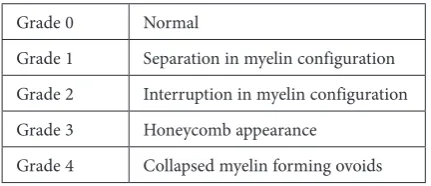
Table 1 Ultrastructural grading system of myelinated axons
Grade 0 Normal
Grade 1 Separation in myelin configuration Grade 2 Interruption in myelin configuration Grade 3 Honeycomb appearance
Results
In the sham group, no rats died in the course of the study. However, 1 rat in the etanercept and 1 in the saline group died during hypoxia.
Electrophysiological Data
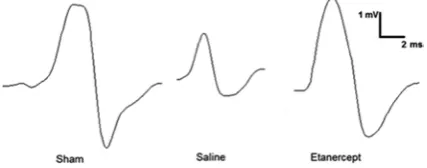
Typical records of CMAP in the sham group, and saline- and etanercept-treated HI groups are shown in Fig. 1. The means and standard devia-tions for amplitude, area, distal motor latency, de-polarization and rede-polarization times of CMAP, and total duration of CMAP in all groups are sum-marized in Table 2. As seen in Table 2, an increase in repolarization duration and motor latency of CMAP, and a decrease in depolarization duration, area and amplitude of CMAP were seen in rats af-ter HI (p < 0.05).
As seen in Table 2, there were no statistically significant differences between the sham and etan-ercept-treated groups regarding CMAP amplitude and area. Also, etanercept applied just after HI prevents the decrease in the amplitude (p < 0.05) but not area of CMAP in rats with HI, showing that CMAP amplitude was only protected by the
treatment with etanercept. In fact, as shown in Table 2, the mean area of the etanercept group rats was higher than the saline group. However, this re-sult was not statistically significant.
In this study, motor dysfunction, defined by a significant increase in DML, was also record-ed. In the saline group, the latency of CMAPs prolonged significantly from 3.39 ± 1.65 ms to 4.77 ± 1.20 ms, when compared to the sham group (p = 0.031). The etanercept treatment did not pre-vent these prolongations, and statistical differenc-es were not found, when compared to the saline group (Table 2).
As seen in Table 2, in the saline group, the du-ration of depolarization of CMAPs decreased sig-nificantly from 1.87 ± 0.72 ms to 1.25 ± 0.28 ms, when compared to the sham group (p = 0.006). The etanercept treatment did not prevent these de-creases, and statistical differences were not found compared to the sham group (Table 2).
Also, as seen in Table 2, in the saline group, the duration of repolarization of CMAPs prolonged significantly from 1.33 ± 0.58 ms to 1.94 ± 0.55 ms, when compared to the sham group (p = 0.034). The etanercept treatment prevented these prolon-gations, and no statistical differences were found when compared to the sham group. Also, as seen in Table 2, there were no statistically significant differences between all groups regarding CMAP total duration (p > 0.05).
The Electron Microscopic
Findings
Qualitative Analysis
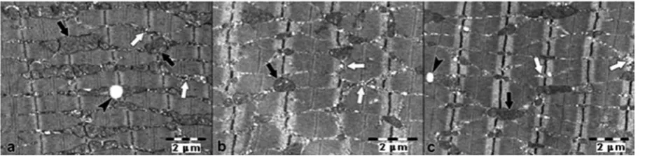
The axons, axonal myelin and Schwann cells of the sciatic nerve showed normal ultrastructural features in the sham group (Fig. 2). Degenerative changes were observed in the axonal myelin in the
Table 2 Descriptive statistics (mean ± SD) for compound motor action potential parameters studied
Experiments Variables
amplitude
(mV) area (mVms) distal motor latency (ms) total duration
(ms)
duration of depolariza-tion (ms)
duration of repolariza-tion (ms)
Sham (sham)
(n = 15) 5.46 ± 2.09 0.0051 ± 0.0025 3.39 ± 1.65 7.17 ± 2.07 1.87 ± 0.72 1.33 ± 0.58 Saline-treated
(saline) (n = 14) 2.26 ± 1.54
a 0.0028 ± 0.0025a 4.77 ± 1.20a 6.54 ± 2.25 1.25 ± 0.28a 1.94 ± 0.55a
Etanercept-treated
(etanercept) (n = 14) 5.51 ± 2.16
b 0.0037 ± 0.0021 5.46 ± 1.17a 6.12 ± 2.37 1.07 ± 0.22a 1.46 ± 0.84
a Significantly different from sham group: p < 0.05. b Significantly different from saline group: p < 0.05.
n – the number of rats in each group.
saline-treated group. There was also axonal shrink-age and endoneurial edema in the saline group (Fig. 3a–b). The grading 0–3 of damage showed that there was no significant difference when comparing the etanercept group to the saline group (Table 3 and Fig. 3c–d), but the amount of myelin damage increased in the etanercept group for grade 4.
As regards the gastrocnemius muscle, fibers have normal morphologic characteristics in all groups (Fig. 4a–c). The nuclei of the cells were reg-ularly outlined and exhibited normal chromatin content. Myofibrils exhibited a regular sarcomere organization. Cytoplasmic organelles and capillary structures between the myofibrils showed a nor-mal structure.
Quantitative Analysis
Table 3 indicates the results of the quantita-tive grading of damage found after 600 myelin-ated axons in 3 groups were examined. All of the
Fig. 2. Electron microscopic image of rat nerve fibers in the sham group. Nerve fibers are normal ultrastruc-turally. Myelin sheath (black arrow), axon (asterisk), Schwann cell (arrowhead), endoneurial connective tis-sue (white arrow) (×3000)
myelinated axons were found to be normal (grade 0: 100.0 ± 0.00%), as expected, when the biopsies tak-en from the sham group were examined. Because the standard deviation of GD0-GD4 scores in the sham group were 0.0, a statistical comparison with the other groups can not be made and p-value can not be given. Therefore, we can say that there was clinical importance in the saline and the etanercept groups according to the sham group. As seen in Table 3, in the saline group, myelin injury devel-oped and the myelin proportion showing normal morphology decreased, while the amount of my-elin damage that has higher grade (i.e., worse) in-creased. No significant differences were found be-tween the biopsies from the etanercept group and the saline group, but the amount of myelin damage showing higher grades increased.
Discussion
In this study, ligation of the right common ca-rotid artery and exposure to hypoxia by a contin-uous flow of 8% oxygen – 92% nitrogen for 2 h resulted in electrophysiological and histological abnormalities in the peripheral nerve of the neona-tal rats. This study demonstrates that HI decreased significantly in the amplitude and area of CMAP in the gastrocnemius muscle, suggesting a partial interruption of the signal through the peripheral nerve fibers, mainly reflecting axonal dysfunction and a significant increase in the DML of CMAP,
mainly reflecting myelin sheath damage (motor dysfunction). However, etanercept, which was ad-ministered shortly after hypoxia, showed a pre-ventive effect on the axonal dysfunction after HI. On the other hand, treatment with etanercept did not have a preventive effect on motor dysfunction after HI.
In addition to electrophysiological abnormal-ities in the peripheral nerve of the neonatal rats, degenerative changes in the electron microscopic evaluationswere observed in the axonal myelins in the saline group. Also, there was axonal shrinkage and endoneurial edema in this group. No signifi-cant differences were found between the biopsies from the etanercept group and the saline group in the grading 0–3 of damage, but the amount of my-elin damage that shows higher (i.e., worse) scores (grade 4) increased in the etanercept group (Ta-ble 3). Thus, it has been found that the electrophys-iological results and detailed analysis made by us-ing the quantitative scorus-ing of the axonal myelin of sciatic nerve tissue apparently supported each oth-er. On the other hand, in this study, no histological changes in the skeletal muscle cells were observed using electron microscopy in the saline group.
A reliable transmission in the central and pe-ripheral nervous systems has saltatory conduction to travel down the myelinated axon. This conduc-tion is defined as an acconduc-tion potential moving in discrete jumps down a myelinated axon. This un-interrupted process is outlined as the charge pas-sively spreading to the next node of Ranvier to
Table 3. Ultrastructural grading scores for the experimental groups
Experimental Groups Grade 0 Grade 1 Grade 2 Grade 3 Grade 4
Sham 100.00 ± 0.00 0.0 ± 0.00 0.0 ± 0.00 0.0 ± 0.00 0.0 ± 0.00 Saline 40.5 ± 20.42a 26.0 ± 19.39a 7.0 ± 4.16a 13.5 ± 8.54a 13.0 ± 18.87a
Etanercept 17.5 ± 9.15a 17.0 ± 19.90a 5.0 ± 2.58a 8.0 ± 3.65a 52.5 ± 18.36a,b
a – clinical importance according to the sham group. b – p < 0.05 compared to the saline group.
depolarize it to threshold which will then trigger an action potential in this region which will then passively spread to the next node and so on. How-ever, the need for a continuous energy supply to maintain appropriate ion concentrations to sup-port uninterrupted action potential generation places central fibers in a potentially vulnerable state [25]. Ion gradients cannot be re-established for proper action potential generation when ener-gy demand exceeds supply under injury conditions such as anoxia/ischemia or trauma. Ionic dysregu-lation within the injured axon occurs causing an influx in Na+ and Ca2+ and an efflux of K+ [26].
Thus, conduction of action potential fails. LoPach-in and LehnLoPach-ing [27] reported an LoPach-increase LoPach-in axo-plasmic Ca2+ in a wide range of experimental nerve
injury models which causes acute or chronic ax-on degeneratiax-on. In our study, possible pathways/ /mechanisms to the alterations seen after HI can be the levels of axoplasmic Ca2+ increase in response
to in vitro ischemia in myelinated axons. This situ-ation needs to be addressed in future research.
Morphological and biochemical changes asso-ciated with HI brain insult have been extensively studied in the experimental model of perinatal as-phyxia [20]. A few studies dealing with the long-term behavioral alterations after HI insult in neo-natal rats have demonstrated obvious sensorimotor deficits, learning and memory impairment [19, 28]. However, peripheral neuronal changes in adult-hood associated with neonatal HI insult have not been studied enough in the experimental mod-el of perinatal asphyxia. Therefore, in our study, the late effects of hypoxia-ischemia on adult rats (17-weeks) were investigated taking into account the action potential parameters and ultrastructure of the sciatic nerve and gastrocnemius muscle.
In this study, the severity of myelin damage to the sciatic nerve was evaluated. The grading 0–3 of damage showed that there was no significant dif-ference between the etanercept group and the sa-line group (Table 3). However, the amount of my-elin damage increased in the etanercept group for grade 4. It is known that the soluble and trans-membrane of TNF has two biological forms. It was reported that transmembrane TNF signaling was essential in preserving myelin integrity and com-paction and, most importantly, in promoting re-myelination [29]. Nevertheless, it was demonstrat-ed that the anti-inflammatory effect of etanercept is not sufficient to stimulate recovery, indicating that it is the protective effect of transmembrane TNF signaling to ultimately drive the positive outcome in chronic disease [29]. In our study, the inhibition by etanercept of transmembrane TNF signaling probably caused myelin dysfunction continuation in rats. In other words, non-selective inhibition of
both forms of TNF with etanercept did not result in myelin preservation as well as remyelination. Therefore, it has been found that the amount of myelin damage increased in the etanercept group compared to the saline group for grade 4.
Hypoxia is a central feature of ischemic, in-flamed, and infected tissues and a principal deter-minant of the pathophysiology of local and gen-eralized systemic inflammatory responses in these conditions [30]. Also, exposures to hypoxia are important components in the pathophysiology of local and systemic inflammatory responses [31]. Indeed, Lahat et al. [32] reported that hypoxia en-hances lysosomal TNF-degradation in mouse peri-toneal macrophages. In addition, Hempel et al. [33] showed that hypoxia increases the release of the cytokines IL-1 (interleukin-1) beta and TNF-α in human alveolar macrophage and that this increase may be due to decreased PGE2 (prostaglandin E2) synthesis during hypoxia.
In this study, it has been found that the expo-sure to hypoxia for 2 h that decreased the amplitude of CMAP by 57.79 ± 25.65% relative to the sham amplitude (p < 0.05), recovered to 57.35 ± 38.80% after treatment with etanercept. Also, in our previ-ous study [1], ligation of the right common carot-id artery and exposure to hypoxia by a continuous flow of 8% oxygen-92% nitrogen for 1 h resulted in electrophysiological abnormalities in the periph-eral nerve of the rats. However, in that study [1], the amplitude of CMAP recorded from the rats treated with saline after hypoxia for 1 h decreased by 21.12 ± 10.29% compared to the sham group (p < 0.05) and recovered to 24.17 ± 18.40% after treatment with a platelet-activating factor (PAF) antagonist (ABT-491). Thus, the more long-term exposure to hypoxia in rats, as expected, is found to increase nerve damage.
In contrast to the central nervous system, nerve fibers of the peripheral nervous system are able to regenerate and reinnervate distal targets [34]. Re-generative and repair processes of the peripheral nerve begin almost immediately after injury [35]. Kato et al. [11] reported that immediate therapy with the TNF-α antagonist etanercept, adminis-tered systemically (i.p.) and locally (epineurially) after peripheral nerve crush injury in adult rats, en-hances the rate of axonal regeneration. Our results confirm that HI has important neuropathic effects on peripheral nerves. This effect was observed espe-cially with axonal myelin and the endoneurium. By using electrophysiological testing, in this study, it has been found that immediate etanercept therapy enhances axonal regeneration after neonatal HI.
durations. Accordingly, the decrease in CMAP de-polarization duration in saline compared to the sham group suggested that the opening-closing ki-netics of Na+ channels accelerated. On the other
hand, treatment with etanercept accelerated this decrease in the depolarization duration more and so it did not have a preventive effect on the Na+
channel kinetic after HI.
In addition, an increase in repolarization du-ration in the saline group compared to the sham group suggested that the opening-closing kinetics of K+ channels slowed down. As we could not find
any difference between the sham and the etanercept
groups regarding repolarization duration, we sug-gest that etanercept treatment alters the opening and closing kinetics of voltage-gated potassium channels. Our results implicate the voltage-gated potassium channels as additional etanercept tar-gets, opening up new perspectives for the pharma-cological prevention of peripheral neuropathy.
The authors concluded that on the basis of the results, this study implies that neonatal HI insult causes axonal and myelin sheath damage to pe-ripheral nerves, but the TNF-α inhibitor etaner-cept has a preventive effect only on axonal dys-function but not motor dysdys-function after HI.
References
[1] Buyukakilli B, Atıcı A, Büyükdereli Z, Taşdelen B, Güneş S, Turhan AH: Protective effects of platelet-activating factor antagonist ABT-491 on the peripheral nerves in hypoxic ischemia-induced neonatal rat model. Turkiye Klinikleri J Med Sci 2011, 31, 1179–1185.
[2] Buyukakilli B, Atici A, Özkan A, Balli E, Güneş S, Turhan AH, Hallioglu O, Kanik A: The effect of tumor necrosis factor-α inhibitor soon after hypoxia-ischemia on heart in neonatal rats. Life Sci 2012, 90, 838–845.
[3] Hendriksen PH, Oey PL, van Veen BK, Wallinga-de Jonge W, Veldman H: Muscle conduction velocity and morphology after prolonged hypoxemia and diabetes in rats. Electromyogr Clin Neurophysiol 1992, 32, 491–497.
[4] Nukada H, van Rij AM, Packer SG, McMorran PD: Pathology of acute and chronic ischaemic neuropathy in atherosclerotic peripheral vascular disease. Brain 1996, 119, 1449–1460.
[5] Nukada H, McMorran PD: Perivascular demyelination and intramyelinic oedema in reperfusion nerve injury. J Anat 1994, 185, 259–266.
[6] Ress AM, Babovic S, Angel MF, Im MJ, Dellon AL, Manson PN: Free radical damage in acute nerve compression. Ann Plas Surg 1995, 34, 388–395.
[7] Schmelzer JD, Zochodne DW, Low PA: Ischemic and reperfusion injury of rat peripheral nerve. Proc Nat Acad Sci USA 1989, 86, 1639–1642.
[8] Clark SM, Basraon SK, Hankins GDV: Intrapartum Asphyxia, Neonatal Encephalopathy, Cerebral Palsy, and Obstetric Interventions in the Term and Near-Term Infant. NeoReviews 2013, 14, 13–21.
[9] Villapol S, Gelot A, Renolleau S, Charriaut-Marlangue C: Astrocyte Responses after Neonatal Ischemia: The Yin and the Yang. Neuroscientist 2008, 14, 339–344.
[10] Wagner R, Myers RR: Schwann cells produce tumor necrosis factor alpha:expression in injured and non-injured nerves. Neuroscience 1996, 73, 625–629.
[11] Kato K, Liu H, Kikuchi S, Myers RR, Shubayev VI: Immediate anti-tumor necrosis factor-a (etanercept) therapy enhances axonal regeneration after sciatic nerve crush. J Neurosci Res 2010, 88, 360–368.
[12] Li F, Fang L, Huang S, Yang Z, Nandi J, Thomas S, Chen C, Camporesi E: Hyperbaric oxygenation therapy alle-viates chronic constrictive injury-induced neuropathic pain and reduces tumor necrosis factor-alpha production. Anesth Analg 2011, 113, 626–633.
[13] Liou JT, Liu FC, Mao CC, Lai YS, Day YJ: Inflammation confers dual effects on nociceptive processing in chronic neuropathic pain model. Anesthesiology2011, 114, 660–672.
[14] Mulleman D, Mammou S, Griffoul I, Watier H, Goupille P: Pathophysiology of disk-related sciatica. I. Evidence supporting a chemical component. Joint Bone Spine 2006, 73, 151–158.
[15] Olmarker K, Rydevik B: Selective inhibition of tumor necrosis factor-alpha prevents nucleus pulposus-induced thrombus formation, intraneural edema, and reduction of nerve conduction velocity. Possible implications for future pharmacologic treatment strategies of sciatica. Spine (Phila Pa 1976) 2001, 26, 863–869.
[16] Burtrum D, Silverstein FS: Hypoxic–ischemic injury stimulates glial fibrillary acidic protein mRNA and protein expression in neonatal rats. Exp Neurol 1994, 126, 112–118.
[17] Towfighi J, Mauger D: Temporal evolution of neuronal changes in cerebral hypoxia-ischemia in developing rats: a quantitative light microscopic study. Dev Brain Res 1998, 109, 169–177.
[18] Vannucci RC, Towfighi J, Vannucci SJ: Hypoxic preconditioning and hypoxic-ischemic brain damage in the immature rat: pathologic and metabolic correlates. J Neurochem 1998, 71, 1215–1220.
[19] Balduini W, Angelis VD, Mazzoni E, Cimino M: Long-lasting behavioral alterations following a hypoxic/isch-emic brain injury in neonatal rats. Brain Res 2000, 859, 318–325.
[20] Spandou E, Papadopoulou Z, Soubasi V, Karkavelas G, Simeonidou C, Pazaiti A, Guiba-Tziampiri O:
Erythropoietin prevents long-term sensorimotor deficits and brain injury following neonatal hypoxia–ischemia in rats. Brain Res 2005, 1045, 22–30.
[22] Rice JE 3rd, Vannucci RC, Brierley JB: The influence of immaturity on hypoxic-ischemic brain damage in the rat. Ann Neurol 1981, 9, 131–141.
[23] Aminoff MJ: Electromyography in Clinical Practice. Churchill Livingstone, New York 1998.
[24] Kaptanoglu E, Palaoglu S, Surucu HS, Hayran M, Beskonakli E: Ultrastructural scoring of graded acute spinal cord injury in the rat. J Neurosurg 2002, 97, 49–56.
[25] Stirling DP, Stys PK: Mechanisms of axonal injury: internodal nanocomplexes and calcium deregulation. Trends Mol Med 2010, 16, 160–170.
[26] Nikolaeva MA, Mukherjee B, Stys PK: Na+-dependent sources of intra-axonal Ca2+ release in rat optic nerve dur-ing in vitro chemical ischemia. J Neurosci 2005, 25, 9960–9967.
[27] LoPachin RM, Lehning EJ: Mechanism of calcium entry during axon injury and degeneration. Toxicol App Pharmacol 1997, 143, 233–244.
[28] Ten VS, Bradley-Moore M, Gingrich JA, Stark RI, Pinsky DJ: Brain injury and neurofunctional deficit in neona-tal mice with hypoxic-ischemic encephalopathy. Behav Brain Res 2003, 145, 209–219.
[29] Brambilla R, Ashbaugh JJ, Magliozzi R, Dellarole A, Karmally S, Szymkowski DE, Bethea JR: Inhibition of soluble tumour necrosis factor is therapeutic in experimental autoimmune encephalomyelitis and promotes axon preservation and remyelination. Brain J Neurol 2011, 134, 736–2754.
[30] Murdoch C, Muthana M, Lewis CE: Hypoxia regulates macrophage functions in inflammation. J Immunol 2005, 175, 6257–6263.
[31] Nathan C: Immunology: Oxygen and the inflammatory cell. Nature 2003, 422, 675–676.
[32] Lahat N, Rahat MA, Kinarty A, Weiss-Cerem L, Pinchevski S, Bitterman H: Hypoxia enhances lysosomal TNF-α
degradation in mouse peritoneal macrophages. Am J Physiol Cell Physiol 2008, 295, 2–12.
[33] Hempel SL, Monick MM, Hunninghake GW: Effect of hypoxia on release of IL-1 and TNF by human alveolar macrophages. Am J Respir Cell Mol Biol 1996, 14, 170–176.
[34] Frisén J: Determinants of axonal regeneration. Histol Histopathol1997, 12, 857–868.
[35] Fawcett JW, Keynes RJ: Peripheral nerve regeneration. Annu Rev Neurosci 1990, 13, 43–60.
Address for correspondence:
Belgin Buyukakilli
Department of Biophysics, Medical Faculty Mersin University Campus Ciftlikköy 33343 Mersin
Turkey
Tel: 90 324 361 06 84
E-mail: [email protected]
Conflict of interest: None declared