Introduction
Main goal of root canal treatment is three-dimensional cleaning, shaping, and proper obturation with the adequate seal of the root canal system as complete debridement with smear layer removal is essential to achieve a successful outcome.[1] Root canal instrumentation is known to produce a smear layer that covers the surfaces of root canal walls. The superficial smear layer of 1-2 μm thickness, and the deeper smear plug having thickness up to 40 μm.[2-4] Smear layer contains both inorganic and organic components, and it has been recommended to remove it as it may be having a mixture of bacteria and their byproducts.[5,6] It also prevents the penetration of irrigants and intracanal medicaments into the dentinal tubules and interferes with the adherence and adaptation of the sealer to the walls.[7-9]
ABSTRACT
The desired outcome of endodontic treatment is to get rid of bacteria in the root canal and to provide a very good seal of root canal filling materials. It has been exhibited that smear layer was found covering the root canal walls after instrumentation of the root canal. The use of herbal alternatives as a root canal irrigant is advantageous as it eliminates the undesirable characteristics of chemical irrigants. Alternative irrigants are proven to be safe and contain active constituents that have beneficial physiologic effect and curative property such as antioxidant, anti-inflammatory, and radical scavenging activity. The aim of this study is to compare the efficacy of ethylenediaminetetraacetic acid, Triphala, and German chamomile on smear layer removal of prepared root canal walls by scanning electron microscopy.
Keywords: Smear layer removal, ethylenediaminetetraacetic acid, Triphala, German chamomile, scanning electron microscopy
Comparative evaluation of the efficacy of smear layer removal
by ethylenediaminetetraacetic acid,
Triphala,
and
German
chamomile
as irrigants - A scanning electron microscopy study
J. Sowjanyaa, Toby
Thomas, C. S. Chandana
Department of Conservative Dentistry and Endodontics, Saveetha Dental College, Saveetha University, Chennai, Tamil Nadu, India
Correspondence: C. S. Chandana, Department of Conservative Dentistry and Endodontics, Saveetha Dental College, Saveetha University, No. 162, Poonamallee High Road, Chennai - 600 077, Tamil Nadu, India. Phone: +91-9600040740. E-mail: [email protected]
Ethylenediaminetetraacetic acid (EDTA) is the frequently used chelator in endodontics.[10] Studies have concluded that the use of a combination of 2.5–5% sodium hypochlorite and 10–17% EDTA is effective in the removal of organic and inorganic debris.[11,12] EDTA is a calcium ion chelating agent and can remove the smear layer. Final flush of EDTA can open the dentinal tubules, and increases the number of lateral canals to be filled.[13]
Triphala is an Indian Ayurvedic herbal formulation consisting of dried and powdered fruits of three medicinal plants Terminalia chebula, Terminalia bellerica, and Emblica officinalis.[14]
The German chamomile has been used for centuries as a medicinal plant mostly for its analgesic, anti-inflammatory, antispasmodic, antimicrobial, and sedative properties. Chamomile has been proven to be effective when used as a mouthwash to treat irritations and minor infections of the mouth and gingivae and is also used in some toothpaste.[15]
The use of herbal alternatives as a root canal irrigant is advantageous as it eliminates the undesirable characteristics of chemical irrigants. Alternative irrigants are proven to be safe and contain active constituents that have beneficial physiologic effect and curative property such as antioxidant, anti-inflammatory, and radical scavenging activity.[16]
How to cite this article: Sowjanyaa J, Thomas T, Chandana CS. Comparative evaluation of the efficacy of smear layer removal by ethylenediaminetetraacetic acid, Triphala, and
German chamomile as irrigants - A scanning electron microscopy study. J Adv Pharm Edu Res 2017;7(3):261-271.
Source of Support: Nil, Conflict of Interest: None declared. Access this article online
Website: www.japer.in E-ISSN: 2249-3379
and G. chamomile on smear layer removal of prepared root canal walls by scanning electron microscopy.
Materials and Methods
In this experimental in vitro study, 30 single-rooted human teeth were selected. The teeth had been recently extracted from patients because of periodontal diseases and prosthetic reasons. The teeth were radiographed to ensure that the teeth had a single canal. The teeth were immersed in isotonic saline solution after cleaning. The samples were then autoclaved. The teeth were decoronated with diamond disc (D & Z, Darmstadt, Germany). The remaining roots lengths were almost 15mm.
The root canals were accessed and the coronal preparation was done initially with Gates-Glidden drills (Dentsply Maillefer, Ballaigues, Switzerland) up to number size 3. The teeth were instrumented with K-files and Protaper Universal rotary files with 2.5% NaOCl irrigation (Novo Dental Product, India) between each file, followed by irrigation with 5 ml of saline. The teeth were instrumented till size F5. They were randomly divided into three groups according to the final irrigation solutions.
Group 1: EDTA,
Group 2: 1:1 ratio of Triphala powder (Impcops, Chennai) and dimethyl sulfoxide,
Group 3: G. chamomile.
Then, samples were irrigated with 5 ml of each irrigant for 1 min. The herbal irrigants were freshly prepared and standardized. The irrigants were delivered with a side vented endodontic irrigating needle (RC Twents, Prime Dental Products, Mumbai, India) until the working length using manual technique into the root canal. Final irrigation was done with 5 ml of distilled water for each sample. The canals were dried with sterile paper points after instrumentation. Each dried specimen was split into two with chisel and mallet along the prepared groove on the buccal and palatal aspect. A half of each specimen was discarded; the other half was prepared for scanning electron microscopic (SEM) examination.[17,18]
The split halves were stored in a 2.5% glutaraldehyde solution. After fixation, the samples were dehydrated in ethanol series (70%, 90%, 95%, and twice at 100%), and then, critical point dried using the dry ice method (BAL-TEC AG, Balzers, Lichtenstein). Each specimen was mounted on an aluminum stub and sputter coated with approximately 20 nm layer of gold, to render a conductive surface. The specimens were examined using a SEM JEOL 6400 (JEOL, Tokyo, Japan) and Cambridge S360 (Cambridge, UK) SEM at a magnification of 2000. The specimens were blind coded. Analysis of the SEM images was accomplished by two investigators who scored the presence of smear layer on the surface of the root canal in the coronal, middle, and apical portion of each canal based on the criteria described by Hulsmann et al. (2002) outlined below:
Score 2: More than 50% of dentinal tubules opened; Score 3: <50% of dentinal tubules opened; and
Score 4: Nearly, all of the dentinal tubules covered with smear layer.
Statistical analysis
The normality tests Kolmogorov–Smirnov and Shapiro–Wilks tests result reveal that the variable does not follow normal distribution. Therefore, to analyze the data nonparametric methods are applied. To compare between groups, Kruskal–Wallis is applied followed by Bonferroni adjusted Mann–Whitney post hoc tests for multiple pairwise comparisons (Table 1). To compare between sides (Apical Coronal and Middle), Friedman repeated measures ANOVA is applied followed by Bonferroni adjusted post hoc tests for multiple pairwise comparisons (Table 2). SPSS version 22.0 is used to analyze the data. Significance level is fixed as 5% (α =0.05).
Results
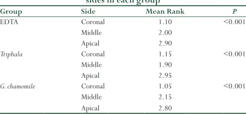
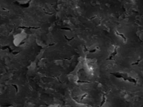
A moderate smear layer was seen in specimens treated with Triphala, especially in the middle and coronal sections [Figures 1 and 2]. In the apical section, there was heavy smear layer [Figure 3]. G. chamomile had substantial smear layer in all sections [Figures 4-6]. No smear layer was noted on the surface of the samples irrigated with EDTA [Figures 7-9].
Triphala was found to be significantly more effective at smear layer removal than G. chamomile (P < 0.005). Statistical analysis revealed that
Table 1: Nonparametric Kruskal–Wallis test to compare between groups in each side
Section Group n Mean rank P
Coronal EDTA 10 8.00 <0.001
Triphala 10 15.00
G. chamomile 10 23.50
Middle EDTA 10 8.30 <0.001
Triphala 10 13.60
G. chamomile 10 24.60
Apical EDTA 10 7.90 <0.001
Triphala 10 15.10
G. chamomile 10 23.50
EDTA: Ethylenediaminetetraacetic acid, G. chamomile: German chamomile
Table 2: Nonparametric Friedman test to compare between sides in each group
Group Side Mean Rank P
EDTA Coronal 1.10 <0.001
Middle 2.00
Apical 2.90
Triphala Coronal 1.15 <0.001
Middle 1.90
Apical 2.95
G. chamomile Coronal 1.05 <0.001
Middle 2.15
Apical 2.80
Figure 1: Group II: Triphala – coronal portion
Figure 2: Group II: Triphala – middle portion
Figure 3: Group II: Triphala – apical portion
Figure 4: Group III: German chamomile – apical portion
Figure 5: Group III: German chamomile – middle portion
Figure 6: Group III: German chamomile – apical portion
Triphala and G. chamomile were not effective in removing the smear layer in comparison with EDTA. There was a significant difference between the EDTA and the other groups at different levels of the root (P < 0.005).
Discussion
standardization and avoid anatomic variation which was confirmed using radiograph.[19]
The smear layer removal action of EDTA can be attributed to its chelation action on the root canal. The moderate smear removal was Figure 7: Group I: Ethylenediaminetetraacetic acid – coronal portion
Figure 8: Group I: Ethylenediaminetetraacetic acid – middle portion
Figure 9: Group I: Ethylenediaminetetraacetic acid – apical portion
of the root canal.[20]
Triphala demonstrated better smear layer removal in the coronal and middle third portion when compared with G. chamomile. Triphala is a traditional Ayurvedic herbal formulation consisting of the dried and powdered fruits of three medicinal plants Phyllanthus emblica, T. chebula, and T. bellerica in equal proportions. P. emblica contains a range of tannins and other phenolic compounds. It also contains ascorbic acid and flavonoids.[14] The smear layer removal efficacy may be attributed to these acid components.
In contrast, it was stated that G. chamomile extracts demonstrated better cleaning in the coronal and middle third portion.[21] The chemical analysis of G. chamomile has revealed its compounds to chamazolene, alpha-bisabolol, and acids such as capric acid, caprylic acid, chlorogenic acid, o-coumaric acid, p-coumaric-acid, dihydroxybenzoic acid, and other components.[22] The cleaning effect of chamomile might be related to these acid components.[21]
The contrast in results can be attributed to the suspension of G. chamomile used in this study. Oil form of G. chamomile would have made it difficult for the smear layer removal.
Conclusion
Within the limitations of this study, it could be concluded that Triphala showed good cleaning efficacy than G. chamomile used in this study. The most effective removal of smear layer occurred with the use 17% EDTA as a final rinse followed by the use of Triphala. G. chamomile did not produce satisfactory results. In future, further investigations are recommended to evaluate the potential use of Triphala as a root canal irrigant.
References
1. Mello I, Kammerer BA, Yoshimoto D, Macedo MC, Antoniazzi JH. Influence of final rinse technique on ability of ethylenediaminetetraacetic acid of removing smear layer. J Endod 2010;36:512-4.
2. Czonstkowsky M, Wilson EG, Holstein FA. The smear layer in endodontic. Dent Clin North Am 1990;34:13-25.
3. Sen BH, Wesselink PR, Türkün M. The smear layer: A phenomenon in root canal therapy. Int Endod J 1995;28:141-8.
4. Torabinejad M, Handysides R, Khademi AA, Bakland LK. Clinical implications of the smear layer in endodontics: A review. Oral Surg Oral Med Oral Pathol Oral Radiol Endod 2002;94:658-66.
5. Yamada RS, Armas A, Goldman M, Lin PS. A scanning electron microscopic comparison of a high volume final flush with several irrigating solutions: Part 3. J Endod 1983;9:137-42.
6. Brännström M, Nyborg H. Cavity treatment with a microbicidal fluoride solution: Growth of bacteria and effect on the pulp. J Prosthet Dent 1973;30:303-10.
7. Economides N, Liolios E, Kolokuris I, Beltes P. Long-term evaluation of the influence of smear layer removal on the sealing ability of different sealers. J Endod 1999;25:123-5.
8. Kennedy WA, Walker WA 3rd, Gough RW. Smear layer removal effects on apical
9. Saunders WP, Saunders EM. The effect of smear layer upon the coronal leakage of gutta-percha fillings and a glass ionomer sealer. Int Endod J 1992;25:245-9. 10. Zehnder M, Schicht O, Sener B, Schmidlin P. Reducing surface tension in endodontic chelator solutions has no effect on their ability to remove calcium from instrumented root canals. J Endod 2005;31:590-2.
11. Paul ML, Mazumdar D, Niyogi A, Baranwal AK. Comparative evaluation of the efficacy of different irrigants including MTAD under SEM. J Conserv Dent 2013;16:336-41.
12. Lottanti S, Gautschi H, Sener B, Zehnder M. Effects of ethylenediaminetetraacetic, etidronic and peracetic acid irrigation on human root dentine and the smear layer. Int Endod J 2009;42:335-43.
13. Nicola MG, Plotino G, Falanga A, Pomponi M, Somma F. Interaction between EDTA and Sodium hypochlorite: A nuclear magnetic resonance analysis. J Endod 2006;32:460-4.
14. Vani T, Rajani M, Sarkar S, Shishoo CJ. Antioxidant properties of the ayurvedic formulation Triphala and its constituents. Int J Pharmacogn 1997;35:313-17.
15. Kamat S, Rajeev K, Prahlad S. Role of herbs in endodontics: An update. Endodontology 2011;23:96-100.
16. Bhargava KY, Aggarwal S, Kumar T, Bhargava S. Comparative evaluation
of the efficacy of three anti-oxidants vs NaOCl and EDTA: Used for root canal irrigation in smear layer removal-SEM study. Int J Pharm Pharm Sci 2015;7:366-271.
17. Aktener BO, Bilkay U. Smear layer removal with different concentrations of EDTA-ethylenediamine mixtures. J Endod 1993;19:228-31.
18. Cengiz T, Aktener BO, Piskin B. Effect of dentinal tubule orientation on the removal of smear layer by root canal irrigants. A scanning electron microscopic study. Int Endod J 1990;23:163-71.
19. Wu MK, Kast’áková A, Wesselink PR. Quality of cold and warm gutta-percha fillings in oval canals in mandibular premolars. Int Endod J 2001;34:485-91. 20. Mahajan VA, Kamra AI, Dahiwale SS. The effect of 17% EDTA and MTAD on
smear layer removal and on erosion of root canal dentin when used as final rinse: An in vitro SEM study. J Int Clin Dent Res Organ 2010;2:113-8. 21. Sadr Lahijani MS, Raoof Kateb HR, Heady R, Yazdani D. The effect of German
chamomile (Marticaria recutita L.) extract and tea tree (Melaleuca alternifolia L.) oil used as irrigants on removal of smear layer: A scanning electron microscopy study. Int Endod J 2006;39:190-5.