This is an open access journal, and articles are distributed under the terms of the Creative Commons Attribution-Non Commercial-ShareAlike 4.0 License, which allows others to remix, tweak, and build upon the work non-commercially, as long as appropriate credit is given and the new creations are licensed under the identical terms.
© 2017 Journal of Advanced Pharmacy Education & Research | Published by SPER Publication
473
Assessment of Prevalence of MRI Findings in Epileptic Patients
of Imam Khomeini Hospital, Urmia, Iran during 2010-2011
Arash Mosarrezaii
1, Abbas Hedayati Asl
2, Ali Asghar Ghafouri
21Department of Neurology, Imam Khomeini Hospital, Urmia University of Medical Sciences, Urmia, Iran, 2Urmia University of Medical Sciences, Urmia, Iran.
Correspondence: Arash Mosarrezaii, Department of Neurology, Imam Khomeini Hospital, Urmia University of Medical Sciences, Urmia, Iran, email:[email protected]
ABSTRACT
Background and aim: Seizure is a common neurological disorder characterized by transient, chronic, unprovoked or spontaneous recurrent disorders due to sudden rush of electrical activity in the brain causing unusual feelings or sensations , loss of consciousness, seizure or a mixture of symptoms. Epilepsy is characterized by recurrent seizures and is a common cause of a periodic loss of consciousness. The present study was aimed to determine the prevalence of MRI-detected brain abnormalities in patients with epilepsy. Materials and methods: This retrospective study was performed to investigate the MRI findings of patients with epilepsy (n
=98). Data on MRI findings and some parameters of the patient’s medical history were collected and then were entered into SPSS software version 21. Findings: According to the results, images of nearly half of the patients had an abnormal MRI finding. History parameter can be useful in predicting the normality or non-normality of MRI findings. Conclusion: The results indicated that nearly half of the subjects had abnormal MRI findings. Therefore, it is recommended to pay close attention to MRI findings in patients with epilepsy.
Keywords: Seizure, epilepsy, MRI finding, pathology
Introduction
Epilepsy is one of the most common neurological disorders causing a lot of damages to community members [1]. Approximately 50 million people worldwide have epilepsy; with new cases of epilepsy occuring most often in infants and the elderly, the vulnerable population groups [2, 3]. About 3 percent of people will experience epilepsy at some point in their lives [4]. In addition to physical and social problems, accidents, sudden unexpected deaths, status epilepticus and suicides constitute a vast majority of cause of death in epileptic patients [5]. People with epilepsy have a risk of premature death due to uncontrolled seizures or underlying neurological disorders. Over 1000 people with epilepsy die each year in the
UK, with this figure rising, despite 42% of epilepsy-related deaths being potentially avoidable [3, 4, 6]. The age of seizure onset commonly ranges between 26 and 28 years old and the ages of 14 and 16 years are the most sensitive age for the disease. Seizures typically disrupt the circadian rhythms through interrupting and impairing natural wake-sleep cycle and pattern, and may trigger or exacerbate the disease through various stimuli such as insomnia, fatigue, alcohol, and stress [7]. Seizures and epilepsy may be triggered by different underlying causes and are considered the most common causes of brain lesions including trauma, tumor, vascular events (hemorrhage, stroke, etc.) in the adults and older people [3]. Seizures and epilepsy in children can be associated with high fever, viral infection, asphyxiation, bacterial toxins caused by Shigella and Campylobacter infections, hyper chemical changes, hyponatremia and hypocalcemia. It is estimated that 15 to 30 percent of these patients have a drug-resistant disease which can lead to social, medical and financial consequences for both individuals and society [8]. Epilepsy is a chronic disease; although there’s no cure for epilepsy, the disorder can be
managed with medications. However, surgery may be a viable treatment option in some epileptic patients if their seizures are not adequately controlled by medication. The percentage of patients considered for epilepsy surgery depends on their brain MRI findings prior to surgery [9-20]. There are a number of Access this article online
Website: www.japer.in E-ISSN: 2249-3379
How to cite this article:Arash Mosarrezaii, Abbas Hedayati Asl, Ali Asghar Ghafouri. Assessment of Prevalence of MRI Findings in Epileptic Patients of Imam Khomeini Hospital, Urmia, Iran during 2010-2011. J Adv Pharm Edu Res 2017;7(4):473-478.
Arash Mosarrezaii, et al.: Assessment of Prevalence of MRI Findings in Epileptic Patients
474 Journal of Advanced Pharmacy Education & Research | Oct-Dec 2017 | Vol 7 | Issue4
studies conducted to detect abnormal brain findings using a variety of paraclinical methods, especially new techniques of brain MRI. Even in epileptic patients who have shown normal MRI scans, there are some brain abnormalities which can be identified with most sophisticated and modern techniques [11,12]. A high percentage of patients have shown microscopic anomalies and quantitative MRI can be utilized as an alternative to tissue studies to detect these brain anomalies [11]. MRI is superior to the other techniques for detection of specific brain abnormalities associated with epilepsy [13]. Therefore, abnormal brain MRI findings in patients with epilepsy are of importance in detection and management of epilepsy disorder. This study was aimed to evaluate the prevalence of abnormal MRI findings in the brain and its relationship with medical history parameters of epileptic patients in Imam Khomeini Hospital in Urmia to estimate the status of patients with epilepsy in West Azarbaijan province, Iran.
Materials and Methods
This retrospective study was performed on patients with epilepsy (n = 98). The MR imaging findings were interpreted by a radiologist to evaluate the presence or absence of abnormal findings such as tumor, gliosis, vascular malformation, neuronal migration disorders, cysts, hippocampal sclerosis (HS), cortical dysplasia, infarction and cortical problems. Then data on MRI findings and some
parameters of the patient’s medical history were collected.
These data included age, sex, history of brain trauma, neurological heart disease, history of MRI or CT, and febrile convulsion. Data were analyzed by SPSS statistical software version 21. Descriptive analyses were performed using descriptive statistics (frequency, percentage) and the relationship between normality and non- normality of MRI findings with medical history parameters was evaluated using Chi-square, ANOVA and T-test .
Results
This study was performed on 98 patients with epilepsy referred to Imam Khomeini Teaching Hospital in Urmia from March 2010 to March 2011 and underwent MRI brain. The patients aged 6 -85 years with a median age of 40 years and 45% (n= 44) were female and 55% (n= 54) male. Of these 98 patients, 55% (n= 54) had normal MRI scan and 45% (n= 44) abnormal MRI scan (Table 1). The relationship between normality and non-normality of MRI finding with some history parameters including age, sex, history of trauma, systemic disease, use of anticonvulsant drug, aura, loss of consciousness and type of seizure were statistically analyzed so that 35 (83.3 %) had normal MRI and 7 (16.7%) had abnormal MRI in the age group less than 30 years. In addition, 5 (38.5%) patients had normal MRI and 8 (61.5%) had abnormality in the age group of 30 years to 40 years. In the age group of 40-50 years, 6 (46.2%) had normal MRI findings and 7 (53.8%) had an abnormal MRI. Among patients older than 50 years of age, MRI was normal in 7 (23.3%) and 23 (76.7%) showed abnormality, considering that there was a significant correlation between age and MRI findings (P <0.001) (Table 1). The association between normality and non-normality of MRI finding regarding the patient's gender showed that among 53 male patients, 27 (50.9%) had normal MRI results and 26 (49.1%) abnormal MRI outcomes. Of 44 female patients, 26 (59.1%) had normal
MRI outcome and 18 (40.9%) abnormal MRI results. The results showed that there is no significant relationship between gender and normal and abnormal MRI results (P = 0.275) (Table 1). In terms of trauma history, out of 17 patients without a history of trauma, MRI results were normal in 15 (88.2%) and abnormal in 2 (11.8%), and out of 18 patients with traumatic history, 6 (33.3%) had normal MRI results and MRI results were abnormal in 12 (66.7%) patients. The results showed a significant relationship between traumatic history and MRI results (P = 0.001) (Table 1). Patients were evaluated for systemic disease so that among 52 patients without systemic disease, 38 (73.1%) had normal MRI and 14 (26.9%) abnormal MRI findings. Moreover, out of 29 patients with systemic disease, 10 (34.5%) had normal MRI results and MRI findings were abnormal in 19 (65.5%) patients. The results showed that there is a significant relationship between the history of systemic disease and MRI findings (P = 0.001) (Table 1). Regarding the patients with anticonvulsant drug history, it was shown that out of 51 patients with epilepsy without history of anticonvulsant therapy, 29 (56.9%) had normal MRI results and 22 (43.1%) had abnormal MRI findings. Out of 23 patients with history of anticonvulsant drug use, normal MRI was seen in 23 (51.1%) and abnormal results in 22 (48.9%) patients. According to the results, there was a significant relationship between the history of anticonvulsant therapy and MRI results (P = 0.06) (Table 1). In terms of aura before seizure, the results showed that out of 79 epileptic patients experienced aura before seizure, 41 patients (51.9%) had normal MRI results and 38 (48.2%) had abnormal MRI results. Of 18 patients had a prior seizure, normal MRI outcomes were observed in 12 (66.7%) and abnormal MRI in 6 (33.3%) patients. It was found that there was no significant correlation between a seizure that occurs prior to an aura and MRI findings (P = 0.192) (Table 1). With regard to loss of consciousness during seizure, the results revealed that out of 36 patients without loss of consciousness during seizure, MRI results were normal in 25 (69.4%) and abnormal in 11 patients (30.6%). Out of 61 patients with loss of consciousness during seizure, MRI scans of 28 patients (45.9%) were normal and abnormal in 33 (54.1%). since P = 0.02, there was a significant relationship between loss of consciousness during seizure and MRI findings (Table 1). Regarding the type of seizure, our findings showed that MRI results were normal among 5 patients with tonic seizure, and in 11 patients with clonic seizure. 6 patients (54.5%) had normal MRI results and MRI results were abnormal in 5 (45.5%) of patients. Of 53 patients with tonic-clonic seizure, normal MRI findings were seen in 27 (50.9%) and abnormal MRI in 26 (49.1%) patients. In addition, of the 17 patients with autonomic type seizure, 10 cases (58.8%) had normal seizure and 7 (41.2%) had abnormal MRI results. Out of 10 patients with other seizures, 4 cases (40%) had normal type of seizure and six (60%) of patients had abnormal MRI results. There was no significant relationship between type of seizure and MRI findings (P = 0.175) (Table 1).
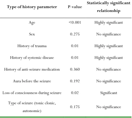
Table 1: The relationship between normality and non- normality of MRI results with history parameters (age, sex, history of trauma, history of systemic disease, etc.)
Variable MRI results Total P value Normal Abnormal
Journal of Advanced Pharmacy Education & Research | Oct-Dec 2017 | Vol 7 | Issue4 475
30-40 5(45.5%) 6(54.5%) 11
40-50 6(40%) 9(60%) 15
<50 7(24.1%) 22(75.9%) 29
Sex Male 27(50.9%) 26(49.1%) 53 0.275 Female 26(59.1%) 18(40.9%) 44
History of trauma No 15(88.2%) 2(11.8%) 17 0.001 Yes 6(33.3%) 12(66.7%) 18
History of systemic disease
No 38(33.1%) 14(26.9%) 52 0.001 Yes 10(34.5%) 19(65.5%) 29
Use of anti-seizure medication
No 29(56.9%) 22(43.1%) 51 0.360 Yes 23(51.1%) 22(48.9%) 45
Aura No 41(51.9%) 38(48.1%) 79 0.192 Yes 12(66.7%) 6(33.3%) 18
Loss of consciousness during
seizure
No 25(69.4%) 11(30.6%) 36 0.02 Yes 28(45.9%) 33(54.1%) 61
Type of seizure
Tonic 5(100%) 0(0%) 5
0.175 Clonic 6(54.4%) 5(45.5%) 11
Tonic-clonic 27(50.9%) 26(49.1%) 53
Autonomic 10(58.8%) 7(41.2%) 17
Other 4(40%) 6(60%) 10
Table 2: P-value in relation to history parameters (age, sex, history of trauma, history of systemic disease, etc.)
Type of history parameter P-value Statistically significant
relationship
Age <0.001 Highly significant
Sex 0.275 No significance
History of trauma 0.01 Highly significant
History of systemic disease 0.01 Highly significant
History of anti-seizure medication 0.360 No significance
Aura before the seizure 0.192 No significance
Loss of consciousness during seizure 0.02 Significant
Type of seizure (tonic clonic,
autonomic) 0.175 No significance
There was a significant relationship between normality or non-normality of MRI findings with parameters of age, history of trauma, history of systemic disease, and loss of consciousness during seizure (Table 2). Accordingly, MRI findings at an early age (average age 31 years) tend to have more normality than
older age (average age 52 years). Regarding the history of trauma, patients with a history of traumatic brain injury demonstrated more abnormal MRI findings than others. In addition, patients with a history of systemic disease revealed more MRI abnormalities than others. With respect to loss of consciousness, our results indicated that patients with a loss of consciousness had more MRI abnormalities than others. According to the age group classification, the MRI results showed normality in 35 (66%) in the age group less than 30 years, followed by 5 (9.4%) in the 30-40 year age group, 6 (11.3%) in the 50 -40 year age group and 7 (13.2%) in the 50 years age group. The tumor was found in 4 (57.1%) in the 30-40 year age group, followed by 2 (28.6%) in the 30-40-50 year age group and 1 (14.3%) in the age group more than 50 years. Gliosis was found in 1 (14.3%) age group less than 30 years old, followed by 1 (14.3%) in the 40-50 year age group and 5 (71.4%) in the age group more than 50 years. Infarction was shown in 1 (5.9%) in the 30-40 year age group followed by 2 (11.8%) in the 40-50 year age group and 14 (82.4%) in the age group more than 50 years. Other MRI findings were found in 5 (38.5%) in the age group less than 30 years old, followed by 3 (23.1%) in the 30-40 year age group, 2 (15.4%) in the 40-50 year age group and 3 (23.1%) in the age group above 50 years old (Table 3).
Table 3: Relationship of MRI findings with age distribution
MRI findings
Age group
Total Less than 30
years old
30-40 years old
40-50 years old
more than 50 years
old
Normal 35(66%) 5(9.4%) 6(11.3%) 7(13.2%) 53
Tumor 0(0%) 4(57.1%) 2(28.6%) 1(14.3%) 7
Gliosis 1(14.31%) 0(0%) 1(14.3%) 5(71.4%) 7
Infarction 0(0%) 1(5.9%) 2(11.8%) 14(82.4%) 17
Other 5(38.5%) 3(23.1%) 2(15.4%) 3(23.1%) 13
Total 41(42.3%) 13(13.41%) 13(13.4%) 30(30.9%) 97
In addition, the relationship between type-MRI finding with age and sex parameters was statistically analyzed. There was a highly significant correlation between age and MRI findings (by type) (p-value <0.001) (Table 3). No significant correlation was found between sex with MRI findings (by type of finding) (p = 0.275) (Table 4). MRI findings were normal in 27 (50.9%) male, and 26 (49.1%) female. The tumor was found in 6 (85.7%) males and 1 in (14.3%) female. Gliosis was found in 5 (7.4%) male and 2 (28.6%) female and infarction in 7 (41.2%) male and 10 (58.8%) female. Other MRI findings were observed in 8 (61.5%) male and 5 (38.5%) female (Table 4).
Table 4: MRI findings and their relationship with sex
Arash Mosarrezaii, et al.: Assessment of Prevalence of MRI Findings in Epileptic Patients
476 Journal of Advanced Pharmacy Education & Research | Oct-Dec 2017 | Vol 7 | Issue4
Male Female
Normal 27(50.9%) 26(49.1%) 53
Tumor 6(85.7%) 1(14.3%) 7
Gliosis 5(7.4%) 2(28.6%) 7
Infarction 7(41.2%) 10(58.8%) 17
Other 8(61.5%) 5(38.5%) 13
Total 53(54.6%) 44(45.4%) 97
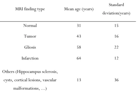
Table 5: The mean and standard deviation of age in the population studied based on the type of MRI found
MRI finding type Mean age (years) Standard deviation(years)
Normal 31 15
Tumor 43 16
Gliosis 58 22
Infarction 64 12
Others (Hippocampus sclerosis, cysts, cortical lesions, vascular
malformations, ...)
13 36
Discussion
The aim of this study was to investigate abnormal MRI and its relationship with the medical history parameters in patients suffering from epilepsy. The wide age range of epilepsy patients from 6 to 85 years old (40 years on average) in this papers reminds us that epilepsy and seizures happens at any age. It was previously said that only two age groups namely children and elder people are suffering from epilepsy [13, 14]. Since children suffering from seizure and epilepsy referred to hospital more than the patients being investigated in this study, it should be noted that lower age, even lower than the range reported in this study (under 6), suffer more from seizure and epilepsy; if these children were included in this study, average age group would be lower. Approximately, half of the patients have abnormal MRI; this is very considerable. Since all these patients have undergone MRI, their doctors probably doubted an underlying pathology and prescribed MRI. Besides, the MRI method in this research is the common method for epileptic patients. However, other studies have used precise and combined imaging methods to detect brain lesions [12, 15]. It was shown that many of epilepsy patients, reported normal by the usual brain imaging methods, were diagnosed with tissue pathology and structural abnormalities using more advanced methods [11, 16]. Thus, the prevalence of abnormal findings by
MRI is more than 45 % found by the present study. Table 4 shows that infarction, tumor and gliosis are the most common ones; other diseases like hippocampus sclerosis, cystic vascular malformation, cortical lesions were less common. [17] reported that among epilepsy patients undergoing surgical operation, hippocampus sclerosis was the most common cause of epilepsy; hypoxemia and postpartum hemorrhage, tumors and vascular malformations ranked next. Infections, migraine-induced anomalies, sclerosis tuberculosis, cortex dysplasia, cysts, and infarcts are recognized as more uncommon causes. Thus, our findings were consistent with these findings and inconsistent with other ones. In contrast to the reports by [17], it was found in this study that infarction was more common because less children and older people were investigated; thus, it is more likely that they have gone through cerebral vascular events before. It was found by the present study that happocamp sclerosis and vascular malformation were less common in contrast to the reports by the afore-mentioned study; because, detecting these symptoms requires special methods that were not utilized by this study [15, 16] and they are less likely to be detected by MRI. They were detected by tissue pathology in that study, which is a very specialized and precise method. Moreover, cases of problems during childbirth and infections were not investigated by the present study. Other studies consistent with the present study and [62] include tumor being common, cysts cortical, and tuberculosis sclerosis being uncommon; however, it should be highlighted that distribution of abnormalities, which are obtained through MRI studies, are different from classifications obtained by surgery [18, 19]. The results of the present study showed that MRI normality or abnormality has a significant relationship with parameters of age, previous trauma history, systemic disease history, and loss of consciousness during seizure. Thus, medical history is instrumental to predict and estimate the normality or abnormality of MRI. According to the results of this study, it can be said that MRI is abnormal at older ages than younger ones. Reviewing neuroscience reference literature shows that idiopathic seizures are more common at younger age. If seizures and epilepsy are started at older age, the underlying causes should be evaluated through tests, imaging methods etc. [20]. The relationship between MRI cases and previous trauma
Journal of Advanced Pharmacy Education & Research | Oct-Dec 2017 | Vol 7 | Issue4 477
Conclusion
According to the findings of the study, nearly half of the patients with epilepsy (patients admitted to the adult hospital) showed abnormal MRI findings using the conventional method. Infarction, tumor and gliosis have been found to be the most common abnormal findings in these patients. History has played an effective and useful role in predicting the normality or non-normality of MRI findings, and thus, older patients with a history of trauma, systemic disease, and loss of consciousness during seizure (complex seizures), abnormalities of MRI findings using conventional can be a stronger predictor for disease diagnosis. Infarction has been found to be the most common MRI finding among the patients, followed by gliosis, tumor, and other findings. No relationship was found between sex (male or female) of the patient and MRI findings.
Suggestions
Given the prevalence of MRI findings in patients with epilepsy in this study, it is recommended to pay close attention to the use of MRI findings in the correct diagnosis of epileptic seizures, especially in patients in old age, history of trauma, history of systemic disease and loss of consciousness during seizure (complex seizures). In addition, the implementation of more accurate studies of brain lesions in patients with epilepsy can be useful for the diagnosis and management of this dangerous disease. For example, the implementation of prospective studies among new cases with epilepsy referred to firstline walk-in clinics and medical centers using advanced and modern imaging techniques along with histological pathology of surgical and autopsy specimens, more accurate assessment of
patient’s history, investigation on epilepsy among children, etc.
can be the other research avenues to explore other important issues in the treatment of the disease.
References
1. Wen X, Han XR, Wang YJ, Wang S, Shen M, Zhang ZF, Fan SH, Shan Q, Wang L, Li MQ, Hu B, Sun CH, Wu DM, Lu J, Zheng YL. MicroRNA-421 suppresses the apoptosis and autophagy of hippocampal neurons in epilepsy mice model by inhibition of the TLR/MYD88 pathway. J Cell Physiol. 2018 Jan 30. doi: 10.1002/jcp.26498.
2. Sander JW. The epidemiology of epilepsy revisited. Curr Opin Neurol. 2003 Apr;16(2):165-70.
3. Wikipedia, The free encyclopedia. Epilepsy. 10
December 2011. http://en. Wikipedia.
Org/wiki/Epilepsy.
4. Hirts D, Thurman DJ, Gwinn- Hardy K, Mohamed M, Chaudhuri AR, Zalutsky R. How Common are the " common" neurologic disorders? Neurology. 2007 Jan 30;68(5):326-37.
5. Goodwin SW, Ferro MA, Speechley KN. Development and assessment of the Quality of Life in Childhood Epilepsy Questionnaire (QOLCE-16). Epilepsia. 2018 Jan 28. doi: 10.1111/epi.14008
6. Walczak TS, Leepik IE, D amelio M, Rarikh J, So E, Ahman P et al. Incidence and risk Factors in sudden unexpected death in epilepsy: a prospective cohort study. Neurology. 2001 Feb 27;56(4):519-25.
7. Kim JH. Grey and White Matter Alterations in Juvenile Myoclonic Epilepsy: A Comprehensive Review. J Epilepsy Res. 2017 Dec 31;7(2):77-88. doi: 10.14581/jer.17013. 8. Bell GS, Sanders JW. The epidemiology of epilepsy: The
size of the problem. Seizure. 2001 Jun;10(4):306-14; quiz 315-6.
9. Xue H, Sveinsson O, Bartek J, Förander P, Skyrman S, Kihlström L, Shafiei R, Mathiesen T, Tomson T. Long-term control and predictors of seizures in intracranial meningioma surgery: a population-based study. Acta Neurochir (Wien). 2018 Jan 11. doi: 10.1007/s00701-017-3434-3.
10. RamachandranNair R, Otsubo H, Shroff MM, Ochi A, Weiss SK, Rutka JT, Snead OC3rd. MEG predicts outcome following surgery for intractable epilepsy in children with Normal or nonfocal MRI findings. Epilepsia. 2007; 48(1): 149-57.
11. Woermann FG, Free SL, Koepp MJ, Sisodiya SM, Duncan JS. Abnormal cerebral Structure in juvenile Myoclonic epilepsy demonstrated with voxel-based analysis of MRI. Brain. 1999; 122(pt11): 2101-8.
12. Perry MS, Donahue DJ, Malik S, Keator CG, Hernandez A, Reddy RK, Perkins FF Jr, Lee MR, Clarke DF. Magnetic resonance imaging-guided laser interstitial thermal therapy as treatment for intractable insular epilepsy in children. J Neurosurg Pediatr. 2017 Dec;20(6):575-582. doi: 10.3171/2017.6. PEDS17158. 13. Brodtkorb E, Nilsen G, Smevik O, Rinck PA. Epilepsy
and anomalies of neuronal Migration: MRI and clinical aspects. Acta neurol Scand. 1992; 86(1): 24-32.
14. Sander JW. The epidemiology of epilepsy revisited. Curr Opin Neurol. 2003 Apr;16(2):165-70.
15. Bernasconi N, Bernasconi A, Caramanos Z, Antel SB, Andermann F, Arnold DL. Mesial Temporal damage in temporal lobe epilepsy: a volumetric MRI study of the hippocampus, Amygdala and parahippocampal region. Brain. 2003; 126(pt2)462-9.
16. Adler S, Hong SJ, Liu M, Baldeweg T, Cross JH, Bernasconi A, Bernhardt BC, Bernasconi N. Topographic principles of cortical fluid-attenuated inversion recovery signal in temporal lobe epilepsy. Epilepsia. 2018 Jan 31. doi: 10.1111/epi.14017.
17. Heinz ER, Crain BJ, Radtke RA, Burger PC, Friedman AH, Djang WT, Wilkinson WE. MR Imaging in patients with temporal lobe seizures: correlation of results with pathologic Findings. AJR Am J Roentgenol. 1990; 155(3): 581-6.
Arash Mosarrezaii, et al.: Assessment of Prevalence of MRI Findings in Epileptic Patients
478 Journal of Advanced Pharmacy Education & Research | Oct-Dec 2017 | Vol 7 | Issue4
temporal lobe epilepsy. Epilepsy Behav. 2017 Dec 26; 79:138-145. doi: 10.1016/j.yebeh.2017.11.040. 19. King MA, Vewton MR, Jackson GD, Fitt GJ, Mitchell
LA, Silvapulle MJ, Berkovic SF. Epileptology of the first-seizure presentation: a clinical, electroencephalographic, and Magnetic resonance imaging study of 300 consecutive patients. Lancet. 1998; 352(9133): 1007-11.