RESEARCH
Detection and molecular characterization
of urinary tract HIV-1 populations
M. L. Mzingwane
1,2*, G. Hunt
3,4, R. Lassauniere
3,5, M. Kalimashe
3, A. Bongwe
3, J. Ledwaba
3, R. E. Chaisson
6,
N. Martinson
7, K. Richter
2,8, S. M. Bowyer
2and C. T. Tiemessen
3,4Abstract
Background: Identification of all possible HIV reservoirs is an important aspect in HIV eradication efforts. The urinary tract has however not been well studied as a potential HIV reservoir. In this pilot study we molecularly characterized HIV-1 viruses in urine and plasma samples to investigate HIV-1 replication, compartmentalization and persistence in the urinary tract.
Methods: Prospectively collected urine and blood samples collected over 12–36 months from 20 HIV-1 infected individuals were analysed including sampling points from prior to and after ART initiation. HIV-1 pol gene RNA and DNA from urine supernatant and urine pellets respectively were analysed and compared to plasma RNA viruses from the same individual.
Results: HIV-1 nucleic acid was detected in urine samples from at least one time point in 8/20 (40%) treatment-naïve subjects compared to 1/13 (7.7%) individuals on antiretroviral treatment (ART) during periods of plasma viral sup-pression and 1/7 (14.3%) individuals with virological failure. HIV-1 RNA was undetectable in urine samples after ART initiation but HIV-1 DNA was detectable in one patient more than 6 months after treatment initiation. There was co-clustering of urine-derived pol sequences but some urine-derived sequences were interspersed among the plasma-derived sequences.
Conclusions: Suppressive ART reduces HIV-1 replication in the urinary tract but HIV-1 DNA may persist in these cells despite treatment. A larger number of sequences would be required to confirm HIV compartmentalization in the urinary tract.
© The Author(s) 2019. This article is distributed under the terms of the Creative Commons Attribution 4.0 International License (http://creat iveco mmons .org/licen ses/by/4.0/), which permits unrestricted use, distribution, and reproduction in any medium, provided you give appropriate credit to the original author(s) and the source, provide a link to the Creative Commons license, and indicate if changes were made. The Creative Commons Public Domain Dedication waiver (http://creativecommons.org/ publicdomain/zero/1.0/) applies to the data made available in this article, unless otherwise stated.
Introduction
Persistent HIV reservoirs harbouring integrated, replica-tion-competent proviral DNA serve as a source of viral rebound on interruption of antiretroviral therapy (ART), resulting in a requirement for lifelong ART. Independent replication and selection pressures at different anatomic sites coupled with restricted genetic exchange may result in distinct compartmentalized variants [1]. Determin-ing the anatomic reservoirs responsible for persistent viremia and viral rebound and the types of cells involved is important in the current efforts for HIV eradication and identification of targets for new drugs.
Lymph nodes have been shown to harbour replica-tion-competent virus in virally suppressed individu-als [2]. Other tissues and organs including the gut, male and female genital tracts and central nervous system have also been identified as potential sources of HIV-1 persistence [3–6]. We have previously reviewed the mechanisms of HIV persistence during ART in these reservoirs [7]. However some potential reservoirs such as the urinary tract, which includes the urethra, blad-der and kidneys, have not been well studied. There is a wide spectrum of HIV-associated renal disease includ-ing HIV-associated nephropathy and HIV immune-com-plex kidney disease, the pathogenesis of which has been linked to HIV-1 infection of renal epithelium [8, 9]. In African countries the prevalence of renal disease associ-ated with HIV ranges from 6 to 45% [10]. HIV infection
Open Access
of renal tubular epithelial cells in patients on suppressive antiretroviral therapy has been reported [11–13], and HIV infection of kidney allografts after transplantation has also been detected in HIV positive recipients with undetectable HIV virus in plasma at the time of trans-plantation [14]. These findings highlight the kidneys as a potential HIV reservoir in virally suppressed individuals on ART. In this pilot study, we isolated and molecularly characterized HIV-1 viruses in urine samples collected prospectively from twenty HIV-1 infected individuals, before and after antiretroviral treatment initiation and compared them to blood-derived viruses to investigate HIV-1 replication, compartmentalization and persistence in the urinary tract.
Methods
Patients and samples
Twenty HIV-1 positive individuals were selected from the parent study, the Soweto Lung Cohort (n = 756) [15] recruited from patients in Soweto, South Africa between November 2008 and October 2012. This sub-study restricted participants to those with detectable viremia after at least 6 months on antiretroviral treatment ini-tiation. Participants had scheduled 6 monthly clinic visits where blood and urine samples and clinical infor-mation were collected (Additional file 1: Table S1). Viral loads and CD4 counts were tested at each visit. If local eligibility criteria for ART initiation were met, patients were referred for initiation of ART. Virological fail-ure was defined as VL > 1000 RNA copies/ml 6 months after starting ART. A total of 97 urine samples from 20 patients were identified and analysed from the archived specimens. Following collection, urine samples were clarified by centrifugation and stored at − 70 °C as super-natants and cell pellets. Virions were concentrated from 2 ml stored urine supernatant by high-speed centrifuga-tion (25,000 rpm for 2 h) and the supernatant removed leaving ~ 200 µl for HIV-1 RNA extraction. Urine pellets were re-suspended in 200 µl phosphate buffered saline before HIV-1 proviral DNA extraction.
HIV‑1 amplification and pol gene sequencing
Total nucleic acid was extracted from 200 µl of sample and eluted in 50 µl of elution buffer using the automated NucliSENS easyMAG extraction system (Biomerieux, Boxtel, The Netherlands) with external lysis, and stored at − 20 °C until required for nucleic acid amplification.
Extracted DNA was quantitated using the NanoDrop 2000 Spectrophotometer (Thermo Scientific). Geno-typing of a 1084 base pair (bp) region of the HIV-1 pol
gene covering the protease (PR) gene and codon 1–250 of the reverse transcriptase (RT) gene was performed using a one step reverse transcription polymerase chain
reaction (RT-PCR) method [16]. Sequences were edited and assembled on Sequencher V 4.5 (Gene Codes Corpo-ration, USA) and submitted to the Stanford HIVdb algo-rithm V 7.0. (http://hivdb .stanf ord.edu/) for subtyping, interpretation of resistance mutations and quality assess-ment. RNA and DNA viruses from urine samples were compared to RNA viruses from plasma samples detected from the same individual using phylogenetic analysis. Alignments were performed in MEGA6 and manually edited in BioEdit v 7.2.5 and neighbour joining trees con-structed under the Kimura 2-parameter model with 1000 bootstrap values.
Results
Characteristics of subjects
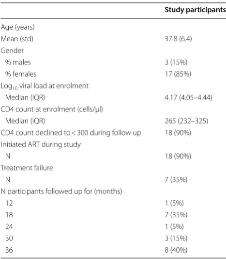
Samples of 20 subjects meeting eligibility criteria were tested for the presence of HIV-1 nucleic acid in their longitudinal urine samples (Table 1). Study participants were followed up for between 12 and 36 months with a mean follow up period of 27 months (std: 8.81 months). Median CD4 count and plasma viral load at enrolment was 265 cells/µl and 4.17 log10 RNA copies/ml,
respec-tively. Eighteen (90%) individuals had progressive HIV-1 disease defined as CD4 decline to < 300 cells/µl of whom 16 were initiated on ART during the study period. The other two individuals initiated ART at CD4 counts of 526 and 420 cells/µl.
Table 1 Clinical and demographical characteristics of 20 study participants
Study participants
Age (years)
Mean (std) 37.8 (6.4)
Gender
% males 3 (15%)
% females 17 (85%)
Log10 viral load at enrolment
Median (IQR) 4.17 (4.05–4.44) CD4 count at enrolment (cells/µl)
Median (IQR) 265 (232–325)
CD4 count declined to < 300 during follow up 18 (90%) Initiated ART during study
N 18 (90%)
Treatment failure
N 7 (35%)
N participants followed up for (months)
12 1 (5%)
18 7 (35%)
24 1 (5%)
30 3 (15%)
Prevalence of HIV‑1 nucleic acid detection in urine samples
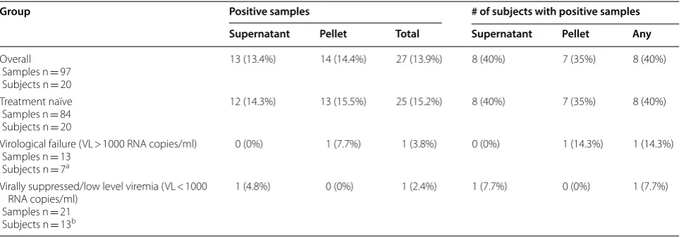
HIV nucleic acid was detected in 27/97 (13.9%) of all urine samples representing 8 (40%) individuals (Table 2). Both HIV-1 RNA and proviral DNA were detected in urine supernatant and urine pellets, respectively, from 7 of the subjects, while only HIV-1 RNA was detected in the urine supernatant from subject 051.
To determine whether the detection of HIV-1 DNA in urine pellets associated with the amount of cells pre-sent in the urine, we compared the quantity of isolated genomic DNA (directly proportional to cell number) between urine pellet samples with and without detectable HIV nucleic acid. The mean DNA quantity was higher for urine pellets with detectable HIV nucleic acid compared to those with no detectable HIV nucleic acid (1.81 micro-grams versus 0.37 μg; P = 0.01). For all urine samples, the
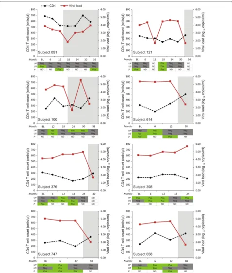
frequency of detectable HIV RNA in supernatants and HIV proviral DNA in pellets were similar (13.4% and 14.4%, respectively). However, the frequency of detection for each individual over time varied substantially (Fig. 1).
HIV‑1 nucleic acid detection in urine samples during treatment period
To investigate if HIV detection in urine samples per-sists during treatment with antiretroviral therapy, we analysed urine samples obtained after treatment ini-tiation. There were 26 urine samples tested (13 super-natant samples and 13 urine pellets) from 7 subjects that corresponded with periods of virological failure (VL > 1000 RNA copies/ml) in blood (Fig. 1). HIV-1 nucleic acid was detected in 1 urine pellet from subject 100. HIV-1 pol gene sequencing of the corresponding plasma samples did not detect resistance-associated markers from these samples. There were 42 samples
tested (21 supernatant samples and 21 urine pellets) from 13 subjects that corresponded with periods of either undetectable viremia (Limit of detection 40 RNA copies/ml) or detectable viremia < 1000 RNA copies/ml in blood. HIV nucleic acid was detected in 1 urine pel-let from subject 100 obtained more than 6 months after therapy initiation and during a period of undetectable viremia in blood.
Comparison of HIV‑1 viral populations in urine to variants in blood
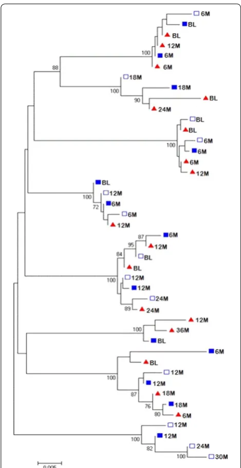
To compare HIV-1 viral populations in the urine to viruses in blood, a phylogenetic tree was constructed using the pol sequences obtained from the urine (super-natant and pellet samples) and from plasma (Fig. 2). Sequences included in the tree were from viruses from individuals where two or more sequences were obtained from urine samples. Co-clustering of urine-derived pol
sequences among the plasma-derived sequences was observed while some urine-derived sequences were interspersed among the plasma-derived sequences. The co-clustering of urine-derived sequences in these individuals occurred between pellet-derived provi-rus sequences and sequences from free viprovi-ruses in the urine supernatant. This is demonstrated in subject 747 for the 6 month sequences (6 M), subject 398 for the 12 month sequences (12 M) and in subject 376 for the 12 month (12 M) sequences (Fig. 2). Interspersing of urine-derived and plasma-derived sequences is shown in most of the sequences exemplified by urine-derived sequence 18 M and plasma-derived 6 M sequence which clustered.
Table 2 HIV-1 nucleic acid detection in urine samples
a Subject IDs with VL > 1000 RNA copies/ml: 038, 051, 100, 121, 662, 664, 732
b Subject IDs with VL < 1000 RNA copies/ml: 030, 051, 074, 100, 121, 614, 236, 747, 658, 664, 668, 705, 732
Group Positive samples # of subjects with positive samples
Supernatant Pellet Total Supernatant Pellet Any
Overall Samples n = 97 Subjects n = 20
13 (13.4%) 14 (14.4%) 27 (13.9%) 8 (40%) 7 (35%) 8 (40%)
Treatment naïve Samples n = 84 Subjects n = 20
12 (14.3%) 13 (15.5%) 25 (15.2%) 8 (40%) 7 (35%) 8 (40%)
Virological failure (VL > 1000 RNA copies/ml) Samples n = 13
Subjects n = 7a
0 (0%) 1 (7.7%) 1 (3.8%) 0 (0%) 1 (14.3%) 1 (14.3%)
Virally suppressed/low level viremia (VL < 1000 RNA copies/ml)
Samples n = 21 Subjects n = 13b
Fig. 1 CD4 T cell counts and viral loads over time in subjects showing detection of HIV nucleic acid in urine samples and plasma. Y-axis left: CD4 (cells/μl), black dotted line; Y-axis right: viral load (log10 copies/ml), red line. Subject IDs: Nucleic acid was detected in urine supernatant and pellet
Discussion
We analysed urine samples for HIV-1 shedding in the uri-nary tract before and after suppressive ART initiation. We have also investigated HIV-1 replication in the urinary
tract, evidenced by detection of HIV-1 RNA in treatment naïve individuals, and HIV-1 compartmentalization by comparing HIV sequences from plasma-derived viruses to sequences derived from viruses in urine samples using phylogenetic analysis. We detected HIV-1 nucleic acid in urine samples from 40% of treatment-naïve subjects compared to 8% in the virally suppressed or low level viremia group and 14% in the virological failure group. Although phylogenetic analysis indicated co-clustering of provirus sequences and sequences from free viruses in the urine supernatant a larger number of sequences will be required to draw conclusions regarding virus com-partmentalization in the urinary tract. We also detected viruses related to those circulating in blood resulting in equilibrium between blood and urinary tract viruses.
The low HIV-1 nucleic acid detection rates in urine samples from individuals on suppressive ART compared to treatment-naïve individuals indicates that suppressive ART is effective in blocking viral replication in the uri-nary tract. HIV-1 nucleic acid detection in urine samples from HIV positive treatment-naïve individuals has been reported in other studies [17, 18], followed by undetect-able levels after treatment initiation [18]. Our data adds more information in that our cohort had a different sub-type (C compared to B) and we analysed longitudinal samples instead of a single time point. In addition we looked at the pol gene instead of the env gene.
We detected HIV-1 RNA in urine pellets more than 6 months after treatment was initiated highlighting that infected cells can be shed into the urinary tract dur-ing periods of both viral suppression and virological failure in blood. The individual had virological failure after a period of viral suppression in blood with detect-able HIV-1 DNA in cells shed in urine. In another study, HIV-1 DNA was detected in the urine samples in 23% of virally suppressed subjects on ART [19]. This raises the question whether HIV-1 DNA in cells of the urinary tract is replication-competent and may be responsible for viral rebound upon treatment cessation or virologi-cal failure during treatment. HIV-1 infection of kidney allografts after transplantation in HIV positive recipi-ents with undetectable HIV-1 virus in plasma has been reported [14] indicating that this HIV-1 DNA may be replication competent and the source of infection in renal transplants.
In individuals with detectable HIV-1 nucleic acid in urine, we noted that detection of the HIV-1 did not occur at all the time points tested regardless of the associated higher plasma viral loads. The frequency of detection for each individual over time varied substan-tially (Fig. 1), which suggests that specific factors may modulate the presence of HIV in urine. Firstly, HIV-1 shedding in the urogenital tract may be intermittent Fig. 2 Phylogenetic tree of the sequenced pol gene from the urine
leading to absence of virus in some samples. Secondly, HIV detection may depend on the urine volumes used. Urine volumes of 2 ml were used for nucleic acid extraction in this study. Larger urine volumes resulted in higher HIV-1 RNA [13] and HIV-1 proviral DNA [17] detection rates. HIV-1 RNA detection rate was 50% in samples with a starting volume ranging from 35 to 630 ml [13] while there was HIV-1 DNA detection rate of 66.25% achieved using cell pellets obtained from 100 ml fresh urine samples [17]. Finally, the presence or degree of renal injury, which may affect the rate of shedding of renal epithelial cells, was unknown in our subjects. This information could help determine why HIV-1 nucleic acid could be detected in some individu-als or at certain times as HIV-associated nephropathy has been associated with HIV-1 replication and com-partmentalization [9, 19]. We however used the amount of DNA in urine pellets after nucleic acid extraction as an indicator of presence and amount of cells shed into the urogenital tract. Our results indicated that urine samples with detectable HIV-1 DNA had significantly higher numbers of cells (larger amount of host DNA extracted) compared to urine samples with no detect-able HIV-1 nucleic acid which may be an indication of renal injury and HIV-1 replication at the time of sample collection. Although HIV-1 virus could have been intermittently shed in urine for some individu-als, HIV-1 RNA and proviral DNA were consistently not detected in longitudinal urine samples taken over 18 months from some treatment-naïve individuals.
In conclusion, we have demonstrated that urine sam-ples can be used as a non-invasive method to study HIV replication, persistence and compartmentalization in the urinary tract. Suppressive ART reduces HIV-1 rep-lication in the urinary tract but HIV-1 DNA can persist and be shed from some cells in the urinary tract despite treatment. Our data suggests HIV-1 replication in the urinary tract but also direct virus importation from blood resulting in equilibrium between blood viruses and viruses shed from the urinary tract but a larger number of sequences would be required to confirm compartmentalization.
Supplementary information
Supplementary information accompanies this paper at https ://doi. org/10.1186/s1294 1-019-0326-9.
Additional file 1: Table S1. Study subjects and time points at which urine
samples were tested.
Abbreviations
DNA: deoxyribose nucleic acid; ART : antiretroviral therapy; RNA: ribonucleic acid; PR: protease; RT: reverse transcriptase; Pol: polymerase; VL: viral load; RT-PCR: reverse transcription polymerase chain reaction.
Acknowledgements Not applicable.
Authors’ contributions
MM, SB, GH and CT conceived and planned the project. CT, RC and NM enrolled study participants, and were responsible for the collection and storage of the blood and urine samples used in the study. MM, MK, AB and JL were responsible for nucleic acid extraction, sequencing and phyloge-netic analysis. RL analysed the data and constructed the figures and tables. MM and KR drafted the manuscript. All authors read and approved the final manuscript.
Funding
This work is based on the research supported by grants awards from South African Research Chairs Initiative of the Department of Science and Technol-ogy and National Research Foundation of South Africa. The Longitudinal Study of HIV-Associated Lung Infections in Soweto was funded by the National Institutes of Health, USA (R01HL090312 and P30AI094189: R. E. Chaisson). The funding bodies had no contribution in the design of the study and collection, analysis, and interpretation of data and in writing the manuscript.
Availability of data and materials
Data generated or analysed during this study are included in this published article and its additional file. Additional datasets used and/or analysed during the current study are available from the corresponding author on reasonable request.
Ethics approval and consent to participate
The parent study was approved by the University of the Witwatersrand Human research ethics committee (Medical); clearance certificate number M140926 and all participants provided informed consent for sample collection.
Consent for publication Not applicable.
Competing interests
The authors declare that they have no competing interests.
Author details
1 Department of Pathology, Faculty of Medicine, National University of Science & Technology, Ascot, P. O Box AC 939, Bulawayo, Zimbabwe. 2 Department of Medical Virology, University of Pretoria, Pretoria, South Africa. 3 Centre for HIV and Sexually Transmitted Infections, National Institute for Commu-nicable Diseases, Johannesburg, South Africa. 4 Faculty of Health Sciences, University of the Witwatersrand, Johannesburg, South Africa. 5 Virus Research and Development Laboratory, Department of Virus and Microbiological Special Diagnostics, Statens Serum Institut, Copenhagen, Denmark. 6 Johns Hopkins University Center for AIDS Research, Baltimore, MD, USA. 7 Perinatal HIV Research Unit (PHRU), SA MRC Soweto Matlosana Collaborating Centre for HIV/AIDS and TB, University of the Witwatersrand, Johannesburg, South Africa. 8 National Health Laboratory Services Tshwane Academic Division, Pretoria, South Africa.
Received: 6 September 2018 Accepted: 16 September 2019
References
1. Nickle DC, Jensen MA, et al. Evolutionary indicators of human immu-nodeficiency virus type 1 reservoirs and compartments. J Virol. 2003;77:5540–6.
2. Rothenberger MK, Keele BF, Wietgrefe SW, et al. Large number of rebounding/founder HIV variants emerge from multifocal infec-tion in lymphatic tissues after treatment interrupinfec-tion. PNAS. 2015;112(10):E1126–34.
•fast, convenient online submission
•
thorough peer review by experienced researchers in your field
• rapid publication on acceptance
• support for research data, including large and complex data types
•
gold Open Access which fosters wider collaboration and increased citations maximum visibility for your research: over 100M website views per year
•
At BMC, research is always in progress.
Learn more biomedcentral.com/submissions
Ready to submit your research? Choose BMC and benefit from:
4. Politch JA, Mayer KH, Welles SL, et al. Highly active antiretroviral therapy does not completely suppress HIV in semen of sexually active HIV-infected men who have sex with men. AIDS. 2012;26:1535–43. 5. Dahl V, Gisslen M, Hagberg L, et al. An example of genetically distinct
HIV type 1 variants in cerebrospinal fluid and plasma during suppressive therapy. J Infect Dis. 2014;209(10):1618–22.
6. Bednar MM, Sturdevant CB, Tompkins LA, et al. Compartmentalization, viral evolution, and viral latency of HIV in the CNS. Curr HIV/AIDS Rep. 2015;12(2):262–71.
7. Mzingwane ML, Tiemessen CT. Mechanisms of HIV persistence in HIV reservoirs. Rev Med Virol. 2017;27:e1924.
8. Wyatt CM, Meliambro K, Klotman PE. Recent progress in HIV-associated nephropathy. Annu Rev Med. 2012;63:147–59.
9. Marras D, Bruggeman LA, Gao F, et al. Replication and compartmen-talization of HIV-1 in kidney epithelium of patients with HIV-associated nephropathy. Nat Med. 2002;8:522–6.
10. Moosa MR, van der Walt I, Naicker S, Meyers AM. Important causes of chronic kidney disease in South Africa. S Afr Med J. 2015;105(4):320. 11. Winston JA, Bruggeman LA, Ross MD, et al. Nephropathy and
establish-ment of a renal reservoir of HIV type 1 during primary infection. N Engl J Med. 2001;344:1979–84.
12. Chen P, Chen BK, Mosoian A, et al. Virological synapses allow HIV-1 uptake and gene expression in renal tubular epithelial cells. J Am Soc Nephrol. 2011;22:496–507.
13. Blasi M, Balakumaran B, Chen P, et al. Renal epithelial cells produce and spread HIV-1 via T-cell contact. AIDS. 2014;28:2345–53.
14. Canaud G, Dejucq-Rainsford N, Avettand-Fenoël V, et al. The kidney as a reservoir for HIV-1 after renal transplantation. J Am Soc Nephrol. 2014;25(2):407–19.
15. Gupte AN, Wong ML, Msandiwa R, et al. Factors associated with pul-monary impairment in HIV-infected South African adults. PLoS ONE. 2017;12(9):e0184530.
16. Zhou Z, Wagar N, DeVos JR, et al. Optimization of a low cost and broadly sensitive genotyping assay for HIV-1 drug resistance surveillance and monitoring in resource-limited settings. PLoS ONE. 2011;6(11):e28184. 17. Li JJ, Huang YQ, Poiesz BJ, Zaumetzger-Abbot L, Friedman-Kien AE.
Detec-tion of human immunodeficiency virus type 1 (HIV-1) in urine cell pellets from HIV-1 seropositive individuals. J Clin Microbiol. 1992;30(5):1051–5. 18. Blasi M, Carpenter JH, Balakumaran B, Cara A, Feng G, Klotman ME.
Iden-tification of HIV-1 genitourinary tract compartmentalization by analysing the env gene sequences in urine. AIDS. 2015;29(13):1651–7.
19. Chakrabarti AK, Caruso L, Ding M, et al. Detection of HIV-1 RNA/DNA and CD4 mRNA in feaces and urine from chronic HIV-1 infected subjects with and without antiretroviral therapy. AIDS Res Ther. 2009;6:20.
Publisher’s Note