HOGG, JENNIFER A., Ph.D. The Influence of Femoral Structure, Hip Capsular Constraints, and Gluteal Muscle Strength and Activation on Temporal Patterns of Functional Valgus Collapse. (2018)
Directed by Dr. Sandra J. Shultz. 252 pp.
Functional valgus collapse (a combination of knee abduction and internal rotation and hip adduction and internal rotation) is a modifiable lower extremity movement pattern commonly associated with anterior cruciate ligament (ACL) injuries in females. Though the gluteus maximus and gluteus medius have frequently been named contributors to functional valgus collapse, evidence supporting their role in lower extremity movement has been inconsistent, and could in part be due to methodological differences between studies and the accepted practice of analyzing discrete variables instead of overall movement patterns. Better elucidation of gluteal muscle influence on lower extremity biomechanics may be a critical step for the reduction of ACL injury rates, as neuromuscular dysfunction is likely more responsive to injury prevention efforts than are other risk factors such as bony anatomy, ligament quality, or hormonal influences, that are more difficult to modify. Therefore, the purpose of this study was to 1) describe the neuromechanical profiles throughout the landing phase of single-leg and double-leg forward landings in males and females, 2) quantify the contributions of gluteal muscle strength and activation to peak angles and moments of functional valgus collapse after controlling for one’s femoral alignment, and 3) explore the association between gluteal muscle function and overall functional valgus collapse throughout the landing phase.
To accomplish this, 45 females and 45 males with no history of knee surgery were measured for femoral anteversion, hip ROM, and hip strength and then underwent biomechanical testing during single-leg and double-leg forward landings to examine muscle activation and 3-dimensional biomechanics. Data were analyzed using conventional group and correlative analyses and also with statistical parametric mapping (SPM), which allowed for a more
comprehensive examination of the entire biomechanical time series. Biomechanical variables of interest included joint angles and moments comprising functional valgus collapse: hip adduction and internal rotation and knee abduction and internal rotation.
In the comparison between single-leg and double-leg landings by sex, sex differences in the frontal plane were task dependent, though females maintained greater absolute knee abduction and hip adduction throughout the landing phases. Sex by task interactions revealed that females landed with smaller knee adduction angles than males, particularly during the single-leg landing (p=.03), while females’ knee abduction excursion was greater than males’, particularly during the
double-leg landing (p=.01). Across task, females displayed 4.1° greater peak knee abduction than males (p=.002), and this was specific to 37-46% of the landing phase (p=.05). Females went through 1.0° more hip abduction than males (p=.05), and used a smaller proportion of their gluteus maximus (p=.01) in both tasks.
Examination of gluteal muscle contribution to individual and overall levels of functional valgus collapse in females revealed that at the 18% and 20% time points during the landing phase, less hip abduction strength and greater gluteus medius activation predicted greater peak hip adduction angles (R2 change = .10; p = .02) and higher external hip adduction moments (R2
change = .14, p = .06). Greater hip extension strength predicted greater peak hip abduction angles (R2 change = .08; p = .05), while greater gluteus maximus activation strengthened the
prediction of greater initial (R2 change = .10, p = .03) and peak (R2 change = .14, p = .01) knee
internal rotation angles. From 7% - 8% of the landing phase, greater external rotation ROM was associated with greater external hip adduction moment (R2 change = .18, p = .01).
In males, less hip abduction strength strengthened the prediction of greater initial (R2
change = .12, p = .01) and peak knee internal rotation angles (R2 change = .14, p = .01), lesser
moments (R2 change = .06, p =.11). Less hip extension strength with greater gluteus maximus
activation predicted greater peak hip external rotation moments (R2 change = .14, p = .01).
Specifically from the 3% - 9% time points of the landing phase, greater hip extension strength was associated with greater knee abduction moment (R2 change = .17, p = .01) and less hip
adduction moment (R2 change = .24, p = .001). At 0% and from 2% - 3% of the landing phase,
greater internal and external rotation ROM were associated with greater knee abduction angle (R2
change = .27, p = .01) and greater hip adduction angle (R2 change = .23, p = .02).
These results indicate that lower extremity biomechanics during a single-leg landing task are appreciably different than those observed during a double-leg landing task, and that a single-leg landing task elicits more profound sex differences, particularly during the early stage of single-leg load acceptance when ACL injuries are thought to occur (30-40ms post initial ground contact). As such, a single-leg landing task may be more appropriate for biomechanical screening of ACL injury risk. Gluteal strength and activation explained a unique proportion of variance in lower extremity biomechanics beyond what was explained by femoral alignment. In females, weaker gluteal muscles predicted riskier frontal plane hip kinematics. In males, gluteal function was more associated with kinetics. This implies that our male cohort used their musculature to create torque about a joint, whereas our female cohort was unable to create torque. Though femoral alignment (total ROM) explained considerably greater proportions of biomechanical variance than did gluteal function, observed associations between gluteal muscle function and biomechanics occurred 10-20ms after associations between femoral alignment and biomechanics. While the gluteal muscles may act mechanically independent of femoral alignment, it is possible that gluteal muscle function could be temporally linked to one’s femoral alignment. With these findings in mind, it may be beneficial for clinicians to implement gluteal strengthening programs
and to encourage gluteal muscle pre-activation in individuals with excessive hip ROM to lessen their propensity for functional valgus collapse.
THE INFLUENCE OF FEMORAL STRUCTURE, HIP CAPSULAR CONSTRAINTS, AND GLUTEAL MUSCLE STRENGTH AND
ACTIVATION ON TEMPORAL PATTERNS OF FUNCTIONAL VALGUS COLLAPSE
by Jennifer A. Hogg
A Dissertation Submitted to the Faculty of The Graduate School at The University of North Carolina at Greensboro
in Partial Fulfillment of the Requirements for the Degree
Doctor of Philosophy Greensboro 2018 Approved by _____________________________________ Committee Chair
ii
APPROVAL PAGE
This dissertation written by JENNIFER A. HOGG has been approved by the following committee of the Faculty of The Graduate School at The University of North Carolina at Greensboro. Committee Chair ________________________________ Committee Members ________________________________ ________________________________ ________________________________ ________________________________ ________________________________ ____________________________ Date of Acceptance by Committee _________________________ Date of Final Oral Examination
iii
TABLE OF CONTENTS
Page
LIST OF TABLES ... vii
LIST OF FIGURES ... ix
CHAPTER I. INTRODUCTION ... 1
Statement of the Problem ... 4
Objective and Hypotheses ... 6
Limitations and Assumptions ... 7
Delimitations ... 8
Operational Definitions ... 9
Independent Variables for Conventional Analyses ... 12
Independent Variables for Statistical Parametric Mapping ... 13
Dependent Variables for Conventional Analyses ... 13
Dependent Variables for Statistical Parametric Mapping ... 15
II. REVIEW OF LITERATURE ... 17
Introduction ... 17
Contributors to ACL Strain ... 17
In Vitro Review ... 18 Retrospective Review ... 20 Prospective Review ... 21 Hip-Knee Coupling ... 23 Sagittal Plane ... 24 Frontal Plane ... 25 Transverse Plane ... 26 Summary ... 27
Factors Influencing Hip and Knee Function ... 27
Bony Alignment ... 28
Hip Capsular Constraints ... 33
Neuromuscular Function... 40
Methodological Considerations ... 49
Interactions Among Bony Alignment, Capsular Constraints, and Neuromuscular Characteristics ... 49
Choice of Landing Task ... 51
Analysis Strategies ... 52
iv
III. METHODS ... 63
Participants ... 63
Procedures ... 64
Anatomical Measures ... 64
Electromyography Sensor Placement ... 65
Maximal Voluntary Isometric Contractions (MVICs) ... 66
Single-Leg Forward Landing ... 68
Data Sampling and Reduction ... 70
Maximal Voluntary Isometric Contractions ... 70
Single-Leg and Double-Leg Forward Landing Biomechanics ... 70
Statistical Approach ... 73 Hypothesis 1a ... 74 Hypothesis 1b ... 74 Hypothesis 2a ... 75 Hypothesis 2b ... 76 Hypothesis 2c ... 76 Power Analysis ... 77
IV. MANUSCRIPT I. NEUROMECHANICAL SEX DIFFERENCES THROUGHOUT THE LANDING PHASES OF SINGLE AND DOUBLE-LEG FORWARD LANDING TASKS ... 80
Abstract ... 80
Content ... 80
Objective ... 80
Design ... 80
Setting ... 80
Patients or Other Participants ... 81
Intervention(s) ... 81
Main Outcome Measures ... 81
Results... 81 Conclusions ... 82 Key Words ... 82 Introduction ... 83 Methods ... 86 Participants ... 86
Surface Electromyography Instrumentation ... 86
Maximal Voluntary Isometric Contractions (MVICs) ... 87
Biomechanical Instrumentation ... 88
Procedure for Single and Double-Leg Forward Landing ... 89
Data Handling and Processing ... 89
Statistical Approach ... 92
Results ... 92
General Linear Model (Conventional) Descriptive Statistics ... 92
Omnibus MANOVA Results ... 98
v
Discussion ... 127
Sex Differences that were Task Dependent ... 127
Sex Difference Main Effects... 130
GLM v. SPM ... 133
Limitations ... 138
Conclusion ... 139
V. MANUSCRIPT II. THE EFFECTS OF GLUTEAL STRENGTH AND ACTIVATION ON THE RELATIONSHIP BETWEEN FEMORAL ALIGNMENT AND FUNCTIONAL VALGUS COLLAPSE ... 141
Abstract ... 141
Content ... 141
Objective ... 141
Design ... 141
Setting ... 141
Patients or Other Participants ... 142
Intervention(s) ... 142
Main Outcome Measures ... 142
Results... 142 Conclusions ... 143 Key Words ... 143 Introduction ... 144 Methods ... 146 Participants ... 146 Anatomical Measures ... 147
Surface Electromyography Instrumentation ... 147
Maximal Voluntary Isometric Contractions (MVICs) ... 148
Biomechanical Instrumentation ... 149
Procedure for Single-Leg Forward Landing ... 150
Data Handling and Processing ... 151
Statistical Approach ... 153
Results ... 154
Frontal Plane Hip Biomechanics in Females ... 155
Transverse Plane Hip Biomechanics in Females ... 159
Frontal Plane Knee Biomechanics in Females... 163
Transverse Plane Knee Biomechanics in Females ... 166
Frontal Plane Hip Biomechanics in Males ... 169
Transverse Plane Hip Biomechanics in Males... 170
Frontal Plane Knee Biomechanics in Males ... 171
Transverse Plane Knee Biomechanics in Males ... 172
Discussion ... 173
Gluteal Influences on Hip and Knee Biomechanics ... 174
The Mediating Effects of Gluteal Muscles on the Relationship between Femoral Alignment and Functional Valgus Collapse ... 176
The Relationship between Femoral Alignment and Functional Valgus Collapse ... 177
vi
Limitations ... 178
Conclusion ... 179
VI. MANUSCRIPT III. A PRELIMINARY MULTIVARIATE APPROACH TO ASSESS THE IMPACT OF GLUTEAL STRENGTH AND ACTIVATION ON FUNCTIONAL VALGUS COLLAPSE DURING A SINGLE-LEG FORWARD HOP LANDING ... 180
Abstract ... 180
Content ... 180
Objective ... 180
Design ... 181
Setting ... 181
Patients or Other Participants ... 181
Intervention(s) ... 181
Main Outcome Measures ... 181
Results... 182 Conclusions ... 182 Key Words ... 182 Introduction ... 183 Methods ... 186 Participants ... 186 Anatomical Measures ... 187
Surface Electromyography Instrumentation ... 187
Maximal Voluntary Isometric Contractions (MVICs) ... 188
Procedure for Single-Leg Forward Landing ... 189
Biomechanical Instrumentation ... 190
Data Handling and Processing ... 191
Statistical Approach ... 193
Results ... 195
Female Kinematic Valgus Collapse ... 195
Female Kinetic Valgus Collapse ... 202
Male Kinematic Valgus Collapse ... 207
Male Kinetic Valgus Collapse ... 213
Discussion ... 218
Functional Valgus Collapse in Females ... 218
Functional Valgus Collapse in Males ... 220
Comparison of Statistical Parametric Mapping and General Linear Model Canonical Correlation Analyses ... 221
Limitations ... 222
Conclusion ... 223
VII. EXECUTIVE SUMMARY ... 224
REFERENCES ... 229
vii
LIST OF TABLES
Page
Table 3.1 Intra-Rater Reliability Statistics for Hip Structural Measures ... 65
Table 3.2 Intra-Rater Reliability Statistics for Hip Strength Using a Handheld Dynamometer ... 67
Table 3.3 Minimal Detectable R2 Values Given Number of Predictors and Sample Size ... 79
Table 4.1 Means ± Standard Deviations (°) for Initial Joint Angles by Sex and by Task. ... 93
Table 4.2 Means ± Standard Deviations (°) for Peak Joint Angles by Sex and by Task. ... 94
Table 4.3 Means ± Standard Deviations (°) for Joint Excursions by Sex and by Task. ... 95
Table 4.4 Means ± Standard Deviations (Nm/N*m) for Peak External Joint Moments by Sex and by Task ... 96
Table 4.5 Means ± Standard Deviations for Gluteal Activation (%MVIC) by Sex and by Task ... 97
Table 4.6 A Summary of Significant (p<.05) Results Yielded from the General Linear Model (GLM) and Statistical Parametric Mapping (SPM) Analyses... 136
Table 5.1 Descriptive Statistics for Independent Variables in Males and Females ... 155
Table 5.2 Within-Sex Bivariate Correlations Among Independent Variables. ... 155
Table 5.3 Final GLM Forward Stepwise Regression Models Detailing the Influence of Control Variables, Anatomical Variables, and Neuromuscular Variables on Frontal Plane Hip Biomechanics ... 157
Table 5.4 Final GLM Forward Stepwise Regression Models Detailing the Influence of Control Variables, Anatomical Variables, and Neuromuscular Variables on Transverse Plane Hip Biomechanics ... 161
Table 5.5 Final GLM Forward Stepwise Regression Models Detailing the Influence of Control Variables, Anatomical Variables, and Neuromuscular Variables on Frontal Plane Knee Biomechanics ... 164
Table 5.6 Final GLM Forward Stepwise Regression Models Detailing the Influence of Control Variables, Anatomical Variables, and Neuromuscular Variables on Transverse Plane Knee Biomechanics ... 167
viii
Table 6.2 Post-Hoc Canonical Correlation Omnibus Results Detailing
the Contributions of Control Variables, Anatomical
Variables, and Neuromuscular Variables to a 4- Component Kinematic Valgus Collapse Combination
at Selected Time Points during the Landing Phase in Females. ... 199 Table 6.3 Univariate Follow-Up Analyses Detailing the Contributions
of Individual Predictors to each Component of Kinematic Valgus Collapse at Selected Time Points during the
Landing Phase in Females. ... 200 Table 6.4 Post-Hoc Canonical Correlation Omnibus Results Detailing
the Contributions of Control Variables, Anatomical
Variables, and Neuromuscular Variables to a 4- Component Kinetic Valgus Collapse Combination
at Selected Time Points during the Landing Phase in Females.. ... 205 Table 6.5 Univariate Follow-Up Analyses Detailing the Contributions
of Individual Predictors to each Component of Kinetic Valgus Collapse at Selected Time Points during the
Landing Phase in Females. ... 206 Table 6.6 Post-Hoc Canonical Correlation Omnibus Results Detailing
the Contributions of Control Variables, Anatomical
Variables, and Neuromuscular Variables to a 4- Component Kinematic Valgus Collapse Combination
at Selected Time Points during the Landing Phase in Males.. ... 210 Table 6.7 Univariate Follow-Up Analyses Detailing the Contributions
of Individual Predictors to each Component of Kinematic Valgus Collapse at Selected Time Points during the
Landing Phase in Males. ... 211 Table 6.8 Post-Hoc Canonical Correlation Omnibus Results Detailing
the Contributions of Control Variables, Anatomical
Variables, and Neuromuscular Variables to a 4- Component Kinetic Valgus Collapse Combination
at Selected Time Points during the Landing Phase in Males.. ... 216 Table 6.9 Univariate Follow-Up Analyses Detailing the Contributions
of Individual Predictors to each Component of Kinetic Valgus Collapse at Selected Time Points during the
ix
LIST OF FIGURES
Page Figure 2.1 A Comparison between Internal Tibial Torque and Valgus Moment
as Contributors to In Vitro ACL Strain (Markolf et al., 1995) ... 20 Figure 2.2 Representative Real-Time Observation of a Female
ACL Injury (Olsen, Myklebust, Engebretsen, & Bahr, 2004) ... 21 Figure 2.3 Schematic Depicting Lower Extremity Kinetic Chain Mechanics
as Theorized by Khamis & Yizhar (2007) ... 28 Figure 2.4 Comparison between Normal Femoral Neck Angle (left) and
Femoral Anteversion and Retroversion (right)
(Hoppenfield, 1976) ... 31 Figure 2.5 Anatomical Arrangement of the Iliofemoral, Pubofemoral,
and Ischiofemoral Ligaments. ... 34 Figure 2.6 Regression Equation Explaining 47% of Knee Abduction
Excursion during a Single-Leg Landing
(Howard, Fazio, Mattacola, Uhl, & Jacobs, 2011) ... 38 Figure 2.7 Example of an Analysis Depicting Temporal Comparison
between Participants with High and Low Knee Laxity
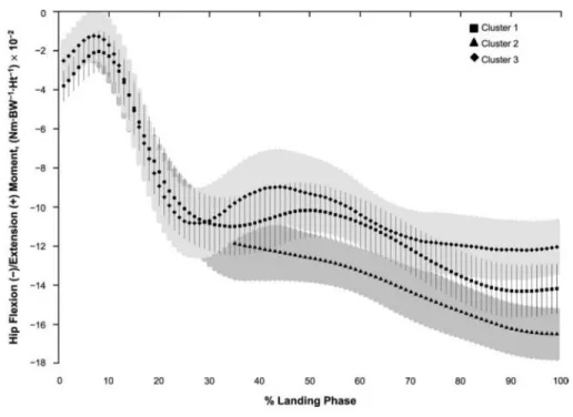
(S. J. Shultz & Schmitz, 2009b) ... 54 Figure 2.8 Example of an Analysis Depicting Temporal Comparison between
Clusters of Subjects Displaying Different Profiles of Lower Extremity Structural Characteristics
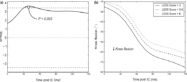
(A.-D. Nguyen, Shultz, & Schmitz, 2015) ... 56 Figure 2.9 Descriptive and Inferential SPM Curves Describing the Relationship
between Less Scores and Knee Flexion during a Functional Task
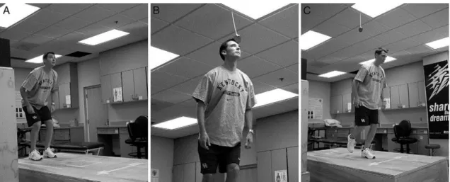
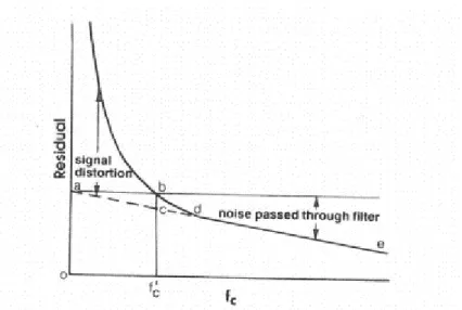
(Fox, Bonacci, McLean, & Saunders, 2016) ... 58 Figure 3.1 Representation of a Single-Leg Forward Landing (Jacobs et al., 2007) ... 69 Figure 3.2 Graphic Representation of a Residual Analysis (Winter, 1990) ... 72 Figure 4.1 Knee Flexion: Univariate Results (1a-c) and SPM Descriptive
and Inferential Results (1d-g)... 100 Figure 4.2 Knee Adduction/Abduction: Univariate Results (2a-e) and SPM
x
Figure 4.3 Knee Rotation: Univariate Results (3a-e) and SPM Descriptive
and Inferential Results (3f-i) ... 104 Figure 4.4 Hip Flexion: Univariate Results (4a-c) and SPM Descriptive and
Inferential Results (4d-g) ... 106 Figure 4.5 Hip Adduction/Abduction: Univariate Results (5a-e) and SPM
Descriptive and Inferential Results (5f-i). ... 108 Figure 4.6 Hip Rotation: Univariate Results (5a-e) and SPM Descriptive and
Inferential Results (5f-i). ... 110 Figure 4.7 Knee Flexion Moment: Univariate Results (6a) and SPM
Descriptive and Inferential Results (6b-e). ... 112 Figure 4.8 Knee Adduction/Abduction Moment: Univariate Results
(8a-b) and SPM Descriptive and Inferential Results (8c-f). ... 114 Figure 4.9 Knee Rotation Moment: Univariate Results (9a-b) and SPM
Descriptive and Inferential Results (9c-f). ... 116 Figure 4.10 Hip Flexion Moment: Univariate Results (10a) and SPM Descriptive and
Inferential Results (10b-e). ... 118 Figure 4.11 Hip Adduction/Abduction Moment: Univariate Results (11a-b) and SPM
Descriptive and Inferential Results (11c-f). ... 120 Figure 4.12 Hip Rotation Moment: Univariate Results (12a-b) and SPM
Descriptive and Inferential Results (12c-f). ... 122 Figure 4.13 Gluteus Maximus EMG Amplitude (%MVIC): Univariate Results (13a)
and SPM Descriptive and Inferential Results (13b-e). ... 124 Figure 4.14 Gluteus Medius EMG Amplitude (%MVIC): Univariate Results (14a)
and SPM Descriptive and Inferential Results (14b-e). ... 126 Figure 5.1 Measurement Position of Anatomical Variables (a), Hip
Extension MVIC (b), and Hip Abduction MVIC (c). ... 149 Figure 5.2 Terminal Position for the Single-Leg Forward Landing Task. ... 151 Figure 6.1 Measurement Position of Anatomical Variables (a), Hip
Extension MVIC (b), and Hip Abduction MVIC (c). ... 189 Figure 6.2 Terminal Position for the Single-Leg Forward Landing Task ... 190
xi
Figure 6.3 Descriptive Curves of Kinematic Functional Valgus Collapse Components (Frontal and Transverse Plane Hip and Knee
Motion) in Females during a Single-Leg Forward Landing Task... 197 Figure 6.4 Inferential Results of SPM Canonical Correlation Analyses:
the Relationship between Individual Predictors and a
4-Component Kinematic Valgus Collapse Combination in Females. ... 198 Figure 6.5 Descriptive Curves of Kinetic Functional Valgus Collapse
Components (Frontal and Transverse Plane Hip and Knee
Moments) in Females during a Single-Leg Forward Landing Task. ... 203 Figure 6.6 Inferential Results of SPM Canonical Correlation Analyses:
the Relationship between Individual Predictors and a
4-Component Kinetic Valgus Collapse Combination in Females... 204 Figure 6.7 Descriptive Curves of Kinematic Functional Valgus Collapse
Components (Frontal and Transverse Plane Hip and Knee
Motion) in Males during a Single-Leg Forward Landing Task. ... 208 Figure 6.8 Inferential Results of SPM Canonical Correlation Analyses:
the Relationship between Individual Predictors and a
4-Component Kinematic Valgus Collapse Combination in Males. ... 209 Figure 6.9 Descriptive Curves of Kinetic Functional Valgus Collapse
Components (Frontal and Transverse Plane Hip and Knee
Motion) in Males during a Single-Leg Forward Landing Task. ... 214 Figure 6.10 Inferential Results of SPM Canonical Correlation Analyses:
the Relationship between Individual Predictors and a
1
CHAPTER I INTRODUCTION
Of the more than 350,000 anterior cruciate ligament (ACL) injuries that occur annually in the United States, an estimated 72% occur through non-contact mechanisms (Moses, Orchard, & Orchard, 2012a; Wojtys & Brower, 2010). It is theorized that functional valgus collapse, a non-contact mechanism comprised of knee abduction, tibial internal rotation, hip adduction, and hip internal rotation, may increase the potential for ACL injury (Hewett et al., 2005; Ireland, 1999). Retrospective videographic studies have consistently reported the presence of a valgus knee collapse during ACL injury, particularly in females, as evidenced by increased pronation, increased medial knee collapse, increased hip adduction, and greater ipsilateral trunk lean (Boden, Torg, Knowles, & Hewett, 2009; Krosshaug et al., 2007). In vitro research has corroborated the injurious nature of functional valgus collapse suggested by videographic evidence. Specifically, the combination of internal tibial rotation and anterior tibial translation increased ACL strain greater than either internal tibial rotation or anterior tibial translation alone (Berns, Hull, & Patterson, 1992; Fukuda et al., 2003; Kiapour et al., 2014; Tron Krosshaug et al., 2007; Markolf et al., 1995). The strain resulting from combined internal tibial rotation and anterior tibial translation was further increased by the addition of a pure frontal plane valgus force (Berns et al., 1992). Of note, in the absence of tibial rotation and anterior tibial translation, a pure valgus force only minimally increased ACL strain, if at all (Berns et al., 1992; Markolf et al., 1995; Y. Oh, Ashton-Miller, & Wojtys, 2011). Conversely, an isolated tibial internal rotation torque, of a magnitude common in athletics, was sufficient to rupture the ACL (Meyer & Haut, 2008). This agreed well with research reporting the ACL to have greater sensitivity to rotational
2
moments than to frontal plane moments (Y. K. Oh et al., 2012). Together, these studies indicate that loads causing ligament rupture likely have a rotational, transverse plane component in addition to frontal plane movements, suggesting that ACL injuries may result from multiplanar loading patterns.
It is accepted that lower extremity movement acts occurs in a kinematics chain fashion, such that anterior pelvic tilt is thought to pair with greater femoral internal rotation, internal tibial rotation, and pronation (Duval, Lam, & Sanderson, 2010; Khamis & Yizhar, 2007). It is also accepted that the knee, hip, and trunk are mechanically coupled via ground reaction forces (Hewett & Myer, 2011; Imwalle et al., 2009). Given a ground reaction force that passes lateral to the knee joint, an adducted hip and an ipsilateral trunk lean become necessary to maintain an upright posture (Timothy E Hewett & Myer, 2011; Sigward & Powers, 2007a). Empirical evidence has demonstrated this coupling, showing that hip adduction alone may account for as much as 25% of the variance in knee abduction during cutting maneuvers (Imwalle, Myer, Ford, & Hewett, 2009). Along with greater vertical GRF and increased hip adduction, increased hip internal rotation has also been shown to contribute to increased knee valgus angles and moments during cutting maneuvers (R2=.36-.62) (Havens & Sigward, 2014; Sigward & Powers, 2007a).
Taking into account an integrated movement strategy and the evidentiary transverse and frontal plane coupling of these joints, controlling adduction and internal rotation of the hip may be an imperative step in the prevention of functional valgus collapse.
Femoral anteversion and passive hip range of motion (ROM) are two anatomical hip characteristics thought to influence dynamic hip adduction and internal rotation (Howard et al., 2011; Nguyen, Shultz, Schmitz, Luecht, & Perrin, 2011), and thus functional valgus collapse. Specifically, increased femoral anteversion and greater internal rotation hip ROM are suggested to bias the femur toward internal rotation and adduction across various functional tasks, thus
3
predisposing one towards greater knee valgus (A.-D. Nguyen et al., 2015; A Nguyen, Cone, Stevens, Schmitz, & Shultz, 2009; Sigward, Ota, & Powers, 2008). Because females are known to have greater amounts of both femoral anteversion and hip internal rotation ROM (Fan, Copple, Tritsch, & Shultz, 2014b; Moreno-Pérez, Ayala, Fernandez-Fernandez, & Vera-Garcia, 2015; A.-D. Nguyen & Shultz, 2007), this may in part account for the valgus collapse mechanism more commonly observed in females (T E Hewett, Torg, & Boden, 2009; Tron Krosshaug et al., 2007).
As the muscles primarily responsible for hip abduction and external rotation, the gluteus medius and gluteus maximus are often considered active restraints to dynamic hip adduction and internal rotation, respectively. As such, they have the potential to mediate the effects of hip range of motion and femoral anteversion. Despite copious literature investigating the gluteal muscles’ contribution to valgus collapse, the evidence is mixed. Varying methodology between studies makes it difficult to compare findings. Both absolute torque generating capacity and
electromyographic (EMG) muscle activation amplitude (as a % of maximal voluntary isometric contraction; MVIC) during functional tasks have been examined for their influence on dynamic hip adduction and internal rotation. At best, greater isometric hip abductor peak torque generation is moderately correlated with less hip adduction and knee valgus excursion (r= -.40 and -.35, respectively) (Jacobs et al., 2007). However, other similarly conducted studies found no significant relationships (Homan et al., 2013; Sigward et al., 2008; Thijs et al., 2007). External rotation isometric strength alone also yields mixed results (Cashman, 2012; Cronstrom, Creaby, Nae, & Ageberg, 2016; Howard, Fazio, Mattacola, et al., 2011). However, when muscle activation is included as a predictor, a more complete picture is rendered. Individuals with weaker hip abductors and external rotators have been shown to use greater percentages of their MVIC to complete a functional task (Homan et al., 2013a). This may explain why another study observed higher gluteal activation amplitude (% of MVIC) in those with greater knee valgus
4
excursion during a single-leg squat (A.-D. Nguyen, Shultz, Schmitz, Luecht, & Perrin, 2011). Therefore, both hip muscle strength and activation may need to be accounted for when analyzing the influence of the gluteal muscles on functional valgus collapse.
While hip internal rotation and adduction appear to be critical components of functional valgus collapse, the combined impact of gluteal strength and activation and femoral anteversion and passive hip ROM has yet to be examined with regard to stabilizing the hip during sport activity. Examining these in combination is important, as the gluteal muscles may have the ability to mitigate potentially negative effects of high internal rotation ROM or femoral anteversion. Therefore, not only is it important to include passive hip ROM and femoral anteversion as predictor variables, but including MVIC values along with muscle activation amplitude may be necessary.
Statement of the Problem
Existing ACL injury prevention programs are designed to improve dynamic lower extremity alignment, and have been successful in reducing ACL injury risk (Taylor, Waxman, Richter, & Shultz, 2015). However, overall rates of ACL injury have remained constant over the past two decades (Arendt & Dick, 1995; Moses, Orchard, & Orchard, 2012b). This suggests that safer dynamic alignment is not being retained after completing an ACL injury prevention
program. This could be the result of underlying structural characteristics, which are not modified by prevention programs. It is also possible that ACL injury prevention programs are targeting the wrong constructs. Because of this, it may be important to account for differences in structural alignment when examining influences of muscle activation on lower extremity biomechanics.
While femoral anteversion, passive hip ROM, and gluteal strength and activation in isolation have the potential to influence hip and knee control, the interaction of these factors to
5
influence movement during a dynamic task has not yet been elucidated. While the existing evidence is inconclusive regarding gluteal influences on functional valgus collapse, previous studies have not analyzed muscle strength and activation in conjunction with femoral anteversion and passive hip ROM, nor have they used single-leg functional tasks to examine these
relationships. Because demands on the lumbo-pelvic-hip complex are greater in a single-leg stance, using a single-leg task may better highlight gluteal contributions to functional valgus collapse. Furthermore, because females are more likely to display functional valgus collapse (T Krosshaug, Slauterbeck, Engebretsen, & Bahr, 2007), sex-specific research designs may be necessary to detect mechanistic patterns. Accounting for differences in transverse femoral alignment and capsular constraints within a sex-specific design may serve to better highlight gluteal impact on functional valgus collapse, and thus provide an avenue to affect biomechanical change in ongoing ACL injury prevention efforts.
Perhaps another reason for the inconclusive findings regarding gluteal influence on functional valgus collapse is that statistical approaches commonly used to analyze these data are limited. Functional valgus collapse is a coupled movement, exhibiting patterns unfolding over the course of a landing or cutting maneuver. Common practice is to collapse this movement pattern to a handful of discrete variables for analysis (e.g. initial contact, peak and excursion values), with each variable representing a single instant in time. Such analyses assume that movement occurs linearly, failing to take into account the possibility that prolonged joint loading or erratic movement may hold importance for ACL injury risk. Few studies have taken the full temporal nature of valgus collapse into account. Those that have were able to better identify loading and timing differences between participants with varying lower extremity alignment and laxity profiles. (A.-D. Nguyen et al., 2015; S. J. Shultz & Schmitz, 2009a). As such, employing a more holistic statistical technique may help to better characterize the impact of hip structure and gluteal
6
muscle function on functional valgus collapse patterns. Understanding these factors and their influences on knee joint loading rates is critical to identifying potential modifiable risk factors to target in ACL injury prevention programs.
Objective and Hypotheses
The objective of this study was to determine the extent to which femoral anteversion, passive hip ROM, and gluteal strength and activation impact patterns of functional valgus collapse during a single-leg forward landing task in separate female and male cohorts.
Aim 1: Examine sex-specific biomechanics throughout the entire landing phases of single-leg and double-leg forward landing tasks.
Hypothesis 1a: Compared to males, females will exhibit greater functional valgus collapse, as exhibited by greater joint angles and external moments associated with knee abduction, knee internal rotation, hip adduction, and hip internal rotation. This pattern will be more pronounced in a single-leg forward landing than in a double-leg forward landing.
Hypothesis 1b: Statistical Parametric Mapping 2x2 ANOVAs, which examine biomechanical differences across the entire landing phase, will identify specific time points at which lower extremity biomechanics differ by task, and by sex, thus providing a more complete analysis than using discrete, singular time point variables.
Aim 2: Determine the extent to which femoral anteversion and passive internal and external rotation hip ROM are associated with functional valgus collapse during a single-leg forward landing task in females and males, and the extent to which these influences are mediated by gluteal muscle strength and activation, and whether these relationships
7
become stronger and more specific once taking into account the timing and temporal nature of functional valgus collapse.
Hypothesis 2a: Greater femoral anteversion and greater internal rotation ROM and lesser external rotation ROM will predict greater movement toward
functional valgus collapse during a single-leg forward landing task, as evidenced by increased joint angles and external moments associated with knee abduction, knee internal rotation, hip adduction, and hip internal rotation.
Hypothesis 2b: The relationship between increased femoral anteversion, increased hip internal rotation ROM, decreased external rotation ROM and components of functional valgus collapse (as evidenced by increased joint angles and external moments associated with knee abduction, knee internal rotation, hip adduction, and hip internal rotation) will be weaker once controlling for the mediating effect of gluteus maximus and gluteus medius strength and activation.
Hypothesis 2c: A statistical parametric mapping canonical correlation analysis, which takes into account the temporal nature of functional valgus collapse, will identify stronger relationships between hip structure and function with functional valgus collapse than will using conventional correlative analyses with discrete, singular time point variables.
Limitations and Assumptions
1. Findings from this dissertation are neither generalizable to populations other than young healthy females and males, nor to tasks other than the single-leg or double-leg forward landing. 2. Three-dimensional motion capture, as represented by The Phase Space IMPULSE motion tracking system, is a valid and reliable tool for measuring biomechanical kinematics.
8
3. Embedded forceplates, as represented by dual Bertec plates, are valid and reliable tools for capturing biomechanical kinetics.
4. Inverse dynamics is an adequate method of computing three dimensional joint forces.
5. Femoral anteversion, as measured by an inclinometer, is a suitable surrogate for radiographic measurement of femoral anteversion.
6. Passive hip ROM, as measured prone with an inclinometer, is representative of capsular restraints of the femoral head.
7. All participants gave a maximal effort during maximal voluntary isometric contraction (MVIC) strength testing.
8. Surface electromyographic amplitude is not analogous to force.
9. Surface electromyography is a valid and reliable method of measuring muscle activity during functional tasks.
10. Surface electromyography signal obtained beneath an electrode is adequately representative of activity throughout the entire muscle.
11. A forward landing task is representative of a movement commonly employed in sport.
Delimitations
1. Only young healthy females and males with no history of lower extremity surgery or lower extremity injury within the immediately preceding six months were included in this study. 2. Femoral anteversion and hip ROM were measured using accepted clinical measurement methods.
3. Biomechanics were measured during the performance of single-leg and double-leg forward landings over a barrier normalized to 15% of each participant’s height.
9
5. Surface electromyography electrode placed over the gluteus maximus is representative of hip external rotation and extension activation.
6. Surface electromyography electrode placed over the gluteus medius is representative of hip abduction activation.
7. Surface electromyography electrode placed over the adductor longus is representative of hip adduction activation.
7. For biomechanical testing, all participants wore standardized clothing and shoes to eliminate between-subject differences related to shoe-surface interactions.
Operational Definitions
Femoral anteversion: The angle (degrees) formed by the tibial diaphysis, as measured on a straight line between the tibial tubercle and the midpoint of the malleoli, and vertical when the participant is prone with the knee flexed to 90 degrees and the greater trochanter at its most lateral position as determined by palpation.
Hip internal rotation ROM (ROMIR): The angle (degrees) formed by the tibial diaphysis, as
measured on a straight line between the tibial tubercle and the midpoint of the malleoli, and vertical when the participant is prone with the knee flexed to 90 degrees and the femur is passively rotated internally until the point of initial sacral tilt as determined by palpation. Hip external rotation ROM (ROMER): The angle (degrees) formed by the tibial diaphysis, as
measured on a straight line between the tibial tubercle and the midpoint of the malleoli, and vertical when the participant is prone with the knee flexed to 90 degrees and the femur is passively rotated externally until the point of initial sacral tilt as determined by palpation. Functional valgus collapse: A lower extremity movement pattern characterized by knee abduction, knee internal rotation, hip adduction, and hip internal rotation angles, and external
10
joint moments associated with knee abduction and internal rotation, and hip adduction and internal rotation.
Single-leg forward landing: A functional task performed by jumping from two legs from a distance equal to 40% of the participant’s height over a barrier equal to 15% of height and landing on the left leg.
Double-leg forward landing: A functional task performed by jumping from two legs from a distance equal to 40% of the participant’s height over a barrier equal to 15% of height and landing on both legs.
Hip extension peak torque: The average maximum hip extension torque produced during two 5-second maximal isometric extension trials against a strap-assisted handheld dynamometer from a prone position with the hip in neutral and the knee flexed to 90°, normalized to the participant’s moment arm (femur length).
Hip external rotation peak torque: The average maximum hip external rotation torque produced during two 5-second maximal isometric external rotation trials against a strap-assisted handheld dynamometer from a seated position with the hip and knee flexed to 90°, normalized to the participant’s moment arm (tibial length).
Hip abduction peak torque: The average maximum hip abduction torque produced during two 5-second maximal isometric hip abduction trials against a strap-assisted handheld dynamometer from a side-lying position with the hip in 10-15° of extension and 10° of external rotation, normalized to the participant’s moment arm (leg length).
Hip adduction peak torque: The average maximum hip adduction torque produced during two 5-second maximal isometric hip adduction trials against a strap-assisted handheld dynamometer from a supine position with the hip and knee extended, normalized to the participant’s moment arm (leg length).
11
Gluteus maximus activation: Gluteus maximus muscle activation is being represented by surface electromyography signal obtained at a location one-third of the distance from the second sacral vertebrae and the greater trochanter. It is expressed as a percentage of EMG activation recorded during maximal voluntary isometric contraction (hip extension and hip external rotation peak torque, respectively).
Gluteus medius activation: Gluteus medius muscle activation is being represented by surface electromyography signal obtained at a location one-third the distance from the most lateral point of the iliac crest to the greater trochanter. It is expressed as a percentage of EMG activation recorded during maximal voluntary isometric contraction (hip abduction).
Adductor longus activation: Adductor longus muscle activation is being represented by surface electromyography signal obtained at a location one-third the distance from the left inferior angle of the pubic symphysis to the left medial femoral condyle. It is expressed as a percentage of EMG activation recorded during maximal voluntary isometric contraction (hip adduction). Initial ground contact: The kinetic parameter defined by the moment at which the vertical ground reaction force (vGRF) exceeds 10N.
Landing phase: During a single-leg forward landing task, the phase commencing with initial ground contact and ending with peak knee flexion.
Healthy: An individual with 1) no history of lower extremity surgery, 2) no history of knee injury affecting ligamentous support or stability (including injuries to the ACL, MCL, PCL, LCL, medial meniscus, or lateral meniscus), 3) no history of lower extremity injury within the previous six months, 4) no presence of cardiovascular disease prohibiting moderate physical activity, and 5) no presence of vestibular condition affecting balance.
12 Independent Variables for Conventional Analyses
Femoral anteversion: Variable representing transverse structural alignment of the femur in relation to the pelvis.
Passive hip internal rotation range of motion (ROMIR): Variable representing transverse capsular
alignment of the femur as limited by the ischiofemoral ligament (Martin et al., 2008).
Passive hip external rotation range of motion (ROMER): Variable representing transverse capsular
alignment of the femur as limited by the iliofemoral ligament (Martin et al., 2008).
Hip extension/external rotation peak torque: Variable representing maximum torque generation capability of the hip extensors and external rotators, respectively.
Hip abduction peak torque: Variable representing maximum torque generation capability of the hip abductors.
Gluteus maximus muscle activation : The peak RMS amplitude of the gluteus maximus from ground contact to maximal knee flexion during five trials of a forward landing normalized to the peak RMS EMG activation recorded during maximal voluntary isometric contraction (hip extension and hip external rotation, respectively).
Gluteus medius muscle activation : The peak RMS amplitude of the gluteus medius during five trials of a forward landing normalized to the peak RMS EMG activation recorded during maximal voluntary isometric contraction (hip abduction).
Sex: Female or male.
13
Independent Variables for Statistical Parametric Mapping
Gluteus maximus muscle activation profile: A time series of RMS sEMG amplitude obtained from the gluteus maximus muscle over the course of the landing phase of five trials of a forward landing, interpolated and normalized to 101 data points.
Gluteus medius muscle activation profile: A time series of RMS sEMG amplitude obtained from the gluteus medius muscle over the course of the landing phase of five trials of a forward landing, interpolated and normalized to 101 data points.
Dependent Variables for Conventional Analyses
Initial Knee Abduction Angle: The frontal plane angle (°) formed by the tibia and femur at initial ground contact.
Peak Knee Abduction Angle: The maximum frontal plane angle (°) formed by the tibia and femur during the landing phase.
Knee Abduction Excursion: The difference, in degrees (°), between initial frontal plane knee angle and peak knee abduction angle.
Initial Knee Rotation Angle: The transverse plane angle (°) formed by the tibia and femur at initial ground contact.
Peak Knee Internal Rotation Angle: The maximum transverse plane angle (°) formed by the tibia and femur during the landing phase.
Knee Internal Rotation Excursion: The difference, in degrees (°), between initial transverse plane knee angle and peak knee internal rotation angle.
Initial Hip Adduction Angle: The frontal plane angle (°) formed by the femur relative to the pelvis at initial ground contact.
14
Peak Hip Adduction Angle: The maximum frontal plane angle (°) formed by the femur relative to the pelvis during the landing phase.
Hip Adduction Excursion: The difference, in degrees (°), between initial frontal plane hip angle and peak hip adduction angle.
Initial Hip Rotation Angle: The transverse plane angle (°) formed by the femur relative to the pelvis at initial ground contact.
Peak Hip Internal Rotation Angle: The maximum transverse plane angle (°) formed by the femur relative to the pelvis during the landing phase.
Hip Internal Rotation Excursion: The difference, in degrees (°), between initial transverse plane hip angle and peak hip internal rotation angle.
Peak Knee Abduction Moment: The maximum external joint moment acting about the anterior-posterior knee joint axis during the landing phase, normalized to height and weight (N·m·BW -1·Ht-1).
Peak Knee Internal Rotation Moment: The maximum external joint moment acting about the axial knee joint axis during the landing phase, normalized to height and weight (N·m·BW-1·Ht-1).
Peak Hip Adduction Moment: The maximum external joint moment acting about the anterior-posterior hip joint axis found during the landing phase, normalized to height and weight (N·m·BW-1·Ht-1).
Peak Hip Internal Rotation Moment: The maximum external joint moment acting about the axial hip joint axis found during the landing phase, normalized to height and weight (N·m·BW-1·Ht-1).
15
Dependent Variables for Statistical Parametric Mapping
Kinematic Knee Adduction/Abduction Profile: A time series of frontal plane knee
adduction/abduction angles during the landing phase, interpolated and normalized to 101 data points.
Kinematic Knee Internal/External Rotation Profile: A time series of transverse plane knee
internal/external rotation angles during the landing phase, interpolated and normalized to 101 data points.
Kinematic Hip Adduction/Abduction Profile: A time series of frontal plane hip
adduction/abduction angles during the landing phase, interpolated and normalized to 101 data points.
Kinematic Hip Internal/External Rotation Profile: A time series of transverse plane hip
internal/external rotation angles during the landing phase, interpolated and normalized to 101 data points.
Kinetic Knee Adduction/Abduction Profile: A time series of frontal plane knee external moments (normalized to height and weight) during the landing phase, interpolated and normalized to 101 data points.
Kinetic Knee Internal/External Rotation Profile: A time series of transverse plane knee external moments (normalized to height and weight) during the landing phase, interpolated and
normalized to 101 data points.
Kinetic Hip Adduction/Abduction Profile: A time series of frontal plane hip external moments (normalized to height and weight) during the landing phase, interpolated and normalized to 101 data points.
16
Kinetic Hip Internal/External Rotation Profile: A time series of transverse plane hip external moments (normalized to height and weight) during the landing phase, interpolated and normalized to 101 data points.
17 CHAPTER II
REVIEW OF LITERATURE
Introduction
The purpose of this literature review is to give an overview of the evidence as it pertains to lumbo-pelvic-hip function and its relationship to functional valgus collapse and ACL injury risk. The aim is to provide a theoretical rationale for the proposed research questions. In keeping with this aim, evidence supporting a valgus collapse ACL injury mechanism will be discussed, as will evidence detailing structural, capsular, and neuromuscular components of the lumbo-pelvic-hip complex and their respective influences on functional valgus collapse and ACL injury risk. In so doing, I will highlight strengths of the literature base, as well as identify gaps to be addressed with future research. Additionally, methodological concerns within the current literature base will be discussed.
Contributors to ACL Strain
In order to investigate underlying causes of functional valgus collapse and their potential influences on ACL injury risk, an understanding of direct contributors to ACL strain is needed. Much research, using a variety of research designs, has been devoted to describing contributors to ACL strain. While cadaveric, in vitro studies provide much of the basis for current thought, retrospective videographic evidence and prospective studies describing potential predictors of ACL injury are also pertinent. Therefore, this section will provide a review of the literature base surrounding contributors to ACL strain and injury, and will be divided into three subsections: in vitro review, retrospective review, and prospective review.
18
In Vitro Review. The anterior cruciate ligament reaches anteriorly and medially from the medial border of the lateral femoral notch to the anteromedial tibial plateau. Because of this positioning, the ACL is thought to limit anterior tibial translation and internal tibial rotation. Indeed, much cadaveric work has been devoted to illustrate this concept. It has been shown that a pure tibial internal rotation torque can increase in-situ ACL strain by 117% and is capable of rupturing the ACL at a failure load of 37.4 kN, a load frequently produced during sport activity (Meyer, Baumer, Slade, Smith, & Haut, 2008; Y. Oh et al., 2011). Similarly, an anterior tibial force has been shown to increase ACL strain in vitro. Near full knee extension, force measured within the anteromedial bundle of the ligament reaches 180N, which equaled 150% of the applied anterior tibial force (Markolf et al., 1995). Furthermore, anterior tibial translation and internal tibial rotation are additive. The greatest amounts of strain within the ACL are induced by an anterior tibial force plus an internal tibial rotation force, with ACL forces in the anteromedial bundle reported to reach nearly 300N in magnitude (Markolf et al., 1995). The additive nature of these movements is important, as these loads likely do not occur in isolation. Due to anatomical constraints, it is thought that anterior tibial translation and internal tibial rotation are coupled motions. For instance, in the event of a more severe lateral posterior tibial slope, the lateral tibial plateau is encouraged to translate anteriorly more than the medial tibial plateau relative to the femur during functional weight-bearing movement, thus inducing internal tibial rotation
(Beynnon et al., 2014; Marouane, Shirazi-Adl, & Hashemi, 2015; Meyer & Haut, 2008; Y. K. Oh et al., 2012) and further straining the ACL.
In the presence of anterior tibial translation and internal tibial rotation coupling, the addition of a frontal plane valgus force has repeatedly been shown to further increase ACL strain, particularly at knee flexion angles less than 30 degrees (Berns, Hull, & Patterson, 1992; Fukuda et al., 2003; Kiapour et al., 2015; Shin, Chaudhari, & Andriacchi, 2011). In the presence of
19
anterior tibial translation and internal tibial rotation under weight-bearing conditions, the knee is more susceptible to a valgus collapse. The axial load introduced in weight-bearing can compress the lateral compartment and tension the MCL, which can then function as an axis of rotation around which the lateral compartment can rotate medially, thus potentially forcing the knee into a valgus position. As the knee moves into greater valgus collapse, the lateral compartment is compressed further (Meyer & Haut, 2008). These events may be exacerbated by the presence of greater lateral posterior tibial slope, which can cause the femur to “fall off” the back of the lateral tibia, inducing even further internal tibial rotation and placing maximal strain on the ACL (Berns et al., 1992). However, it is interesting to note that in the absence of internal tibial rotation and anterior tibial force, a pure valgus force only minimally increases ACL strain, if at all (Berns et al., 1992; Markolf et al., 1995; Oh et al., 2011). The argument for a multiplanar injury
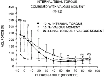
mechanism is made stronger by evidence showing that the ACL is less robust to torque applied in the transverse plane than in the frontal plane (Kiapour et al., 2015; Y. K. Oh et al., 2012). Figure 1 shows that when using similar amounts of torque, a rotary force induces greater strain within the ligament than a frontal plane moment, and that these moments are additive at knee flexion angles of 20-50° (Markolf et al., 1995). This would indicate that tibial rotation and anterior translation may be necessary components for an injurious valgus force to occur. Without these components occurring concomitantly, an isolated valgus force may be rendered impotent.
20
Figure 2.1. A Comparison between Internal Tibial Torque and Valgus Moment as Contributors to In Vitro ACL Strain (Markolf et al., 1995)
Retrospective Review. Retrospective videographic studies consistently indicate the presence of valgus knee collapse during ACL injury (Boden, Torg, Knowles, & Hewett, 2009; Krosshaug et al., 2007), as represented in Figure 2. Compared with sex-matched controls, injured males and females progressively moved into greater valgus collapse, with the injured cohort displaying frontal plane knee angles ten degrees greater than uninjured controls at the assumed moment of injury (66ms after initial contact) (Boden et al., 2009; Hewett, Torg, & Boden, 2009). In further videographic evidence, up to 53% of females display visible knee valgus at time of injury, compared with 17% of males (Krosshaug et al., 2007). This may indicate that females are more likely to injure their ACLs via valgus collapse mechanisms, whereas males may be more prone to alternative, more sagittal plane, injury mechanisms (Quatman & Hewett, 2009).
21
Figure 2.2. Representative Real-Time Observation of a Female ACL Injury (Olsen, Myklebust, Engebretsen, & Bahr, 2004)
Prospective Review. There is also prospective evidence supporting the relationship between functional valgus collapse and ACL injury. In 2005, Hewett et al. screened 205 female adolescent athletes (aged 15-16) during preseason using 3D motion capture of a drop vertical jump. Of the 205 screened, 9 went on to sustain ACL tears. Participants with ruptured ACLs were reported to display knee valgus angles 8° greater than uninjured counterparts. The primary variable of interest was peak knee abduction moment, which was shown to predict ACL injury status with 78% specificity and 73% sensitivity (Hewett et al., 2005). This study does have limitations. Nine ACL-injured athletes is a relatively small sample size, made smaller by the presence of an extreme outlier. Removing this outlier substantially weakens the relationship between peak knee abduction moment and ACL injury. Secondly, peak knee abduction moment refers to a pure frontal plane force, and this runs counter to cadaveric work indicating that a pure
22
frontal plane valgus torque isn’t likely to injure the ACL (Berns et al., 1992; Markolf et al., 1995; Oh et al., 2011).
In a replication of Hewett’s original work, 710 elite female soccer and handball athletes,
aged 21±4 years, were also screened using the drop vertical jump and tracked for 1-4 years (Krosshaug et al., 2016). Forty-two noncontact ACL injuries occurred that were suitable for analysis. Medial knee displacement was statistically different between injured and non-injured groups, though the mean difference was only half a centimeter (OR=1.40). Peak knee abduction moment and knee valgus at initial contact were not statistically different between groups. This could possibly be explained by the reported reliability of these characteristics. In a subset of the sample, test-retest reliability of motion capture was measured with 1-4 years between sessions, and the reliability of peak knee abduction moment is quite poor (ICC=.25). Because the average time between data collection and injury was 1.5±1.3 years, this study cannot conclusively claim that peak knee abduction moment is or is not associated with ACL injury.
Another recent publication detailed the relationship between 2D knee separation during a drop jump and general knee injury (OKane et al., 2016). While not aiming to explicitly predict ACL injury, this study has relevant implications. The sample consisted of females aged 11-14. Interestingly, the postmenarchal females in this cohort had a relative risk ratio of 3.62, indicating that females with the 10% most extreme valgus angles at maximum knee flexion were 3.62 times more likely to sustain a knee injury, whereas no such relationship existed in premenarchal females (OKane et al., 2016).
Collectively, these prospective studies indicate that functional valgus collapse may have a mild to moderate influence on ACL injury risk. Taking into account the varying methodology and populations between studies, drawing definitive conclusions is not possible. However, one explanation may be that the effect of functional valgus collapse on ACL injury risk could be a
23
function of maturity and skill level, creating an inverted-U phenomenon. In premenarchal females and in elite, well-trained females, functional valgus collapse may only have a minimal impact on ACL injury risk. In lesser trained adolescent or college age females, functional valgus collapse may pose more of a threat. Further research using more homogenous methods and populations is needed to examine this potential effect.
One theme consistent throughout this literature base is that valgus collapse entails more than pure frontal plane movement and moments. It represents a number of factors colliding at the knee and forming what we call dynamic, or functional, knee valgus. Valgus collapse is more of a lower extremity profile, rather than a single joint motion as a risk factor. While it includes the knee motions of anterior tibial translation, internal tibial rotation, and valgus torque (knee abduction), it also encompasses hip adduction and internal rotation (Berns et al., 1992; T E Hewett et al., 2009; Ireland, 1999; T Krosshaug et al., 2007). To that end, many investigators have looked proximally to the hip for factors contributing to functional valgus collapse.
Hip-Knee Coupling
Globally, it is accepted that movement is generated proximally and transferred distally (Duval et al., 2010; Khamis & Yizhar, 2007; Reiman, Bolgla, & Lorenz, 2009). Accordingly, internal rotation and adduction of the femur is followed by internal tibial rotation and knee abduction. The reverse is also true: external femoral rotation predisposes one to external tibial rotation (Duval et al., 2010; Khamis & Yizhar, 2007). From this line of thinking has come the idea that the hip may be a key to better understanding knee motion. Paired with the dual-joint nature of functional valgus collapse, this has given birth to a body of literature detailing the ways in which hip movement couples with knee motion to potentially influence ACL injury risk.
24
Sagittal Plane. There has been evidence to suggest that decreased hip flexion upon landing may be a potentiator of knee valgus. Greater hip flexion upon landing and cutting allows for the absorption of ground reaction forces by contractile tissue. When sagittal plane hip flexion is insufficient to absorb energy, the resulting stiff-legged landing strategy is thought to cause the ground reaction force to spill over into the frontal plane, thus leading to ligamentous absorption of forces and potential functional valgus collapse (Hewett, Paterno, & Myer, 2002). Moreover, it has been suggested that insufficient hip flexion in the presence of ample knee flexion may increase the potential for shear forces between the femur and tibia (Hashemi et al., 2011), which in turn may increase anterior tibial translation, already described as a joint movement thought to contribute to knee valgus (S. J. Shultz & Schmitz, 2009b). These concepts have been
corroborated in a number of research studies. Speaking to the presence of a landing strategy relying on ligamentous absorption of force, Schmitz et al (2007) reported that females who adopt a more stiff-legged landing than their male counterparts also elicit more ground reaction forces and absorb less energy with contractile tissue during a single-leg landing task, thus forcing inert (ligamentous) tissue to absorb the surplus energy, encouraging functional knee valgus (Schmitz, Kulas, Perrin, Riemann, & Shultz, 2007). There is also evidence that makes a more direct
association between decreased hip flexion and increased functional valgus collapse across various tasks. For instance, females previously shown to display greater dynamic valgus angles than male counterparts also exhibited decreased hip flexion during a side-step cutting maneuver (Pollard, Sigward, & Powers, 2007). No within-sex comparisons were made, thus making it difficult to determine how much of the differences were attributable to sex. However, the same group later made within-sex comparisons using a drop landing task and found that females displaying low hip flexion angles also exhibited increased knee valgus angles (Pollard, Sigward,
25
& Powers, 2010). Males were not included in this study, therefore it remains unknown if this mechanism is true in both sexes.
There are limitations within this body of evidence surrounding sagittal plane hip
kinematics and their effect upon knee biomechanics. Specifically, the extent to which hip flexion couples with knee flexion hasn’t been well described. Of the three studies reviewed, only one
(Pollard, Sigward, & Powers, 2010) quantified both hip and knee kinematics concurrently. Examining hip and knee kinematics together would aid in determining the extent of hip-knee coupling in the sagittal plane, which may in turn provide an avenue for biomechanical intervention. Another limitation is the lack of within-sex research designs. Due to evidence suggesting knee valgus could be a sex-specific injury mechanism (Quatman & Hewett, 2009), it may not be appropriate to include both sexes in the same analyses. By comparing males to females, one cannot be certain whether knee movement strategies are truly due to proximal factors or of simply being male or female. As such, making between-sex or combined-sex comparisons may not be as beneficial as within-sex analyses.
Frontal Plane. Hip adduction is a chief component of functional valgus collapse (Ireland, 1999). It is accepted that increased hip adduction upon cutting and landing increases frontal plane knee load and in turn, valgus collapse (Timothy E Hewett & Myer, 2011; Imwalle, Myer, Ford, & Hewett, 2009). In a study of female soccer athletes, hip adduction was the only significant predictor of knee abduction during both 45 and 90 degree cuts (Imwalle et al., 2009), accounting for 25% of the variance in knee valgus angles during both cutting conditions. Also employing a 45 degree cutting task, Sigward & Powers (2007) found that females with excessive internal valgus moments demonstrated greater hip abduction at initial contact than females with lesser internal valgus moments (12.8±6.5 v. 7.7±6.4 degrees). Though this finding seems
26
counterintuitive, it is likely due to the nature of the cutting task. In preparation for a side-step cut, the trunk moves toward the intended direction and away from the plant limb. As a result, the stance limb is abducted in preparation for push-off. Thus, during a sidestep cutting maneuver in which the trunk leans toward the planned direction, greater hip abduction at initial contact may be warranted in order to stay upright and successfully complete the maneuver. To confirm the task-specific nature of this strategy however, research using an alternative functional task is needed.
To more completely describe the extent of hip-knee coupling within the frontal plane, future research needs to include a greater variety of tasks. To date, the primary work in this area has been conducted within the purview of cutting maneuvers. To rule out the influence of trunk position, tasks such as drop jumps or forward landings would be beneficial to verify that hip-knee coupling relationships also exist in sagittal plane tasks and are comparable to those found in cutting tasks. Furthermore, this work is exclusively in females. Though it is necessary and useful to have within-female comparisons, thus avoiding sex-related confounds, it is noteworthy that these patterns have not been validated in an all-male cohort. As previously stated, exploring these relationships in a male cohort would aid in determining how much of this movement strategy is sex-specific.
Transverse Plane. Although empirical research is limited, hip internal rotation is also considered a key component of knee valgus collapse. During side-step and cutting maneuvers, females displayed increased hip internal rotation during the early deceleration phase of the task when compared to males (Imwalle et al., 2009; Pollard et al., 2007). Within sex, females
possessing greater knee valgus also had more hip internal rotation at initial contact during a side-step cutting task than females displaying normal valgus alignment (Sigward & Powers, 2007), further suggesting that the hip and knee may be coupled joints.
27
Similar limitations exist in the transverse plane literature as in the previous sagittal and frontal plane literature. Females have been shown to have an increased propensity for dynamic hip internal rotation, which is a component of dynamic knee valgus. However, this pattern has been validated exclusively within cutting tasks. Perhaps it is a function of the task instead of faulty biomechanics specifically. Further work needs to explore the extent of hip-knee coupling during other tasks in which the ACL is typically injured, such as landing tasks.
Summary. In summary, evidence for hip-knee coupling is mixed across planes. In the sagittal plane, research suggests that decreased dynamic hip flexion is associated with greater knee valgus, but this remains to be validated across tasks. Additionally, there is a need for sex-stratified research in this area. While evidence for hip-knee coupling is strongest in the frontal plane, cutting tasks have primarily been used to examine hip adduction’s effects upon knee abduction. Though it’s generally accepted that greater hip adduction is linked with more dynamic knee abduction, further research is needed to determine if its effect is task-dependent, or if it holds true in non-cutting tasks as well. In the transverse plane, while it is established that females exhibit more dynamic hip internal rotation than males, only one study observed that greater dynamic hip internal rotation is associated with an excessive valgus moment (Sigward & Powers, 2007b). Similar to the sagittal and frontal planes, more work is needed to confirm this transverse plane relationship in various tasks and in both sexes.
Factors Influencing Hip and Knee Function
From the perspective that the hip and knee move as a system coupled via the ground reaction force (Timothy E Hewett & Myer, 2011), factors that impact hip motion have the potential to impact knee motion, thus increase functional valgus collapse. To this end, there are
28
multiple factors capable of influencing hip movement. They fall into three broad categories: hip structure and alignment, capsular restraints, and neuromuscular characteristics.
Bony Alignment. From a kinetic chain perspective, anterior pelvic tilt and femoral anteversion are two variants in the lumbo-pelvic-hip complex which have the potential to
influence hip, and thus knee, motion. In theory, an increase in anterior pelvic tilt induces an acute internal femoral rotation, followed by internal tibial rotation (Figure 3) (Duval et al., 2010; Khamis & Yizhar, 2007). Acting along the same mechanistic lines, anteversion also represents an internally rotated femur and by extension an internally rotated tibia. There is evidence that both anterior pelvic tilt and an anteverted femur may contribute to functional valgus collapse.
Figure 2.3. Schematic Depicting Lower Extremity Kinetic Chain Mechanics as Theorized by Khamis & Yizhar (2007). Subtalar Pronation Sequentially Leads to Internal Tibial Rotation, Internal Femoral Rotation, and Finally Anterior Pelvic Tilt.
29
Anterior Pelvic Tilt. In theory, increased anterior pelvic tilt is thought