_____________________________________________________________________________________________________ *Corresponding author: E-mail: [email protected];
www.sciencedomain.org
Bilateral Sagittal Split Osteotomy: Is Anything Else
to Facilitate the Technique?
Mohammad Hassan Samandari
1, Navid Naghdi
1and Milad Etemadi Sh
1*1
Department of Oral and Maxillofacial Surgery, School of Dentistry, Isfahan University of Medical Sciences, Isfahan, Iran.
Authors’ contributions
This work was carried out in collaboration between all authors. All authors read and approved the final manuscript.
Article Information
DOI: 10.9734/BJMMR/2016/26880 Editor(s): (1) Panagiotis Korovessis, Chief Orthopaedic Surgeon, Orthopaedic Department,
General Hospital “Agios Andreas” Patras, Greece. Reviewers: (1) Fatih Gunhan, Izmir Katip Celebi University, Turkey. (2)Kishor Patil, SMBT Dental College and Hospital, Amrutnagar, Maharashtra, India. (3)Srikanth Gunturu, Drs S&NR’s Siddhartha Institute of Dental Sciences, Andhra Pradesh, India. (4)Bhushan P. Mundada, Datta Meghe Institute of Medical Sciences, Wardha, India. (5)Cristian Statkievicz, Aracatuba Dental School, Universidade Estadual Paulista (UNESP), Brazil. Complete Peer review History:http://www.sciencedomain.org/review-history/15580
Received 7th May 2016 Accepted 10th July 2016 Published 3rd August 2016
ABSTRACT
Nowadays bilateral sagittal split osteotomy (BSSO) is the most common technique for treatment of mandibular skeletal deformities. One of the most sensitive stages in BSSO is recognition of inferior alveolar nerve entrance. This technical note presents a noble and safe approach for medial osteotomy in BSSO which is based on anatomy of mandible during the surgery. In this approach osteotomy initiates at the junction of buccal and lingual cortices of ramus and will continue parallel to buccal cortex and in the same direction with sagittal cut. Compared to conventional BSSO technique, less neurosensory complications, risk of condylar sagging and bad splitting are expected in suggested modification. The operation time, healing and recovery periods are shorter, which cause more convenience for both surgeon and patient.
Keywords:Bilateral sagittal split osteotomy; orthognathic surgery.
1. INTRODUCTION
Nowadays bilateral sagittal split osteotomy (BSSO) is the most common technique for
treatment of mandibular skeletal deformities [1-3]. Despite its advantages such as mandibular
3-D relocation, appropriate bony contact and using rigid fixation [4], it has several disadvantages and complications. Some of the most common complications are bad splitting, neurosensory disturbance and inferior alveolar nerve injuries [3-5].
Since introduction of BSSO technique by Obwegeser and Trauner in 1957 till now [6], there were lots of technical modifications with purpose of decreasing the risk of unwanted complications during and after surgery. Some of these modifications are based on alteration of osteotomy cut lines [7]; other modifications are through employing new devices and instruments such as piezosurgery, endoscope, separator and bone splitter [4-8]. Some individuals use para-clinical examinations (CT-scan, radiography, etc.) or anatomical landmarks for performing safer osteotomy during surgery and reducing complications [9,10].
Medial dissection of ramus for recognition of inferior alveolar nerve is one of the most precise stages in BSSO. Risk of injury to inferior alveolar nerve is the highest in this stage. The more extensive medial dissection and nerve exploration result in more neurosensory complications and longer nerve recovery period [11-13].
This technical note presents a noble and safe approach for medial osteotomy in BSSO which is based on anatomy of mandible with least dissection during the surgery.
2. METHODS
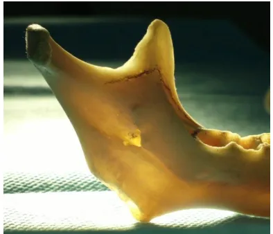
Based on the normal anatomy of mandible, from the region of the upper molar toward mandibular ramus, lingual cortex and buccal cortex gradually come closer together and bone marrow spaces decrease. Before sigmoid notch, these two cortices join together, so in this region ramus contains cortical bone purely except in condylar neck. This junction area can easily be recognized during surgery (Fig. 1).
In this approach, after subperiostal elevation of oral mucosa from external oblique ridge and exposure of coronoid process, medial dissection
will be continued until reaching to junction area of two cortices. No attempt performs to expose sigmoid notch or mandibular foramen in this approach (Fig. 2).
Fig. 1. Light emission pattern in a dry mandible. The more volume of bone marrow spaces, the less translucency of bone and the
darker view. The modified osteotomy line is drawn at the junction of buccal and lingual
cortices
Fig. 2. Exposure of coronoid process and junction of buccal and lingual cortices. No attempt has been done to expose sigmoid
notch and mandibular foramen
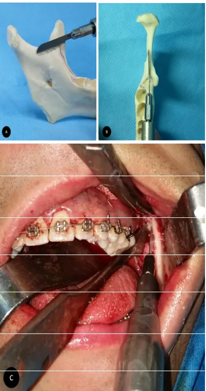
After recognition of junction area, osteotomy cut will be done parallel to buccal cortex and in the same direction with sagittal cut applying reciprocating saw in 25mm depth (Fig. 3). After
bone, sagittal osteotomy will be continued limited to cortical bone thickness in downward direction (Fig. 4). Then buccal cut will be done. Mandibular splitting will be accomplished easily by applying two osteotomes at the beginning and ending of sagittal cut (Fig. 5).
Fig. 3. A and B, direction and antero-posterior dimension of osteotomy. Note the paralleling of saw with buccal cortex of mandible and the
relation between saw and mandibular foramen. C, Osteotomy cut begins at top of
the junction of buccal and medial cortices and will be done parallel to buccal cortex
3. DISCUSSION
In conventional protocol of BSSO, the first step is to determine the location of mandibular foramen and exploration of neurovascular bundle. The
amount of manipulation of soft tissue in medial mandibular ramus is related to post-surgical neurosensory deficits [11-13]. In the current modification, lower risk of nerve damages is anticipated. The nerve exposure is not required in this method; thus less soft tissue dissection and less neurosensory disorders are expected.
Fig. 4. The similar direction of medial and sagittal osteotomy lines
Fig. 5. The splitted ramus. Note the same thickness of bone in all length of buccal
cortex
During medial ramus osteotomy in conventional BSSO, over approximation of horizontal osteotomy line to lingual or buccal cortical plates may cause bad splitting. In proposed technique, paralleling of medial osteotomy with buccal cortex of ramus results in lower risk of bad splitting.
Incomplete osteotomy in the crossing point of medial and sagittal cuts is a significant problem in common BSSO procedure. Possible outcomes include stress accumulation, resistance in splitting, and bad split. However in current modification, the medial and sagittal osteotomy lines are aligned in a similar direction. Thus the stress accumulation and resistance in splitting are reduced and faster osteotomy and splitting is achievable.
When conventional BSSO procedure is considered for superior repositioning or counter-clockwise rotation of distal part of mandible, a small bony fragment should be removed superior to the medial osteotomy line. It will cause passive repositioning of distal part without any interference to proximal segment [16]. Since in proposed technique the medial and sagittal osteotomy lines are in a similar pathway, removal of bony interference is not required. Thus it will facilitate passive repositioning of segments and will reduce the probability of condylar sagging. In addition, because of telescopic splitting, large bony contact and fewer gaps between the segments will achieve.
All of the osteotomies in current modification are made with saw and no rotary devices are required. Thus the risk of overheating and twisting the soft tissue around rotary devices are resolved. Also the risk of nerve transection and neurosensory damages are reduced because of reciprocating motion of device along the nerve pathway.
However because of less exposure of medial table of ramus, the access to surgical site is more difficult in proposed technique. On the other hand, the control of saw is not as simple as rotary devices, especially for novice surgeons.
Although the junction of buccal and medial cortices of ramus is far lower than the top of coronoid process, high medial cut might be a deterrent to mandibular functionality, especially in cases of huge mandibular advancement or correction of severe long face. So that great anterior and upward relocation of distal segment
of mandible might interfere with maxillary molar region and zygomatic buttress.
4. CONCLUSION
Compared to conventional BSSO technique, less neurosensory complications, risk of condylar sagging and bad splitting are expected in suggested modification. The operation time, healing and recovery periods are shorter, which cause more convenience for both surgeon and patient.
CONSENT
It is not applicable.
ETHICAL APPROVAL
The present study was considered as a technical note; thus no potentially harmful intervention was done in patients or animals.
DISCLAIMER
This manuscript Title was presented in the conference “OMFS Specialty Program” 14th International Congress of Iranian Society of Oral & Maxillofacial Surgeons.
Available link is:
“http://www.omfscongress.org/en/index.php/scien tific-schedule/omfs-specialty-program”
(16-19 February, 2016).
COMPETING INTERESTS
Authors have declared that no competing interests exist.
REFERENCES
1. Plooij JM, Naphausen MT, Maal TJ, Xi T, Rangel FA, Swennnen G, de Koning M, Borstlap WA, Bergé SJ. 3D evaluation of the lingual fracture line after a bilateral sagittal split osteotomy of the mandible. Int J Oral Maxillofac Surg. 2009;38(12):1244-9.
2. Tsuji Y, Muto T, Kawakami J, Takeda S. Computed tomographic analysis of the position and course of the mandibular canal: Relevance to the sagittal split ramus osteotomy. Int J Oral Maxillofac Surg. 2005;34(3):243-6.
morphology before and after sagittal split ramus osteotomy. J Oral Maxillofac Surg. 2010;68(8):1795-801.
4. Böckmann R, Schön P, Frotscher M, Eggeler G, Lethaus B, Wolff K-D. Pilot study of modification of the bilateral sagittal split osteotomy (BSSO) in pig mandibles. J Cranio-Maxillofac Surg. 2011;39(3):169-72.
5. Barakat AA-M, Abou-ElFetouh A, Hakam MM, El-Hawary H, Abdel-Ghany KM. Clinical and radiographic evaluation of a computer-generated guiding device in bilateral sagittal split osteotomies. J Cranio-Maxillofac Surg. 2014;42(5):e195-e203.
6. Muto T, Akizuki K, Tsuchida Y. Technical modification designed to facilitate sagittal split ramus osteotomy. J Oral Maxillofac Surg. 2008;66(7):1542-4.
7. Al-Nawas B, Kämmerer PW, Hoffmann C, Moergel M, Koch FP, Wriedt S, Walter C. Influence of osteotomy procedure and surgical experience on early complications after orthognathic surgery in the mandible. J Cranio-Maxillofac Surg. 2014;42(5):e284-8.
8. Mensink G, Gooris PJJ, Bergsma JE, Wes JT, van Merkesteyn JPR. Bilateral sagittal split osteotomy in cadaveric pig mandibles: Evaluation of the lingual fracture line based on the use of splitters and separators. Oral Surg Oral Med Oral Pathol Oral Radiol. 2013;116(3):281-6.
9. Ylikontiola L, Moberg K, Huumonen S, Soikkonen K, Oikarinen K. Comparison of three radiographic methods used to locate the mandibular canal in the buccolingual direction before bilateral sagittal split
osteotomy. Oral Surg, Oral Med Oral Pathol Oral Radiol Endod. 2002;93(6):736-42.
10. Yu IH, Wong YK. Evaluation of mandibular anatomy related to sagittal split ramus osteotomy using 3-dimensional computed tomography scan images. Int J Oral Maxillofac Surg. 2008;37(6):521-8.
11. Teerijoki-Oksa T, Jääskeläinen SK, Forssell K, Forssell H, Vähätalo K, Tammisalo T, Virtanen A. Risk factors of nerve injury during mandibular sagittal split osteotomy. Int J Oral Maxillofac Surg. 2002;31(1):33-9.
12. Westermark A, Bystedt H, Von Konow L. Inferior alveolar nerve function after sagittal split osteotomy of the mandible: Correlation with degree of intraoperative nerve encounter and other variables in 496 operations. Br J Oral Maxillofac Surg. 1998;36(6):429-33.
13. Panula K, Finne K, Oikarinen K. Neurosensory deficits after bilateral sagittal split ramus osteotomy of the mandible— influence of soft tissue handling medial to the ascending ramus. Int J Oral Maxillofac Surg. 2004;33(6):543-8.
14. Hetson G, Share J, Frommer J, Kronman JH. Statistical evaluation of the position of the mandibular foramen. Oral Surg, Oral Med Oral Pathol. 1988;65(1):32-4.
15. Hayward J, Richardson ER, Malhotra SK. The mandibular foramen: Its anteroposterior position. Oral Surg, Oral Med Oral Pathol. 1977;44(6):837-43. 16. Reyneke JP. Essentials of orthognathic
surgery. 2nd ed. Hanover Park, IL: Quintessence Publishing Co Inc; 2010.
_________________________________________________________________________________ © 2016 Samandari et al.; This is an Open Access article distributed under the terms of the Creative Commons Attribution License (http://creativecommons.org/licenses/by/4.0), which permits unrestricted use, distribution, and reproduction in any medium, provided the original work is properly cited.
Peer-review history: