Medical Devices: Evidence and Research
Dove
press
O R i g i n a l R E s E a R c h open access to scientific and medical research
Open access Full Text article
Video abstract
Point your SmartPhone at the code above. If you have a QR code reader the video abstract will appear. Or use:
http://dvpr.es/1c54kqh
Power port contrast medium flushing
and trapping: impact of temperature,
an in vitro experimental study
Gérard Guiffant1 Jean Jacques Durussel1 Patrice Flaud1
laurent Royon1 Pierre Yves Marcy2 Jacques Merckx1,3
1University Paris Diderot, Paris, France; 2Radiodiagnosis and
Interventional Radiology Department, Caen, France; 3University Teaching Hospital Necker-Enfants Malades, Paris, France
Correspondence: Gérard Guiffant University Paris Diderot, MSC, UMR CNRS 7057, 10 rue Alice, Domon et Léonie Duquet, 75205 Paris, Cedex 13, France Fax +33 01 5727 6211
Email [email protected]
Purpose: The use of totally implantable venous access devices (TIVADs) certified as “high pressure resistant” or “power port” has begun to spread worldwide as a safe procedure for power contrast injection. Owing to the thermo-rheological properties of the contrast media, the primary aim of this work is to present an in vitro experimental impact study concerning the impact of the temperature level on flushing efficiency after contrast medium injection. Moreover, we report experimental data that confirms the role of needle bevel orientation. The secondary aim is to answer the following questions: Is there significant device contrast medium trapping after contrast medium injection? Is saline flushing efficient? And, finally, is it safe to inject contrast medium through an indwelled port catheter?
Results: The experimental results show that in addition to hydrodynamics, temperature is a key parameter for the efficiency of device flushing after contrast medium injection. It appears that this is the case when the cavity is incompletely rinsed after three calibrated flushing volumes of 10 mL saline solution, even by using the Huber needle bevel opposite to the port exit. This leads to a potentially important trapped volume of contrast medium in the port, and conse-quently to the possibility of subsequent salt precipitates and long term trisubstituted benzene nuclei delivery that might impair the solute properties, which may be further injected via the power port later on.
Conclusion: We thus suggest, in TIVADS patients, the use of a temporary supplementary intravenous line rather than the port to perform contrast medium injections in daily radiology routine practice.
Keywords: contrast medium, implantable ports, totally implantable venous access devices (TIVADs), flushing, obstruction, prevention, central lines
Introduction
For more than two decades, totally implantable venous access devices (TIVADs) have been in use, allowing repeated injection or perfusion for drug administration as well as blood collection. These devices have proven to be safe and effective in overcoming problems of repeated venous access. Furthermore, their characteristics, risks, benefits as well as their usage setting and maintenance are well described and detailed in vari-ous protocols and papers in the literature.1–12
More recently, the use of TIVADs for power contrast injection has begun to spread as a safe procedure,13–17 and is presented as highly desirable from both the patient and
the radiologist’s point of view.17 Besides infectious and thrombotic complications17
that are common to all TIVADs, mechanical complications due to the high pressure administration of contrast medium (CM) by automatic injectors tend to become less
Medical Devices: Evidence and Research downloaded from https://www.dovepress.com/ by 118.70.13.36 on 24-Aug-2020
For personal use only.
Number of times this article has been viewed
This article was published in the following Dove Press journal: Medical Devices: Evidence and Research
Dovepress Guiffant et al
important, thanks to the specification and label “high pres-sure resistant” of the so-called “power ports”. One other complication of injecting CM into the power port lies in the possibility of obstruction of the TIVAD. In spite of the vari-ous typical rinsing protocols, the high level of viscosity of the injected CM induces persistence on the endoluminal wall of the TIVADs. The CM settles on the device wall proteins, and absorbs, in turn, part of the chemical or biological infused products. Thus, the definition of dedicated protocols for the indwelled device flushing is clearly of crucial importance.
The primary aim of this paper is to report experimental data regarding the hydrodynamic efficiency on the TIVAD flushing after CM injection, with special attention paid to the effect of temperature on this process.
We showed in a preceding paper that the dynamic of the device flushing flow plays an essential role18 in the
effective-ness of rinsing. Moreover, in the particular case of using implantable venous access catheter port devices, we have previously shown19 that the orientation of the Huber point
needle bevel was a determinant for flushing. The present work is a continuation of this approach.
Materials and methods
Hydrodynamics and temperature were retained as two key parameters to be assessed to qualify flushing efficiency of TIVADs after CM injection.
The ports being used in the present study included: first, high pressure Polysite® Perouse Medical 4008 (ISP
Perouse Medical, Ivry le Temple, France) ports (internal diameter = 13 mm and internal volume = 0.55 mL) con-nected to an outlet 25 cm long 8F silicone catheter (1.4 mm of inner diameter and 2.7 mm of outer diameter) and; sec-ond, Polysite® 4017 (ISP Perouse Medical) (internal
diam-eter = 13 mm and internal volume = 0.55 mL) connected to a polyurethane 7F catheter (1.7 mm of inner diameter and 2.7 mm of outer diameter) to test and compare between silicone and polyurethane catheters.
Two classes of non-ionic CM were used in the present study. Guerbet (Villepinte France) XENETIX®350 mg/mL
has a viscosity of 21 mPa.s at 20°C and of 10 mPa.s at 37°C; and Guerbet XENETIX®300 mg/mL has a viscosity
of 11 mPa.s at 20°C and of 6 mPa.s at 37°C. These CM were representative of sixth generation non-ionic monomers. Visipaque® (GE Healthcare, Aulnay sous bois, France)
320 mg/mL has a viscosity of 26.6 mPa.s at 20°C and of 11.8 mPa.s at 37°C, and Visipaque®270 mg/mL has a
viscosity of 12.7 mPa.s at 20°C and of 6.3 mPa.s at 37°C. These CM were representative of seventh generation
non-ionic dimers. Guerbet XENETIX®350 mg/mL CM
associated with Polysite® Perouse Medical 4008 (ISP Perouse
Medical) port device was taken as the “standard” reference for the different comparisons with the other “experimental” CMs or materials.
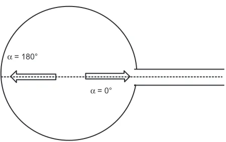
In accordance with the objective of the study, two tem-peratures of port device flushing (saline 0.9% sodium chlo-ride) were tested namely at 37°C (central body temperature) as well as at 23°C (ambient temperature), and two direc-tions of port device flushing including α= 0° and α= 180°
(Figure 1). The Huber needle (19G) was manually inserted at the center of the port with the bevel opening oriented facing the exit channel α= 0° or towards the diametrically opposite point of the port, α = 180°. The experimentation was performed in a thermostated enclosure, which maintains power port at 37°C.
The experimentation was conducted in the follow-ing way: the port was first perfused usfollow-ing 100 mL of CM (37°C) at a flow rate of 2 mL/second. Then, rinsing with saline solution was performed using a series of calibrated saline fractions of 10 mL at flow rates 0.5 mL/second17,18
that were previously thermalized either at 37°C or 23°C. There was a 1-minute time delay between the two saline solution injections. Each experimentation was conducted using a new port thus without any pre-existing pollution. The content of each syringe was then titrated using an ultraviolet spectrophotometer at 254 nm (Gilson 112 UV/ VIS detector; Gilson, Middleton, WI, USA). The results (ie, CM remaining in the power port catheter device after saline solution flushing) were calculated on the percentage basis of the total quantity of contrast.
The rheological measurements were performed using a cone plan Haake viscometer allowing the variation of the sample temperature level.
α = 180°
α = 0°
Figure 1 Bidimensionalschematic representation of a port indicating the two directions of the needle bevel orientation.
Medical Devices: Evidence and Research downloaded from https://www.dovepress.com/ by 118.70.13.36 on 24-Aug-2020
Dovepress The effect of temperature on contrast medium flushing
The measurements of the thermal conductivity of the CM at room temperature and at 37°C were performed using the TPS 500 hot disk thermal constants analyzer technique,20
manufac-tured by Perkin Elmer Company (Waltham, MA, USA).
statistical analysis
The Mann–Whitney U test was used on means (N = 6). Statistical significance thresholds are shown on the figures using the classical correspondence: * for 0.01 , P , 0.05; ** for 0.001 , P , 0.01; *** for P , 0.001 The symbol ns indicates not significant.
Experimental results
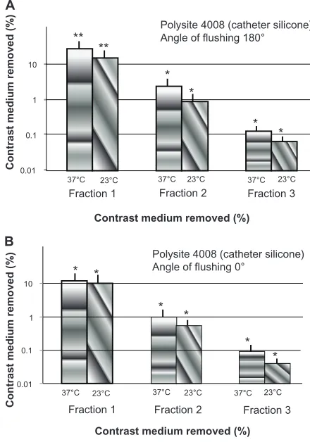
T h e r e s u l t s r e g a r d i n g t h e s t a n d a r d G u e r b e t XENETIX®350 mg/mL experimentation are reported in
Figure 2A and B. Table 1 shows the results of a series of three successive calibrated syringe fractions of 10 mL at flow rates 0.5 mL/second. Each column represents the mean (N = 6) per-centage of extracted CM. A logarithmic scale was utilized to clarify result presentation regarding the removed percentages of CM into the device. Only the first flushing syringe fraction
gave significant values. As device flushing was repeated, subsequent fractions gradually led to smaller values of CM removal until they finally dropped below the detector limit for the last fraction. To facilitate the understanding of the comparisons between experimentation results, the percentage values are reported in Table 1 together with the total amount of CM removed after ten rinsing fractions.
The results presented in Figure 2A and B were obtained while testing ports associated with silicone catheters. It was then reasonable to investigate the impact of the nature of the port catheter on the efficacy of power port flushing. The results reported in Figure 3 give the percentages of CM removed after three rinsing fractions of 10 mL in the most favorable conditions; namely, at 37°C temperature and with a needle bevel orientation α= 180°. The material distinction between silicone and polyurethane catheters was found to be non-significant. Although the situation is not clinically relevant, the result obtained without the output catheter has been reported for comparison in Figure 3 as an additional indication. This shows that the CM is mainly trapped into the port itself. As an example, we display a typical image (Figure 4) of the inside (after removing the septum) of a power port chamber, shown after ten rinsing fractions of
Table 1 Percentage of removed XENETIX®350 after successive
flushing fractions of 10 mL for the two temperatures (37°c and 23°C) and the two orientations of the needle bevel (α= 180°
and α= 0°)
Fraction 10 mL
37°C,
a=180°
% removed 37°C,
a=0°
% removed 23°C,
a=l80°
% removed 23°C,
a=0°
% removed
1 28 ± 2 12 ± 1 15 ± 1 9.6 ± 0.5 2 2.3 ± 0.2 1.1 ± 0.1 0.9 ± 0.1 0.57 ± 0.06 3 0.12 ± 0.02 0.09 ± 0.01 0.06 ± 0.01 0.04 ± 0.01 4 to 10 0.05 ± 0.01 0.14 ± 0.02 0.05 ± 0.01 0.13 ± 0.03
10 **
* *
* *
* * **
* *
* * 1
0.1
0.01
37°C
Fraction 1
Fraction 1 Fraction 2 Fraction 3 Fraction 2
Polysite 4008 (catheter silicone) Angle of flushing 0°
Polysite 4008 (catheter silicone) Angle of flushing 180°
Fraction 3
37°C 37°C
23°C
37°C 37°C 37°C
10
Contrast medium removed (%) Contrast medium removed (%)
1
0.1
0.01
23°C 23°C 23°C
23°C 23°C
A
B
Contrast medium removed (%)
Contrast medium removed (%)
Figure 2 semilogarithmic representation of the percentage of removed XENETIX®350 after three successive flushing fractions of 10 mL, for two flushing temperatures flushing (37°c and 23°C) and two needle bevel orientations: α= 180° (A) and α= 0° (B).
Note: statisticalsignificance is represented by * for 0.01 , P , 0.05; ** for 0.001 ,P, 0.01.
Polysite 4008 (catheter silicone) Polysite 4017 (catheter polyurethane)
Contrast medium removed (%) Silicone
without catheter Polyurethane 0
10 20 30 40
ns ns
Contrast medium removed (%)
Figure 3 Percentageof removed XENETIX®350 after three flushing fractions of 10 ml at 37°c and α= 180° for the needle bevel orientation and for two types of catheter: silicone and polyurethane.
Abbreviation: ns,not significant.
Medical Devices: Evidence and Research downloaded from https://www.dovepress.com/ by 118.70.13.36 on 24-Aug-2020
Dovepress Guiffant et al
10 mL. Indeed, an important quantity of CM is still remaining in the port chamber. The effect of varying the flushing flow level is shown in Figure 5. It was found that the percentage of CM removed after three rinsing fractions of 10 mL is not significantly different when comparing the two flows of flushing that were respectively tested at 0.5 mL/second and 1 m L/second rates.
Taking XENETIX®350 as the standard reference CM, a
comparison was made with different CM. Figure 6 shows the results obtained when comparing the XENETIX®350
to XENETIX®300; no significant difference was found
concerning the rinsing efficiency of the CM after three suc-cessive flushing fractions of 10 mL under the most favorable conditions. The results clearly show that even repeating ten flushing fractions were still ineffective in clearing out the trapping CM.
As mentioned above, XENETIX®350 was taken as
rep-resentative of the sixth generation of non-ionic monomers of CM commonly used in daily radiology practice. Then, a comparison was carried out by experimenting two
repre-sentative CM of the seventh generation of non-ionic dimers namely Visipaque®320 and Visipaque®270. Figure 7A
and B give the efficiency of the flushing after three succes-sive flushing fractions of 10 mL under the most favorable conditions. Figure 7A shows that no significant difference was found regarding the flushing between XENETIX®350 Figure 4 Magnifiedpicture of the interior of a chamber perfused with XENETIX®350
after ten flushing fractions of 10 mL at 37°c and α= 180°.
0
1 mL/second 0.5 mL/second 10
20 30
ns
ns
Polysite 4008 (catheter silicone)
Contrast medium removed
(%)
Figure 5 Percentageof removed XENETIX®350 after three flushing fractions of 10 ml at 37°c and α= 180° for two different values of the flushing flow rate.
Abbreviation: ns,not significant.
Contrast medium removed (%)
Polysite 4008 (silicone) ns
30
20
10
0
XENETIX®350 XENETIX®300 ns
Figure 6 Percentageof removed contrast media after three flushing fractions of 10 ml at 37°c and α= 180° for XENETIX®350 and XENETIX®300.
Abbreviation: ns,not significant.
0
XENETIX®350 Visipaque®320
10 20 30
ns
ns 40
A
B
Contrast medium removed
%
0
XENETIX®350 Visipaque®270
10 20 30
**
**
Contrast medium removed
%
Figure 7 Percentageof removed contrast after three flushing fractions of 10 mL at 37°c and α = 180°; comparison of XENETIX®350/Visipaque®320 (A) and comparison of XENETIX®350/Visipaque®270 (B).
Note: Statistical significance is represented by ** for 0.001 , P , 0.01.
Abbreviation: ns, not significant.
Medical Devices: Evidence and Research downloaded from https://www.dovepress.com/ by 118.70.13.36 on 24-Aug-2020
Dovepress The effect of temperature on contrast medium flushing
and Visipaque®320 while flushing Visipaque®270 was found
to be slightly different from flushing XENETIX®350.
Discussion
Taking into account the marked effects of both hydrodynam-ics and temperature on the efficiency of power port flushing after CM injection, the extreme results (Figure 2A and B) are reported in Figure 8. It is noticeable that the effectiveness of rinsing at 37°C and α= 180° is found to be highly significant with respect to rinsing at 23°C and using a power port needle bevel orientation α= 0°.
The first point to be noted is that according to previous results19 it is always preferable to direct the bevel of the Huber
port needle towards the opposite of the outlet (α = 180°; Figure 1). This orientation leads to a better distribution of the shear rate of fluid injected into the port chamber thus permitting a better flushing.
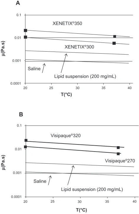
The second point to be noted is that temperature plays a determinant role in the efficiency of the flushing. It appears highly preferable to use flushing fluids at 37°C (Figure 8) rather than at 23°C. The reasons for such behavior are likely related to the thermo-rheological properties of the CM. As a matter of fact, from a rheological point of view, CM are Newtonian fluids (the viscosity is independent of the flow level) and present high values of viscosity. The dependence of viscosity with respect to the temperature is indeed classical; these measurements are reported in Figure 9A and B. The values of viscosity given by the manufacturers are reported on the figures and are in accordance with the measured values in the present study. The temperature dependence of the viscosity regarding two classical fluids currently used in human pathology, namely saline solution and lipid suspen-sion (200 mg/mL), is reported in the Figure 9A and B for
37˚C 180˚ 0˚
Fraction 1 0.01
0.1 1 10
*** ***
*** ***
*** *** Polysite 4008 (catheter silicone)
Contrast medium removed
(%)
Fraction 2 Fraction 3 23˚C 37˚C
180˚ 0˚
23˚C 37˚C 180˚ 0˚
23˚C
Figure 8 semilogarithmic representation of the comparison of the percentage of removed XENETIX®350 after three successive flushing fractions of 10 mL, for two temperatures of flushing (37°c and 23°C) and two needle bevel orientations.
Note: Statistical significance is represented by *** for P, 0.001.
0.1
µ
(Pa.s
)
µ
(Pa.s)
0.01 0.1
0.01 0.001
0.001 0.0001
0.0001 20
A
B
25 T(°C)
T(°C)
Lipid suspension (200 mg/mL)
Lipid suspension (200 mg/mL) Saline
Saline
XENETIX®300
Visipaque®320
Visipaque®270 XENETIX®350
30 35 40
20 25 30 35 40
Figure 9 semilogarithmic representation of the measured temperature dependence of the dynamic viscosity of the XENETIX® (A) and Visipaque® (B) and comparison with saline and a lipid suspension for injection.
Note: indicates the values given in the literature.
Abbreviation: T,temperature.
comparison. It is clear that heating the CM is an absolute necessity to reduce its viscosity thus permitting easier injec-tion. However, even when heating the flushing liquid, viscos-ity variations between the flushing liquid and the CM still remain important. Consequently, the subsequent shear stress produced against the CM interface remains insufficient to ensure effective rinsing as shown in the experimental results, even under better temperature and hydrodynamic conditions, port flushing efficiency remains definitively low (Figure 8).
Indeed, the rheological properties of the CM are clearly responsible for the difficulty in implementing effective flushing of the power port. The understanding of the impact of temperature on the flushing process can be clarified by examining the thermal exchange process that exists in the port chamber. The thermal conductivities of the CM at 37°C have been measured and the results obtained are reported in the Table 2.
Medical Devices: Evidence and Research downloaded from https://www.dovepress.com/ by 118.70.13.36 on 24-Aug-2020
Dovepress Guiffant et al
Moreover, the CM thermal conductivity is found to be directly dependent on the iodine concentration, as shown in Figure 10. In both Table 2 and Figure 10, the values of the thermal conductivity of the saline solution at ambi-ent temperature have been reported for comparison. The important point to be noted is that in all cases the CM conductivity was found to be higher than the saline solution conductivity. Next, the behavior of the interface that exists at 37°C between the CM and the saline solution intended to be injected initially at ambient temperature is described in Figure 11. This clearly shows the temperature distribution in the vicinity of the interface 2 seconds after rinsing. The temperature profiles are obtained by means of a numerical simulation using COMSOL®. Figure 11 shows that the main
effect of the “cold” (23°C) rinsing fluid is to generate a superficial colder layer of CM with a thickness magnitude of 1 mm. As CM viscosity is temperature dependent (Fig-ure 9A and B), this superficial layer will be of higher viscos-ity, and thus a supplementary major handicap to improve flushing. All rheological and thermal data concerning the CM and saline solution are summarized in Figure 12. It is then easy to visualize the significant differences on both
5 20˚C
T(˚C) 37
Saline XENETIX®350
0 5 e (mm)
Interface
Figure 11 calculatedprofile of temperature at the interface of XENETIX®350 at 37°C and injected saline at 20°c.
Abbreviation: T,temperature.
2 0.5
(saline, 20˚C) (saline, 37˚C)
(270)
Visipaque®
µ(mPa.s)
λ
(W/m/K)
XENETIX®
(320) (350) (300)
0.7 0.9
4 6 8 10 12 14
× ×
Figure 12 Dependenceof the thermal conductivity of contrast medium as a function of the dynamic viscosity. The corresponding values for the saline are reported for comparison.
Table 2 Measured values of the thermal conductivity of the contrast medium retained in this study (XENETIX® and
Visipaque®)
Thermal conductivity (W/m/K)
XENETIX®350 (37°c) 0.90 ± 0.01
XENETIX®300 (37°c) 0.80 ± 0.01
Visipaque®270 (37°c) 0.75 ± 0.02
Visipaque®320 (37°c) 0.84 ± 0.02
Saline (23°c) 0.60 ± 0.02
Note: The value of the thermal conductivity of saline is reported for comparison.
the rheological and thermal parameters of the CM and the flushing liquid.
Two types of TIVADs are classically reported in the literature: the central chest ports that are inserted on the patient’s chest, and the upper periphery ports inserted in the arm or forearm.24,25 Nowadays, it is tempting to use them
in daily radiology practice. Some radiologist teams have already started to perform the “power contrast enhanced computerized tomography (CT)” examinations, namely in patients presenting with cardiovascular and oncology conditions, by using “power port” CM injections.17,21,22
Preventing patient pain or discomfort related to the super-ficial venipuncture (to inject CM before CT examination) is indeed essential. The development of “high pressure” device components that are resistant to viscous CM high flow rates and thermostated to reduce the viscosity and
250 0.4 0.5 0.6 0.7 0.8 0.9 1
270 290 310 330 350 370
V®270
λ (saline 20˚C)
λ
(W/m/K) at 37˚C
Iodine (mg/mL)
X®300 V®320 X ®350
Figure 10 Measuredthermal conductivity of the contrast medium as a function of the iodine concentration. The thermal conductivity of the saline at 20°C is given for comparison.
Medical Devices: Evidence and Research downloaded from https://www.dovepress.com/ by 118.70.13.36 on 24-Aug-2020
Dovepress The effect of temperature on contrast medium flushing
facilitate the flow, thus allows “safe power CM injection” during a patient’s CT examination. The referring radiologist performs the Huber port needle puncture of the power port septum, under strict aseptic conditions, and securely fastens the needle through the port overlying skin. Though a little time-consuming, this innovative technique seems promis-ing and prevents risks of CM extravasation.21–23 However,
the present study shows significant CM retention in the chamber, less into the connected catheter (Figure 3), and no significant difference in the catheter component (sili-cone or polyurethane) used. Thus, the results of the present study suggest that the persistence of endoluminal CM is, undoubtedly, a “potential seat of pollution”. As a matter of fact, CM can have a pH ranging from 6.8 to 7.7 according to the iodine concentration. In a physico-chemical point of view, any further power port injection of basic drugs such as antibiotics and oncology products (whose pH ranges from 3.5 to 6) would potentially involve local acid basic reactions between those drugs and the trapping CM. This might lead to the possibility of device lumen soluble salt formation in situ. In addition, even in the best flushing conditions tested (see Figure 8 and Table 1), the persistence of endoluminal power device CM, might contribute to long-term delivery of trisubstituted benzene nuclei, whose potential toxic impact is unknown in human patients.
As in vivo means to identify venous port device CM trapping are very limited, quantification implementation was performed in vitro. A significant proportion of CM (around 70%) was found to be retained in the port chamber, and less in the catheter lumen (due to its lower intraluminal surface), with no difference according to device material type (silicone versus polyurethane). The present study suggests that heating the flushing saline solution fraction increases the efficiency of the device flushing. However, even by flushing a larger quantity of saline solution (100 mL), device flushing quality still looks very ineffective. Considering the routine use of CM injection throughout the so called “power port” device and the potential use of power port CM injection in daily radiology practice worldwide, we claim that power ports are not properly flushed out even under the best conditions tested. The persistence of CM in the device lumen as high as 70% to 90% may cause a “potential seat of pollution”, leading to the production device lumen salt precipitate and long-term delivery of trisubstituted benzene nuclei into the patient’s circulation. Moreover, any further supplementary CM injection will increase CM trapping and thus increase the risk of subsequent partial occlusion and malfunction of the device.
Conclusion
The work presented here is part of the global questioning17
posed by the injection of CM throughout TIVADs. Clearly, poor CM flushing is related to both the physico-chemical and rheological properties of the CM and is thus unavoidable. The secondary risk of CM catheter port obstruction is particu-larly relevant regarding high osmolarity and high-viscosity of CM. The present in vitro experimental data do not argue in favor of this type of use with central venous catheter ports. This concept is applicable to any type of catheter including Peripheral inserted central catheter (PICC) lines, as well. We advocate for using a supplementary intravenous line to be removed after extemporaneous CT examination, except in selected patients presenting with limited venous access and seemingly no superficial vein. Following a recent study,11
our findings suggest that CM injection into TIVADs should be used with circumspection in exceptional cases. In such cases, the flushing solution (0.9% NaCl) should be injected after heating to 37°C, via an implanted Huber needle whose bevel is oriented opposite the power port catheter exit. This might contribute to limit CM trapping and thus decrease the subsequent risk of clogging and mechanical malfunction of the TIVAD.
Acknowledgments
The authors thank Perouse Medical for providing the TIVADs necessary for the study. The authors thank Guerbet France for providing XENETIX®350. The authors thank FRESENIUS
KABI France for providing SMOF lipids.
Disclosure
The authors report no conflicts of interest in this work.
References
1. Adler A, Yaniv I, Steinberg R, et al. Infectious complications of implantable ports and Hickman catheters in paediatric haematology-oncology patients. J Hosp Infect. 2006;62(3):358–365.
2. Ahmadi J, Izadyar M, Ashjaei B, et al. Study of advantages and disad-vantages of totally implanted venous access device. Acta Med Iranica. 2006;44(3):199–202.
3. Hall P, Cedermark B, Swedenborg J. Implantable catheter system for long-term intravenous chemotherapy. J Surg Oncol. 1989;41(1): 39–41. 4. Herrmann KA, Waggershauser T, Sittek H, Reiser MF. Liver intraarterial
chemotherapy: use of the femoral artery for percutaneous implantation of catheter-port systems. Radiology. 2000;215(1):294–299.
5. Hirota T, Yamagami T, Tanaka O, et al. Brain infarction after percutaneous implantation of port-catheter system via the left subclavian artery. Br J
Radiol. 2002;75(898):799–804.
6. Yamagami T, Kato T, Iida S, Hirota T, Nishimura T. Management of end hole in placement of port-catheter system for continuous hepatic arterial infusion chemotherapy using the fixed catheter tip method. AJR Am J
Roentgenol. 2005;184(4):1332–1339.
Medical Devices: Evidence and Research downloaded from https://www.dovepress.com/ by 118.70.13.36 on 24-Aug-2020
Medical Devices: Evidence and Research
Publish your work in this journal
Submit your manuscript here: http://www.dovepress.com/medical-devices-evidence-and-research-journal
Medical Devices: Evidence and Research is an international, peer-reviewed, open access journal that focuses on the evidence, technology, research, and expert opinion supporting the use and application of medical devices in the diagnosis, treatment and management of clini-cal conditions and physiologiclini-cal processes. The identification of novel
devices and optimal use of existing devices which will lead to improved clinical outcomes and more effective patient management and safety is a key feature. The manuscript management system is completely online and includes a quick and fair peer-review system. Visit http://www. dovepress.com/testimonials.php to read real quotes from authors.
Dovepress
Dove
press
Guiffant et al
7. Yamagami T, Terayama K, Yoshimatsu R, Matsumoto T, Miura H, Nishimura T. Use of N-butyl cyanoacrylate in implantation of a port-catheter system for hepatic arterial infusion chemotherapy with the fixed-catheter-tip method: is it necessary? AJR Am J Roentgenol. 2008;191(5):1523–1529.
8. Berger P. La bonne gestion des chambres implantables. [The good management of implantable ports]. ANAES, lettre circulaire DH/EM1. October 28, 1996:96–6225. French.
9. Matillon Y. Evaluation de la qualité de l’utilisation et de la surveillance des chambres à cathéters implantables. [Quality assessment of the use and monitoring of implantable ports]. ANAES. January 16, 2001:1–57. 10. Baskin JL, Pui CH, Reiss U, et al. Management of occlusion and
throm-bosis associated with long-term indwelling central venous catheters.
Lancet. 2009;374(9684):159–169. French.
11. Pittiruti M, Hamilton H, Biffi R, MacFie J, Pertkiewicz M; ESPEN. ESPEN Guidelines on Parenteral Nutrition: central venous catheters (access, care, diagnosis and therapy of complications). Clin Nutr. 2009;28(4):365–377.
12. Ragni MV, Journeycake JM, Brambilla DJ. Tissue plasminogen acti-vator to prevent central venous access device infections: a systematic review of central venous access catheter thrombosis, infection and thromboprophylaxis. Haemophilia. 2008;14(1):30–38.
13. Rigsby CK, Gasber E, Seshadri R, Sullivan C, Wyers M, Ben-Ami T. Safety and efficacy of pressure-limited power injection of iodinated contrast medium through central lines in children. AJR Am J Roentgenol. 2007;188(3):726–732.
14. Vescia S, Baumgärtner AK, Jacobs VR, et al. Management of venous port systems in oncology: a review of current evidence. Ann Oncol. 2008;19(1):9–15.
15. Biffi R, Orsi F, Pozzi S, et al. Best choice of central venous insertion site for the prevention of catheter-related complications in adult patients who need cancer therapy: a randomized trial. Ann Oncol. 2009;20(5): 935–940.
16. Goltz JP, Machann W, Noack C, Hahn D, Kickuth R. Feasibility of power contrast injections and bolus triggering during CT scans in oncologic patients with totally implantable venous access ports of the forearm.
Acta Radiol. 2011;52(1):41–47.
17. Bonciarelli G, Batacchi S, Biffi R, et al; Gruppo Aperto di Studio Accessi Venosi Centrali a Lungo Termine (Study Group on Long-Term Central Venous Access). GAVeCeLT* consensus statement on the correct use of totally implantable venous access devices for diagnostic radiology procedures. J Vasc Access. 2011;12(4):292–305.
18. Guiffant G, Durussel JJ, Merckx J, Flaud P, Vigier JP, Mousset P. Flushing of intravascular access devices (IVADs) – efficacy of pulsed and continuous infusions. J Vasc Access. 2012;13(1):75–78.
19. Guiffant G, Durussel JJ, Flaud P, Vigier JP, Merckx J. Flushing ports of totally implantable venous access devices, and impact of the Huber point needle bevel orientation: experimental tests and numerical computation.
Med Devices (Auckl). 2012;5:31–37.
20. Al-Ajlan SA. Measurement of thermal properties of insulation materials by using transient plane source technique. Applied Thermal Engineering. 2006;26(17–18):2184–2191.
21. Schummer C, Sakr Y, Steenbeck J, Gugel M, Reinhart K, Schummer W. Risk of extravasation after power injection of contrast media via the proximal port of multilumen central venous catheters: case report and review of the literature. Rofo. 2010;182(1):14–19.
22. Marcy PY, Thariat J, Figl A. Power injection via a venous port: a new challenge for radiologists. Rofo. 2010;182(6):536–537.
23. O’Sullivan P, Brown M, Hartnett B, Mayo JR. Central line pump infusion and large volume mediastinal contrast extravasation in CT.
Br J Radiol. 2006;79(944):e75–e77.
24. Marcy PY, Magné N, Castadot P, et al. Is radiologic placement of an arm port mandatory in oncology patients?: analysis of a large bi-institutional experience. Cancer. 2007;110(10):2331–2338.
25. Marcy PY. Central venous access: techniques and indications in oncology. Eur Radiol. 2008;18(10):2333–2344.
Medical Devices: Evidence and Research downloaded from https://www.dovepress.com/ by 118.70.13.36 on 24-Aug-2020