O R I G I N A L R E S E A R C H
Egr1 Knockdown Combined with an ACE Inhibitor
Ameliorates Diabetic Kidney Disease in Mice:
Blockade of Compensatory Renin Increase
This article was published in the following Dove Press journal:
Diabetes, Metabolic Syndrome and Obesity: Targets and Therapy
Fang Hu 1
Rui Xue2 Xiaohong Wei1 Zheng Wang1 Shunkui Luo1 Jianghong Lin1 Zhixiang Yan3 Liao Sun1
1Department of Endocrinology and Metabolism, The Fifth Affiliated Hospital Sun, Yat-Sen University, Zhuhai, Guangdong, People’s Republic of China; 2Department of Cardio-Thoracic Surgery, Zhuhai Hospital of Integrated Traditional Chinese Western Medicine, NanFang Medical University, Zhuhai, Guangdong, People’s Republic of China; 3Key Laboratory of Biomedical Imaging of
Guangdong Province, Guangdong Provincial Engineering Research Center of Molecular Imaging, The Fifth Affiliated Hospital of Sun Yat-Sen University, Zhuhai, Guangdong, People’s Republic of China
Background:Increased compensatory intrarenal renin diminishes the efficacy of
angioten-sin-converting enzyme inhibitors (ACEIs) and angiotensin receptor blockers (ARBs) in the treatment of diabetic kidney disease (DKD). Early growth response-1 (Egr1) is a crucial transcriptional factor in the progress of DKD and is a potential transcription factor of intrarenal renin according to bioinformatic analysis. However, whether inhibition of Egr1 can suppress compensatory renin increase in DKD is unclear.
Methods:We generated a high-fat diet (HFD)/streptozotocin (STZ)-induced diabetic mouse
model. The mice were treated with either enalapril (an ACEI) or enalapril combined with a shEgr1 plasmid, and age-matched DKD mice were used as controls. Urine microalbumin, urinary renin and kidney TGF-β1 were determined by enzyme-linked immunosorbent assay (ELISA). Hematoxylin and eosin (H&E) and Masson staining were used to determine renal pathological changes. Egr1, renin, TNF-α, and FN were measured by real-time quantitative PCR, Western blot, and immunohistochemistry. The SV40-MES13 murine mesangial cell line was transfected with pENTER-Egr1 plasmid and siEgr1.
Results:Our results showed that enalapril increased the renin level of urinary and renal in
DKD mice, while shEgr1 attenuated this effect. In addition, enalapril treatment reduced the levels of urinary microalbumin, TNF-α, TGF-β1 and FN, and alleviated the pathological changes, while shEgr1 strengthened these effects. The protein and mRNA expression of renin in the SV40 MES13 cells was upregulated and downregulated following overexpres-sion and silence of Egr1, respectively.
Conclusion: Silence of Egr1 could alleviate renal injury in DKD by downregulating
intrarenal renin.
Keywords:diabetic kidney disease, early growth response-1, renin-angiotensin system
Introduction
The incidence of diabetic kidney disease (DKD) has increased as the number of people with diabetes has increased,1and is the primary cause of end-stage nephro-pathy in China.2The renin-angiotensin-system is a crucial pathogenic factor in the development of DKD, and has long been considered as a therapeutic target.3Despite this, there is only 30% remission in DKD patients treated with angiotensin-converting enzyme inhibitors (ACEIs), angiotensin receptor blocker (ARBs), or a combination of the two.4This is mainly due to the increased compensatory intrarenal renin that is produced following blocking of the angiotensin-angiotensin I-angiotensin II loop by ACEI or ARB.5,6 The increase in renin stimulates the conversion of Ang I, and Correspondence: Zhixiang Yan
Key Laboratory of Biomedical Imaging of Guangdong Province, Guangdong Provincial Engineering Research Center of Molecular Imaging, The Fifth Affiliated Hospital of Sun Yat-Sen University, Zhuhai, Guangdong, People’s Republic of China Tel +86 13680373940
Fax +86 7562528741
Email [email protected]
Liao Sun
Department of Endocrinology and Metabolism, The Fifth Affiliated Hospital Sun Yat-Sen University, Zhuhai, Guangdong, People’s Republic of China Tel/Fax +86 7562528741
Email [email protected]
Diabetes, Metabolic Syndrome and Obesity: Targets and Therapy
Dove
press
open access to scientific and medical researchOpen Access Full Text Article
Diabetes, Metabolic Syndrome and Obesity: Targets and Therapy downloaded from https://www.dovepress.com/ by 118.70.13.36 on 24-Aug-2020
ultimately Ang II, which largely limits the efficacy of RAS inhibition.7,8In addition, it is possible for renin to bind to the (pro)renin receptor and activate Mitogen-activated pro-tein kinase signal pathway, and ultimately promote kidney fibrosis independent of Ang II.9,10 Therefore, blocking the renin increase during ACEI and ARB treatment has become the primary focus of DKD treatment.
Whole-genome microarray gene expression data for glomeruli from diabetic male OVE26 mice, as well as for glomeruli from nondiabetic male FVB mice revealed that Egr1 was one of the most differentiating factor.11It mainly activates the transcription of target genes by recognizing and binding to the specific DNA sequence 5ʹ -CGCCCCCGC-3ʹ(Egr1 site).12Currently, the target genes of Egr1 have been reported to include collagen 1α1 (COL1A1), cartilage oligomeric matrix protein, periostin, matrix metalloproteinase 2 (MMP2), tissue metalloprotei-nase inhibitor 1 (TIMP1), and osteopontin (OPN).13 Our previous studies confirmed that Egr1 gene and protein expression in DKD rats and mice were increased compared to the non-DKD group.14,15 Furthermore, it has been demonstrated that Egr1 can promote the development of DKD by binding to the TGF-βand NOX4 promoters.14,15 Furthermore, transient expression of Egr1 has been shown to be increased in HK-2 cells after TGF-β1 stimulation, and high glucose is known to promote Egr1 expression by downregulating microRNA-181a-5p, thereby promoting DKD renal fibrosis.16 Klotho inhibits DKD renal fibrosis by inhibiting Egr1.17 In summary, Egr1 is an important regulator of renal fibrosis and plays an important role in the progression of DKD. It has been shown that Egr1 knock-out mice have improved kidney pathology in an adenine-enriched diet induced tubulointerstitial nephritis mouse model.18 We found that Egr1 could bind to the promoter of the renin gene by bioinformatic analysis
(http://jaspar.genereg.net/); thus, we speculated that Egr1
might positively regulate the gene expression of renin, and inhibition of Egr1 could decrease intrarenal renin expres-sion at the transcription level.
In order to explore whether silencing of Egr1 could downregulate intrarenal renin, and in an attempt to deter-mine the possible mechanism, we used male C57BL/6 mice treated with a high-fat diet (HFD) and streptozotocin (STZ) as a DKD model, and SV40 MES 13 as a cell model. First, we observed renin expression after enalapril (an ACEI) treatment or combination therapy (enalapril and shEgr1 plasmid). Second, hematoxylin and eosin (H&E) staining and masson staining were performed on the
tissues, and TNF-α, TGF-β1 and FN expression were examined among the control, enalapril treatment, and combination therapy groups in order to determine the level of renal injury. Third, we measured the expression of renin following overexpression and silence of Egr1 in SV40 MES13 cells to explore the possible mechanism.
Methods and Materials
Animal Models
C57BL6/J male mice (Animal Center of Guangdong pro-vince, 3–4 w, 15–16 g) were used to construct the DKD model as described previously.15 In total, 24 DKD mice were randomly assigned to four groups at 12 w. Our pre-vious study showed that DKD mice models could be conducted successfully, and the gene expression of Egr1 was significantly higher in DKD mice than in controls at 12 w.15 E-DM were treated with oral enalapril (5 mg/ 150 mL water). EG-DM were treated with enalapril and pGPU6-shEgr1 plasmid, GV-DM were treated with pGPU6 vector plasmid, and the remainder were controls. The pGPU6-shEgr1 plasmid (1μg/g, Genepgarma, China, 5′-GCTGCTTCATCGTCTTCCTCT-3′) was rapidly injected into the tail vein through by hydrodynamic gene delivery every week for 4 weeks.15,19,20All interventions lasted for 4 weeks (12–16w). The mice were housed in separate cages in a temperature-controlled room with a 12-h light-dark cycle in a specific pathogen free envir-onment. At the end of the study, the 24 h urine samples were collected in the metabolism cage. Blood samples were collected from the orbital sinus after inhalation of CO2 and 8 h fasting. The kidneys were removed and weighed, and the renal cortex was collected, quickly fro-zen in liquid nitrogen, and then stored at−80°C for later analysis. The other was fixed in 4% formalin for patholo-gical assessment. Plasma, glucose, creatinine, urinary albumin, and glycated hemoglobin (HbA1c) were detected by ELISA (R&D Systems, UK or Bethyl Laboratories Inc., Montgomery, TX). Urinary renin and kidney TGF-β1 were determined by ELISA (YuanMu Biological Technology, China). All animal experiments were per-formed in accordance with the National Institutes of Health Guide for the Care and use of Laboratory Animals (NIH Publications No. 8023, revised 1978). In addition, This study was approved by the Ethics Committee of The Fifth Affiliated Hospital, Sun Yat-sen University. All efforts were made to minimize the suffer-ing of animals.
Diabetes, Metabolic Syndrome and Obesity: Targets and Therapy downloaded from https://www.dovepress.com/ by 118.70.13.36 on 24-Aug-2020
Cell Culture and Transfection
SV40 MES 13 cells (Chinese Academy of Sciences cell bank) were cultured in Dulbecco’s modified Eagle low-glucose medium (containing 5.5 mmol/L D-low-glucose) sup-plemented with 5% fetal bovine serum (Gibco, Australia) in a humidified 5% CO2 incubator at 37°C, and passaged every 2–3 days. Cells were treated with recombinant human TGF-β1 (10 ng/mL; Gibco, New York, USA) and transfected with pENTER-Egr1 plasmid (2 μg; Vigene Biosciences, Shandong, China) and siEgr1 (50 nM, Ribobio, Guangzhou, China) depending on the experiment. Transfections were performed using Lipofectamine™3000 reagent (Invitrogen, USA).
Real-Time Quantitative PCR
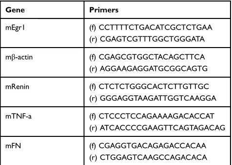
Total RNA was extracted from mice kidney cortices and SV40 MES 13 cells. cDNAs were synthesized from 0.5μg of total RNA in a 10μL reaction using MML-V reverse transcriptase (Invitrogen). Real-time quantitative PCR was performed in a Biosystems 4800 Real-Time PCR System as described previously.15The relative expression of each gene was esti-mated using the comparative 2−ΔΔCtmethod, withβ-actin as the reference. The PCR primers used in this study are listed in
Table 1.
Western Blot
Total protein was extracted from kidney tissue and SV40 MES 13 cells using RIPA lysis buffer (KeyGENbioTECH, China). The target protein was quantified using a previously described protocol,15using antibodies against TNF-α(1:400 dilution; Santa Cruz Biotechnology) and FN (1:500 dilution; Santa Cruz Biotechnology). Fluorescence
was obtained using an Odyssey Infrared Imaging System (LI-COR) and quantified by Gelpro32 software.
Kidney Histology and
Immunohistochemistry
Kidney tissue was embedded in paraffin and cut into 4μm sections and stained as outlined previously15using TNF-α -specific antibodies (1:400 dilution; Santa Cruz biotechnol-ogy) and FN (1:500 dilution; Santa Cruz Biotechnolbiotechnol-ogy). Sections were also stained with H&E and Masson’s Trichrome (Maiwei, Xiamen, China) using a standard pro-tocol. All sections were analyzed using an Olympus B ×40 upright light microscope (Olympus, Tokyo, Japan).
Statistical Analysis
Data values are described as mean ± SD. SPSS 25.0 was used for statistical analysis. The Student’st-test was used in the comparison of two groups, and p≤ 0.05 was con-sidered statistically significant.
Results
Inhibition of Egr1 Downregulated
Intrarenal Renin Was Increased by ACEI
in HFD/STZ-Induced DKD Mice
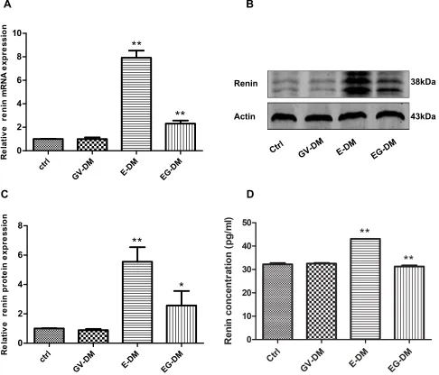
A DKD mouse model was successfully generated with the general characteristics shown inTable 2. The blood glucose levels of all groups were above 16.7 mmol/L. The glucose level, body weight, kidney weight index (kidney weight equal body weight), and creatinine level showed no inter-group difference (P> 0.05) (Table 2). After 4 weeks intervention, the renin mRNA and protein level increased 7.9-fold and 5.6-fold, respectively in the enalapril treatment group com-pared to the control group (P< 0.01) (Figure 1). Addition of shEgr-1 to the enalapril treatment reduced the renin protein and mRNA level by 70.8% and 53.7%, respectively (P< 0.05) (Figure 1). Urinary renin was found increased in the kidneys of DKD mice treated with enalapril vs controls Moreover, there was less urinary renin in the combined treatment group com-pared to the enalapril treatment group (Figure 1D).
Combination Therapy Alleviated Renal
Injury Further in HFD/STZ-Induced DKD
Mice
Egr1 mRNA expression decreased by 79% following injec-tion of the shEgr1 plasmid into the mouse tail (hydrody-namic gene delivery) every week for 4 weeks (Figure 2A), indicating that the Egr1 knockdown DKD model was
Table 1Sequences of Primers for Quantitative RT-PCR
Gene Primers
mEgr1 (f) CCTTTTCTGACATCGCTCTGAA
(r) CGAGTCGTTTGGCTGGGATA
mβ-actin (f) CGAGCGTGGCTACAGCTTCA
(r) AGGAAGAGGATGCGGCAGTG
mRenin (f) CTCTCTGGGCACTCTTGTTGC
(r) GGGAGGTAAGATTGGTCAAGGA
mTNF-a (f) CTCCCTCCAGAAAAGACACCAT
(r) ATCACCCCGAAGTTCAGTAGACAG
mFN (f) CGAGGTGACAGAGACCACAA
(r) CTGGAGTCAAGCCAGACACA
Diabetes, Metabolic Syndrome and Obesity: Targets and Therapy downloaded from https://www.dovepress.com/ by 118.70.13.36 on 24-Aug-2020
successful. We measured kidney injury among the four groups and observed that enalapril treatment reduced urin-ary microalbumin compared to the control group (35.8 ±
1.3/24 h vs 69.6 ± 3.7 μg/24 h) (P < 0.01). Combination therapy with shEgr1 plasmid and enalapril further alleviated urinary microalbumin by 39.6% (P < 0.01) (Figure 2B).
Renin
Actin
38kDa
43kDa
A
B
C
D
Figure 1Renin expression in DKD mice treated with oral enalapril (5 mg/150 mL water) and combined treatment (enalapril and pGPU6-shEgr1 plasmid). (A) Expression level of renin mRNA among the four groups of DKD mice. (BandC) Expression level of renin protein measured by Western blotting. (D) Urinary renin measured using ELISA. The results are expressed as fold change over baseline (control group). Values are represented as mean ± SD. *P < 0.05, **P < 0.01 vs ahead group by Student’st-test. Abbreviations:E-DM, Mice treated with oral enalapril (5 mg/150 mL water); EG-DM, mice treated with enalapril and pGPU6-shEgr1 plasmid; GV-DM, mice treated with pGPU6 vector plasmid (n = 6).
Table 2Metabolic Profile Analysis of Mouse Parameters
Parameters Ctrl GV-DM E-DM EG-DM
Glucose, mM 21.20±1.34 22.38±1.13 21.20±1.34 22.38±1.13
HbA1c, % 9.31±0.40 9.83±0.56 9.31±0.40 9.83±0.56
Renal weight index, g/kg 9.14±0.45 9.23±0.82 8.52±0.78 8.07±0.43
Body weight, g 24.31±1.97 23.30±1.46 24.570±1.26 25.03±1.06
Creatinine, M 177.67±12.54 174.15±2.31 180.23±6.37 164.29±4.26
Abbreviations:E-DM, mice treated with oral enalapril (5mg/150mL water); EG-DM, mice treated with enalapril and pGPU6-shEgr1 plasmid; GV-DM, mice treated with pGPU6 vector plasmid; Means±SD, n=6.
Diabetes, Metabolic Syndrome and Obesity: Targets and Therapy downloaded from https://www.dovepress.com/ by 118.70.13.36 on 24-Aug-2020
Quantitative RT-PCR assays showed that the mRNA level of TNF-α, a widely recognized inflammatory indicator of DKD, decreased by 33% (P < 0.01) in the enalapril treat-ment group compared to the control group, and decreased by 45% (P < 0.01) after combing shEgr1 with enalapril treatment (Figure 2C). FN, a widely recognized indicator of renal fibrosis in DKD, decreased by 31% in the enalapril treatment group compared to the control group, and decreased by 62% (P< 0.05) after combining shEgr1 with enalapril treatment (Figure 2C). H&E staining showed that the glomeruli volume and mesangial matrix appeared reduced in the enalapril treatment group compared to the control, and silence of Egr1 further improved this effect
(Figure 2C). Masson staining revealed obvious tubular
interstitial collagen deposition in both the control group and the enalapril treatment group, but thefibrosis alleviated after silence of Egr1 (Figure 2D). Immunohistochemistry
revealed that the protein expression of TNF-αand FN was downregulated in the enalapril treatment group, and further decreased after including shEgr1 treatment (Figure 2D). FN protein expression was confirmed further by Western blot
(Figure S1A). ELISA revealed that kidney TGF-β1 was also
downregulated in enalapril treatment group and further decreased after adding shEgr1 treatment (Figure S1B).
Upregulated and Downregulated Renin
Following Overexpression and Silence of
Egr1 in SV40 MES13 Cells
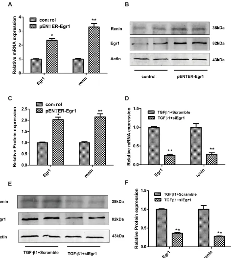
Egr1 mRNA and protein expression increased 2.3- and 2.0-fold after transfection of the SV40 MES13 cells with the Egr1 plasmid, respectively (P < 0.05) (Figure 3A–C), while renin mRNA and protein expression increased 3.3-and 2.2-fold, respectively (P < 0.01) (Figure 3A–C).
A
B
C
D
Ctrl
GV-DM
E-DM
EG-DM
TNF-α
TNF-α
Masson FN
H&E
Figure 2Kidney injury in DKD mice treated with oral enalapril (5 mg/150 mL water) and combined treatment (enalapril and pGPU6-shEgr1 plasmid). (A) Expression level of Egr1 mRNA among the three groups of DKD mice. The results are expressed as fold change over baseline (control group). (B) Urinary microalbumin concentration among the four groups of DKD mice. (C) Expression level of TNF-αand FN mRNA among the four groups of DKD mice. The results are expressed as fold change over baseline (control group). (D) H&E, Masson staining, and immunohistochemical staining of TNF-αand FN among the four groups of mice. Values are represented as mean ± SD. *P < 0.05, **P < 0.01 vs ahead group by Student’st-test.
Abbreviations:E-DM, Mice treated with oral enalapril (5 mg/150 mL water); EG-DM, mice treated with enalapril and pGPU6-shEgr1 plasmid; GV-DM, mice treated with pGPU6 vector plasmid (n = 6).
Diabetes, Metabolic Syndrome and Obesity: Targets and Therapy downloaded from https://www.dovepress.com/ by 118.70.13.36 on 24-Aug-2020
Transfection with siEgr1 reduced the mRNA and protein expression of Egr1 by 75% and 64%, respectively (P < 0.01) (Figure 3D–F), and reduced the renin mRNA and protein expression by 72%, respectively (P < 0.01) (Figure 3D–F).
Discussion
Diabetes is a major public issue worldwide. DKD is one of the most common and serious complications of diabetes and is the leading cause of end-stage renal disease (ESRD).21,22 Although ACEI and ARB, as renin-angiotensin-system blockage, have been used in the treatment of DKD for many years, the compensatory increase of intrarenal renin after ACEI and ARB treatment limits their positive effects. Many factors have been explored to inhibit the feedback increase in renin, but there is no clear breakthrough so far. Our study offers a new method to target renin feedback by demonstrating that inhibition of Egr1 could downregulate intrarenal renin in DKD mice. Enalapril treatment increased intrarenal renin expression in DKD mice, and silencing Egr1 combined with enalapril treatment decreased intrare-nal renin expression vs the eintrare-nalapril treatment group. Moreover, enalapril treatment alleviated renal injury, and silencing Egr1 combined with enalapril treatment alleviated renal injury further. Finally, the renin levels were upregu-lated and downreguupregu-lated following overexpression and silencing of Egr1 in SV40 MES13 cells, respectively. Because Egr1 may bind to the renin gene promoter, as determined by bioinformatic analysis (http://jaspar.genereg. net/), we speculated that silencing of Egr1 may inhibit renin expression at the transcriptional level.
The increase in renin is related to the affinity of ACEI to tissue ACE (ACE includes both plasma and tissue types).23Quantitatively, more than 90% of ACE is tissue-bound, and plasma ACE accounts for less than 10% of the total ACE.24The limitation of the efficacy of ACEI is also partly due to the lack of inhibition of tissue ACE. Many previous studies have found that the relative rank order of tissue potency of ACEI is as follow: quinaprilat = bena-zeprilat > ramiprilat > perindoprilat > lisinopril > enala-prilat > fosinoenala-prilat > captopril.25
Many previous studies have attempted to develop methods to inhibit the expression of intrarenal renin. A study by Zhongyi Zhang showed that losartan could increase local AngII and renin in the kidney of DKD mice induced by STZ, although the degree of renal fibrosis was only slightly alleviated. Following the addition of Vitamin
D analogs (paricalcitol, 19-nor-1, 25-dihydroxyvitamin D2), the expression of renin was decreased, renalfibrosis was significantly alleviated, proteinuria was prevented, the glomerular filtration barrier was restored, and glomerular sclerosis was significantly reduced. Vitamin analogs were thought to inhibit renin production at the transcriptional level.26 A study by Xiaoyue Tan showed that the use of doxorubicin alone could increase the renin in mice kidney with obstructive renal disease by unilateral ureteral liga-tion. Following addition of the vitamin D analog parical-citol, renin expression, interstitial fibrosis, and inflammation were reduced.6Zhoulili found that ICG-001 (a small-molecule b-catenin inhibitor) could reduce expression of renin-angiotensin-system, including angio-tensin, renin, ACE, and angiotensin type 1 Receptor in a mouse model of nephropathy induced by Adriamycin.27 Renin antagonists-aliskiren was once used as a clinical therapy, but was removed from the market due to its risk of nonfatal stroke, kidney complications, high blood potas-sium, and low blood pressure in people with diabetes and kidney impairment.28 In this study, we found that knock-down of Egr1 could reduce the expression of intrarenal renin. Our study extends the previous study and provides a method to inhibit intrarenal renin in DKD mice.
Tumor necrosis factor a (TNF-a) is a type II transmem-brane protein that plays an important role in the progress of DKD. TNF-a can induce expression of adhesion molecules and chemokines,29 promote cytotoxicity and apoptosis/ necrotic necrosis of sensitive cells,30 alter intraglomerular blood flow and GFR, increase endothelial permeability,31 and induce oxidative stress.32Moreover, TNF-a has increas-ingly become a biomarker in clinical and experimental DKD patients, as well as a therapeutic target.33FN, an important component of the extracellular matrix (ECM), is a marker of DKD.34Accordingly, we used both TNF-a and FN as mar-kers of kidney injury of DKD. Masson staining was used to display tissuefibers (collagen, blue; muscle, red) by mixing with two or three anionic dyes, and could accurately reflect renalfibrosis in DKD. Proteinuria was an important marker of DKD and could also be used for diagnosis and judging the DKD stage.35In the present study, we observed that enala-pril treatment could downregulate TNF-a, FN, and protei-nuria, as well as improve the pathology. When combined with the shEgr1 plasmid, enalapril could downregulate TNF-a, FN, and proteinuria further, as well as improve the kidney pathology to a greater extent.
In summary, enalapril treatment induced compensa-tory intrarenal renin, reducing the efficacy of
renin-Diabetes, Metabolic Syndrome and Obesity: Targets and Therapy downloaded from https://www.dovepress.com/ by 118.70.13.36 on 24-Aug-2020
angiotensin-system inhibition. Egr1 was an important transcriptional factor in the progress of DKD, and silen-cing Egr1 could downregulate renin feedback and alle-viate renal injury. In addition, renin increased and
decreased following Egr1 overexpression and silencing in SV40 MES 13, respectively. Therefore, silencing Egr1 might suppress compensatory renin increase at the transcriptional level.
38kDa
43kDa 82kDa Renin
Actin Egr1
control pENTER-Egr1
Renin
Actin Egr1
TGF-β1+Scramble
38kDa
43kDa 82kDa
TGF-β1+siEgr1
A
C
E
F
D
B
Figure 3Renin expression following either overexpression or knockdown of Egr1 in SV40 MES 13 cells. (A) Cells were treated with either a pENTER-Egr1 overexpression plasmid or with a pENTER vector for 48 h, and the mRNA levels of Egr1 and renin were measured by RT-qPCR. (BandC) The protein levels of Egr1 and renin were measured by Western blotting. (D) Cells were either silenced with siEgr1 or treated with a scrambled control RNA for 48 h prior to exposure to TGF-β1 (10 ng/mL) for 24 h. The mRNA levels of Egr1 and renin were measured using RT-qPCR. (EandF) The mRNA levels of Egr1 and renin were measured by Western blotting. The results are expressed as fold change over baseline. Values are represented as mean ± SD. *P < 0.05, **P < 0.01 vs control group by Student’st-test (n = 3).
Diabetes, Metabolic Syndrome and Obesity: Targets and Therapy downloaded from https://www.dovepress.com/ by 118.70.13.36 on 24-Aug-2020
Conclusion
Egr1 is an important transcriptional factor in DKD, and inhibition of Egr1 may delay DKD injury by downregulating the compensatory renin increase at the transcriptional level.
Acknowledgments
We thank Shenqiao Li in the Department of Traditional Chinese Medicine at the Fifth Affiliated Hospital Sun Yat-Sen University (Guangdong, China) for Guidance in writ-ing. This work was supported by the Natural Science Foundation of Guangdong Province (grant number: 2018A0303130263) and Guangdong Medical Scientific Research Project (grant number: A2018142).
Disclosure
The authors have no conflicts of interest to report.
References
1. Wang L, Gao P, Zhang M, et al. Prevalence and ethnic pattern of diabetes and prediabetes in China in 2013.JAMA.2017;317:2515. doi:10.1001/jama.2017.7596
2. Xue R, Gui D, Zheng L, Zhai R, Wang F, Wang N. Mechanistic insight and management of diabetic nephropathy: recent progress and future perspective.J Diabetes Res.2017;2017:1–7. doi:10.1155/2017/1839809 3. Yacoub R, Campbell KN. Inhibition of RAS in diabetic nephropathy.
Int J Nephrol Renovasc Dis.2015;8:29–40. doi:10.2147/IJNRD.S37893 4. St PW, Odum LE, Whaley-Connell AT. To RAS or not to RAS? The
evidence for and cautions with renin-angiotensin system inhibition in patients with diabetic kidney disease. Pharmacotherapy. 2013;33: 496–514. doi:10.1002/phar.1232
5. Nakamura T, Fujiwara N, Kawagoe Y, Sugaya T, Ueda Y, Koide H. Effects of telmisartan and enalapril on renoprotection in patients with mild to moderate chronic kidney disease. Eur J Clin Invest.
2010;40:790–796. doi:10.1111/j.1365-2362.2010.02319.x
6. Tan X, He W, Liu Y. Combination therapy with paricalcitol and trandolapril reduces renal fibrosis in obstructive nephropathy.
Kidney Int.2009;76(12):1248–1257. doi:10.1038/ki.2009.346 7. Borghi C, Boschi S, Ambrosioni E, Melandri G, Branzi A,
Magnani B. Evidence of a partial escape of renin-angiotensin-aldosterone blockade in patients with acute myocardial infarction treated with ACE inhibitors. J Clin Pharmacol. 1993;33:40–45. doi:10.1002/j.1552-4604.1993.tb03901.x
8. Roig E, Perez-Villa F, Morales M, et al. Clinical implications of increased plasma angiotensin II despite ACE inhibitor therapy in patients with congestive heart failure.Eur Heart J.2000;21:53–57. doi:10.1053/euhj.1999.1740
9. Sakoda M, Ichihara A, Kaneshiro Y, et al. (Pro)renin receptor-mediated activation of mitogen-activated protein kinases in human vascular smooth muscle cells. Hypertens Res. 2007;30:11 39–1146. doi:10.1291/hypres.30.1139
10. Huang Y, Noble NA, Zhang J, Xu C, Border WA. Renin-stimulated TGF-beta1 expression is regulated by a mitogen-activated protein kinase in mesangial cells.Kidney Int.2007;72:45–52. doi:10.1038/ sj.ki.5002243
11. Wang WN, Zhang WL, Zhou GY, et al. Prediction of the molecular mechanisms and potential therapeutic targets for diabetic nephropa-thy by bioinformatics methods.Int J Mol Med.2016;37:1181–1188. doi:10.3892/ijmm.2016.2527
12. Pagel J, Deindl E. Disease progression mediated by Egr-1 Associated signaling in response to oxidative stress. Int J Mol Sci.
2012;13:13104–13117. doi:10.3390/ijms131013104
13. Zcharia E, Atzmon R, Nagler A, et al. Inhibition of matrix metalloproteinase-2 by halofuginone is mediated by the Egr1 tran-scription factor.Anticancer Drugs.2012;23:1022–1031. doi:10.1097/ CAD.0b013e328357d186
14. Wang D, Guan M, Zheng Z, et al. Transcription factor Egr1 is involved in high glucose-induced proliferation and fibrosis in rat glomerular mesangial cells. Cell Physiol Biochem. 2015;36:20 93–2107. doi:10.1159/000430177
15. Hu F, Xue M, Li Y, et al. Early Growth Response 1 (Egr1) Is a Transcriptional activator of NOX4 in oxidative stress of diabetic kidney disease. J Diabetes Res. 2018;2018:3405695. doi:10.1155/ 2018/3405695
16. Xu P, Guan M, Bi J, Wang D, Zheng Z, Xue Y. High glucose down-regulates microRNA-181a-5p to increase pro-fibrotic gene expression by targeting early growth response factor 1 in HK-2 cells.Cell Signal.2017;31:96–104. doi:10.1016/j.cellsig.2017.01.012 17. Li Y, Hu F, Xue M, et al. Klotho down-regulates Egr-1 by inhibiting TGF-β1/Smad3 signaling in high glucose treated human mesangial cells. Biochem Bioph Res Co. 2017;487:216–222. doi:10.1016/j. bbrc.2017.04.036
18. Ho L, Sung J, Shen Y, et al. Egr-1 deficiency protects from renal inflammation andfibrosis.J Mol Med.2016;94:933–942. doi:10.10 07/s00109-016-1403-6
19. Suda T. Hydrodynamic gene delivery its principles.Mol Ther.2007. doi:10.1038/sj.mt.6300314
20. Zhou L. Klotho ameliorates kidney injury andfibrosis and normalizes blood pressure by targeting the renin-angiotensin system. Am J Pathol.2015;185:3211–3223. doi:10.1016/j.ajpath.2015.08.004 21. Saran R, Li Y, Robinson B, et al. US renal data system 2014 annual
data report: epidemiology of kidney disease in the United States.Am J Kidney Dis.2015;66:S1–S305. doi:10.1053/j.ajkd.2015.05.001 22. Hill CJ, Fogarty DG. Changing trends in end-stage renal disease due
to diabetes in the United kingdom. J Ren Care. 2012;38:12–22. doi:10.1111/j.1755-6686.2012.00273.x
23. Mizuiri S. ACE and ACE2 in kidney disease. World J Nephrol.
2015;4:74. doi:10.5527/wjn.v4.i1.74
24. Cushman DW, Cheung HS. Concentrations of angiotensin-converting enzyme in tissues of the rat. Biochim Biophys Acta. 1971; 250:261–265. doi:10.1016/0005-2744(71)90142-2
25. Dzau VJ, Bernstein K, Celermajer D, et al. Pathophysiologic and ther-apeutic importance of tissue ACE: a consensus report. Cardiovasc Drugs Ther.2002;16:149–160. doi:10.1023/A:1015709617405 26. Zhang Z, Zhang Y, Ning G, Deb DK, Kong J, Li YC. Combination
therapy with AT1 blocker and vitamin D analog markedly amelio-rates diabetic nephropathy: blockade of compensatory renin increase.
Proc Natl Acad Sci U S A. 2008;105:15896–15901. doi:10.1073/ pnas.0803751105
27. Zhou L, Li Y, Hao S, et al. Multiple genes of the renin-angiotensin system are novel targets of Wnt/beta-catenin signaling. J Am Soc Nephrol.2015;26:107–120. doi:10.1681/ASN.2014010085 28. Pantzaris N, Karanikolas E, Tsiotsios K, Velissaris D. Renin
Inhibition with Aliskiren: a decade of clinical experience. J Clin Med.2017;6:61. doi:10.3390/jcm6060061
29. Ortiz A, Bustos C, Alonso J, et al. Involvement of tumor necrosis factor-alpha in the pathogenesis of experimental and human glomer-ulonep hritis.Adv Nephrol Necker Hosp.1995;24:53–77.
30. Boyle JJ, Weissberg PL, Bennett MR. Tumor necrosis factor-alpha promotes macrophage-induced vascular smooth muscle cell apoptosis by direct and autocrine mechanisms.Arterioscler Thromb Vasc Biol.
2003;23:1553–1558. doi:10.1161/01.ATV.0000086961.44581.B7 31. McKenzie JA, Ridley AJ. Roles of Rho/ROCK and MLCK in
TNF-alpha-induced changes in endothelial morphology and permeability. J Cell Physiol.2007;213:221–228. doi:10.1002/jcp.21114
Diabetes, Metabolic Syndrome and Obesity: Targets and Therapy downloaded from https://www.dovepress.com/ by 118.70.13.36 on 24-Aug-2020
32. Radeke HH, Meier B, Topley N, Ge J F, Habermehl GG, Resch K. Interleukin 1-alpha and tumor necrosis factor-alpha induce oxygen radical production in mesangial cel ls.Kidney Int.1990;37:767–775. doi:10.1038/ki.1990.44
33. Barutta F, Bruno G, Grimaldi S, Gruden G. Inflammation in diabetic nephropathy: moving toward clinical biomarkers and targets for treatment.Endocrine.2015;48:730–742. doi:10.1007/s12020-014-0437-1 34. Jia Y, Zheng Z, Guan M, et al. Exendin-4 ameliorates high glucose-induced fibrosis by inhibiting the secretion of miR-192 from injured renal tubular epithelial cells.Exp Mol Med.2018;50 (5):1–13. doi:10.1038/s12276-018-0084-3
35. Aziz KM. Correlation of urine biomarkers: microalbuminuria and spot urine protein among diabetic patients. application of spot urine protein in diabetic kidney disease, nephropathy, proteinuria estimation, diagnosing and monitoring. Recent Pat Endocr Metab Immune Drug Discov.
2015;9:121–133. doi:10.2174/1872214809666150708111022
Diabetes, Metabolic Syndrome and Obesity: Targets and Therapy
Dove
press
Publish your work in this journal
Diabetes, Metabolic Syndrome and Obesity: Targets and Therapy is an international, peer-reviewed open-access journal committed to the rapid publication of the latest laboratory and clinicalfindings in the fields of diabetes, metabolic syndrome and obesity research. Original research, review, case reports, hypothesis formation, expert opinion
and commentaries are all considered for publication. The manu-script management system is completely online and includes a very quick and fair peer-review system, which is all easy to use. Visit http://www.dovepress.com/testimonials.php to read real quotes from published authors.
Submit your manuscript here:https://www.dovepress.com/diabetes-metabolic-syndrome-and-obesity-targets-and-therapy-journal
Diabetes, Metabolic Syndrome and Obesity: Targets and Therapy downloaded from https://www.dovepress.com/ by 118.70.13.36 on 24-Aug-2020