Programmed Cell Death during Heart
Development
Pundrique R. Sharma
A thesis submitted for the degree o f Doctor o f Philosophy in the
University o f London
2001
Neural Development Unit,
Institute o f Child Health,
ProQuest Number: 10016046
All rights reserved
INFORMATION TO ALL USERS
The quality of this reproduction is dependent upon the quality of the copy submitted. In the unlikely event that the author did not send a complete manuscript and there are missing pages, these will be noted. Also, if material had to be removed,
a note will indicate the deletion.
uest.
ProQuest 10016046
Published by ProQuest LLC(2016). Copyright of the Dissertation is held by the Author. All rights reserved.
This work is protected against unauthorized copying under Title 17, United States Code. Microform Edition © ProQuest LLC.
ProQuest LLC
789 East Eisenhower Parkway P.O. Box 1346
A
c k n o w l e d g e m e n t s
This project would have been impossible without three people: Professor Andy Copp, without whose personal efforts this project would not have even got off the ground, and who has always made time whenever I ’ve needed help or advice; Professor Bob Anderson, whose passion for the heart first inspired me to study cardiac
development, and, of course. Dr. Deb Henderson who is a supervisor o f the very highest quality and has put up remarkably well with the daily trauma o f supervising me.
My thanks also go to all the members, past and present, o f the Neural Development Unit for making my time in the group most enjoyable. I would particularly like to thank Dr. Viccy Reed, for “heart-to-heart” conversations. Dr. Dorothea Stiefel, for trying (unsuccessfully) to culture me, and Dr. Andrew Hynes for our Friday runs. I’d also like to thank Dr. Nick Greene, Dr. Jonathan Ham, Dr. Jenny Murdoch and Sarah Reid for their advice in writing this thesis.
For their help with specific experimental procedures I wish to thank Dr. Takashi Shibata, Louisa Dunlevy and Dr. Maria Kolatsi-Joannou in the Neural Development Unit, and Brian Young and Professor David Landon at the Institute of Neurology, London. Also, for the provision o f the F4/80 antibody, I would like to thank Professor Siamon Gordon at Oxford and Dr. Paul Martin at University College London (through whose fault, in part. I’ve wound up doing this PhD). The F ADD dominant negative embryos and the human F ADD antibody were kindly provided by Dr. Anne Odile- Hueber, who currently works in Nice, France.
Et, Marie-Pierre, merci pour tout, et en particulier ton soutien et pour m ’avoir rappelé qu’il y a “un lumière au bout du TUNEL”.
A
b s t r a c t
Programmed cell death has been demonstrated to be of importance in
mammalian organogenesis. In this thesis, the distribution, regulation and function of programmed cell death are investigated during mouse cardiac septation. During this process, the four chambers and two outflow vessels of the heart become separated from one another. In the mouse, this occurs between embryonic days 10.5 to 13.5. The study commences with a detailed spatio-temporal analysis of programmed cell death during cardiac septation. This demonstrates specific, reproducible foci of apoptotic activity, which are grouped into low intensity and high intensity, the latter occurring in the endocardial cushions as septation completes. This qualitative investigation is then supplemented, and supported, by a quantitative analysis. Immunohistochemical
techniques demonstrate which cell types undergo apoptosis during cardiac septation and show that macrophages are not clearing apoptotic debris. The question of an
association between cardiac neural crest cells and programmed cell death during the process of septation is addressed using double-labelling for a cardiac neural crest marker, a-smooth muscle actin, and an apoptosis marker, TUNEL, and by investigation of apoptosis in the mutant mouse, in which neural crest cell migration to the
cardiac outflow tract is impaired. The results suggest that neural crest cells are involved in apoptosis, and may indeed be dying, within the cardiac outflow tract cushions. The molecular mechanisms that lead to programmed cell death in the heart are then
A
b b r e v i a t i o n s
MHC = myosin heavy chain ADP = adenosine di-phosphate
AIDS = acquired immunodeficiency syndrome ANOVA= analysis of variance
ANF = atrial natriuretic factor AP-1 = activating protein -1
Apaf-1= apoptotic protease-activating factor-1 ATP = adenosine tri-phosphate
Bel = B-cell lymphoma BH = Bcl-2 homology
BMP = bone morphogenetic protein bp = base pairs
C. elegans = Caenorhabditis elegans
CAD = caspase activated DNAse
DAB = diaminobenzadine tetrahydrochloride DAPI = 4,6-diamindino-2-phenyl indole DD = death domain
DED = death effector domain
DISC = death inducing signalling complex DMEM = Dulbecco’s Modified Eagles Medium DMSO = dimethyl sulphoxide
E = embryonic day
EDTA = ethylenediamine-tetra-acetic acid F ADD = Fas associated death domain FCS = fetal calf serum
FLICE = FADD-like IL -lp converting enzyme FLIP = FLICE inhibitory protein
HIV = human immunodeficiency virus LAP = inhibitor of apoptosis protein I-kB = inhibitor o f NF-kB
IKK = I-kB kinase IL = interleukin
IMS = industrial methylated spirits JNK = Jun amino-terminal kinase JNKK = JNK kinase
JPEG = Joint Photographic Experts Group Msx = muscle segmental homeobox-2 NCAM = neural cell adhesion molecule NF = Neurofibromin
NF-KB = nuclear factor kB
Pax = Paired box
PARP = poly-ADP ribose polymerase PBS = phosphate buffered saline PCD = programmed cell death PCR = polymerase chain reaction PFA = paraformaldehyde
RAR = retinoic acid receptor RIP = receptor interacting protein RNA = ribonucleic acid
RXR = retinoid X receptor
SERCA2a = sarcoplasmic reticulum Ca^"^ ATPase Shh = sonic hedgehog
TdT = terminal deoxynucleotidyl transferase TAB = Tris-acetate EDTA
TBS-TX = Tris buffered saline/Triton-Xl 00 TESPA = 3-aminopropyltriethoxy silane TGF = transforming growth factor TIE = tagged image format
TNF = tumour necrosis factor
TNFR = tumour necrosis factor receptor TRADD = TNFR-associated death domain
TUNEL = TdT-mediated dUTP nick end labelling VCAM-1 = vascular cell adhesion molecule WT = Wilm’s tumour
T
a b l e o fC
o n t e n t sAc k n o w l e d g e m e n t s 3
Ab s t r a c t 4
Ab b r e v ia t io n s 5
Ta b l eo f Co n t e n t s 8
Lis to f Ta b l e s 18
Lis to f Fig u r e s 20
CHAPTER 1 Ge n e r a l In t r o d u c t io n 25
1.1 Programmed cell death 27
1.1.1 Overview and history of programmed cell death 28 1.1.2 Characteristic features of PCD and their molecular basis 31 1.1.3 Molecular pathways leading to PCD 34
1.1.3.1 PCD following cellular stress 34
1.1.3.2 Death receptor-mediated PCD 39
1.1.3.3 Cross-talk between stress-mediated and
death receptor-mediated apoptosis 43
1.1.4 Clearance of apoptotic cells 44 1.1.5 Necrosis and apoptosis 46 1.1.6 Programmed cell death in development 46 1.1.7 Programmed cell death in pathology and physiology 48
1.2 Cardiac development 50
1.2.1 Comparative time-line of cardiac development 51 1.2.2 Formation of the primary heart tube and cardiac looping 52 1.2.3 Genetic pre-specification of the cardiac compartments:
1.2.5 Re-alignm ent o f the cardiac channels 60
1.2.6 M orphological changes during cardiac septation 62
1.2.6.1 A trial septation 62
1.2.6.2 Ventricular septation 65
1.2.6.3 Atrioventricular septation: the central m esenchym al mass 68
1.2.6.4 Outflow tract - definitions 71
1.2.6.5 Outflow tract septation 73
1.2.6.6 Other events in outflow tract septation 80
1.2.7 The role o f epicardial cells in cardiac developm ent 81
1.3 Program m ed cell death during cardiac septation 84
1.3.1 The study o f cardiac PCD to date 84
1.3.2 Spatio-tem poral location o f PCD during cardiac septation 86
1.3.3 Cell types undergoing apoptosis during cardiac septation 88
1.3.4 Clearance o f apoptosis during cardiac developm ent 90
1.3.5 The m olecular controls and the effects o f abnorm al PCD
during cardiac septation 91
1.4 Aims 95
CHAPTER 2 Ma t e r ia l sa nd Me th o d s 96
2.1 General considerations 97
2.2 M aintenance of mouse colonies 97
2.2.1 C D l and Splotch (Sp^^) mice - colony conditions 97
2.2.2 Breeding o f CD 1 and Sp^^ mice 97
2.2.3 FADD dom inant negative mice 98
2.3 Dissection, storage and staging of embryos 99
2.3.2 Dissection of embryos 99
2.3.3 Fixation, storage and paraffin embedding of embryos 99
2.3.4 Fixation and preparation of embryos for cryosectioning 100 2.3.5 Staging of embryos 101 2.3.6 Division of embryos into somite groups 101
2.4 Genotyping of embryos 103
2.4.1 PCR of DNA from embryos 103 2.4.2 Electrophoresis on agarose gels 105 2.4.3 PCR of FADD dominant-negative embryos 105
2.5 Histology, Immunohistochemistry and TUNEL 108
2.5.1 TESPA-coating of slides 108 2.5.2 Sectioning of wax embedded tissue 108
2.5.3 Cryosectioning 108
2.5.4 Methyl green staining 110 2.5.5 Immunohistochemistry procedures 110
2.5.6 TUNEL 114
2.5.7 Double-labelling 115
2.6 Open yolk sac, whole embryo culture 117
2.6.1 Preparation of rat serum 117 2.6.2 Dissection of embryos for culture 117 2.6.3 In vitro culture conditions 120
2.7 Image capture and presentation 122
2.7.1 Digital image capture 122 2.7.2 Digital capture of fluorescence 122
2.8 Data summation and analysis 124
2.8.1 Templating 124
2.8.2 Cell counting 127 2.8.3 Length of the interventricular septum 130 2.8.4 Templating of FADD dominant negative embryos 132 2.8.5 Blind analysis of interventricular septal development
following embryo culture 132 2.8.6 Statistical analysis and graphs 133 2.9 Transmission electron microscopy 134 CH APTER 3 Sp a t io-t e m p o r a ld is t r ib u t io no fp r o g r a m m e d
CELL DEATH DURING CARDIAC SEPTATION 135
3.1 Introduction 136
3.1.1 Detection of apoptosis 137
3.2 Results 139
3.2.1 Positive and negative controls 139 3.2.2 High levels of PCD occur in the endocardial cushions
atE12.5toE13.5 141
3.2.2.1 PCD in the atrioventricular cushions 142
3.2.2.2 PCD in the outflow tract cushions 145
3.2.3 Confirmation of high intensity PCD within the outflow
tract cushions by transmission electron microscopy 149 3.2.4 Confirmation of high intensity PCD within the
atrioventricular cushions by cleaved-caspase-3
immunohistochemistry 150 3.2.5 Low intensity PCD foci occur throughout the heart during
cardiac septation 153
3.2.5.1 Atrial PCD 153
3.2.5.2 Ventricular PCD 161
3.2.5.3 Outflow tract PCD 168
3.3 Discussion 173
3.3.1 TUNEL is an appropriate method for detection of PCD
in this study 173
3.3.2 The templating technique is a useful tool for the summation
of data in regions where levels of PCD are low 176 3.3.3 The distinction between high and low intensity foci of PCD 178 3.3.4 High intensity foci of PCD in the atrioventricular region
develop as the central mesenchymal mass forms 179 3.3.5 High intensity PCD in the proximal outflow tract
cushions occurs as this structure is myocardialised and fuses 182 3.3.6 Low intensity PCD foci are frequently associated with
septal structures 183
5.3.6.1 Low intensity PCD and the morphogenesis o f septal structures 184 3.3.6.2 Low intensity PCD and outflow tract morphogenesis 186
3.3.6.3 Symmetry o f low intensity PCD 187
3.4 Summary 188
CHAPTER 4 Qu a n t it a t iv ea n a l y s iso fp r o g r a m m e d c e l ld e a t h
DURING CARDIAC SEPTATION 189
4.1 Introduction 190
4.2 Results 192
4.2.1 Difference between estimated and actual cell number, and estimated and actual PCD index in the
4.2.2 Cell number and PCD index changes during cardiac septation 194 4.2.3 Statistical analysis of PCD indices 198 4.2.4 Comparisons of PCD indices 204
4.2.4.1 General observations 204
4.2.4.2 Atria 204
4.2.4.3 Ventricles 206
4.2.4.4 Outflow tract 208
4.2.4.5 Endocardial cushion tissues 209
4.2.4.6 The outflow tract and ventricular walls 209
4.3 Discussion 210
4.3.1 Quantitation of cell numbers using estimates from
templates is accurate 210 4.3.2 Quantitation confirms trends observed by
templating and detects previously unobserved trends 211 4.3.3 Relationship of cell number to PCD index 213 4.3.4 Is outflow tract shortening related to myocardialisation
of the outflow tract septum? 216
4.4 Summary 219
CHAPTER 5 Pr o g r a m m e dc e l ld e a t hino u t f l o w
TRACT SEPTATION 220
5.1 Introduction 221
5.2 Results 224
5.2.1 Controls 224
5.2.2 Imaging of double-labelled sections 226 5.2.3 PCD in the outflow tract walls occurs in the epicardium
and the myocardium 229
5.2.4 High intensity PCD in the proximal outflow tract
cushions occurs in non-myocardialised cells 229 5.2.5 A diffuse group o f cells labelled with a-smooth
muscle actin co-localises with high intensity PCD
within the outflow tract cushions at E13.5 235 5.2.6 Transmission electron microscopy of the septation complex 239 5.2.7 PCD in the atrioventricular cushions of Sp^^/Sp^^ embryos
is normal 239
5.2.8 High intensity PCD foci are absent from the outflow tract cushions o f Sp^^/Sp^^ embryos at E l2.5, and abnormally positioned within the proximal outflow tract
cushions of Sp^^/Sp^^ embryos at E13.5 242 5.2.9 Macrophages do not localise to foci of PCD during
cardiac septation 248
5.3 Discussion 252
5.3.1 Apoptotic cells are likely to have been phagocytosed by
neighbouring cells in the same cell layer 252 5.3.2 A potential role for PCD in the shortening of the outflow tract
wall 254
5.3.3 Myocardialisation has a complementary association with
high intensity PCD in the outflow tract cushions 255 5.3.4 Distribution of neural crest cells in the outflow tract cushions 256 5.3.5 Neural crest cells are related to PCD in the
outflow tract cushions 256 5.3.6 Are neural crest cells dying in the outflow tract cushions? 259 5.3.7 Possible relationship of PCD, myocardialisation and
cardiac neural crest cells 262 5.3.8 Clearance of PCD during cardiac septation is by local
cells and not by macrophages 263
5.4 Sum m ary 267
CH A PTER 6 De a t hr e c e p t o r-m e d ia t e da p o p t o s is
DURING CARDIAC SEPTATION 268
6.1 Introduction 269
6.2 Results 271
6.2.1 Controls 271
6.2.2 Location of the FADD dominant-negative construct 273 6.2.3 Cardiac morphology of FADD dominant-negative
transgenic mice 273
6.2.4 Reduction in apoptosis within low intensity PCD foci in the hearts of embryos expressing the FADD
dominant-negative construct 273 6.2.5 Quantification of apoptosis confirms a reduction in PCD
in the hearts of embryos expressing the dominant-negative
construct 277
6.2.6 During septation, Fas is expressed in the heart, but
not in the endocardial cushions 281 6.2.7 Fas ligand is expressed in regions in which low intensity
foci of PCD occur 285
6.3 Discussion 288
6.3.1 Death receptor-mediated PCD is required for cardiac
apoptosis in development 288 6.3.2 A role for Fas and Fas ligand in apoptosis during
cardiac septation 293
6.4 Summary 295
CHAPTER 7 Th er o l eo fp r o g r a m m e d c e l ld e a t h int h e e a r l y
DEVELOPMENT OF THE INTERVENTRICULAR SEPTUM 296
7.1 Introduction 297
7.2 Results 300
7.2.1 Controls 300
7.2.2 Epicardial cells do not become trapped within the
interventricular septum 300 7.2.3 Early changes in interventricular septal length 302 7.2.4 The early interventricular septum consists of myocardium
and endocardial channels, both of which contain PCD 302 7.2.5 Embryos grown in vitro develop normally 307 7.2.6 Cardiac morphology of control embryos grown
in vitro is normal 311
7.2.7 PCD inhibition by zVAD-fmk 313 7.2.8 Abnormal development of the interventricular septum
in embryos following inhibition of PCD by zVAD-fmk 315
7.3 Discussion 319
7.3.1 The morphology and histology of the early development o f the interventricular septum is consistent with a
trabecular-fusion model 319 7.3.2 PCD in the interventricular septum: a role in the
removal of endocardial channels 321 7.3.3 General development of embryos cultured in zVAD-fmk
is grossly normal 322
7.3.4 Inhibition o f PCD in embryo culture perturbs
development of the interventricular septum 323
7.4 Summary 326
CHAPTER 8 Ge n e r a l Dis c u s s io n 327
8.1 Possible improvements and sources of error 328
8.1.1 Sample numbers 328 8.1.2 Primary myocardium versus working myocardium 329 8.1.3 Cleaved-caspase-3 immunohistochemistry of
cultured embryos 329
8.1.4 Negative controls 330
8.2 Future work 330
8.2.1 Programmed cell death and outflow tract development 330 8.2.2 The control of apoptosis in low intensity
foci of PCD 333
8.2.3 PCD in avian and human hearts 333
8.3 Programmed cell death outside the heart 334
8.3.1 Apoptosis adjacent to the heart 334 8.3.2 Apoptosis in the tracheal diverticulum 336
8.4 Concluding remarks 338
Bib l io g r a p h y 33 9
L
i s t
o f
T
a b l e s
Table Page
Table 1.1 Studies o f apoptosis during cardiac development 85 Table 2.1 Reagents, and their sources, used in PCR mix 104 Table 2.2 Details and sources of antibodies used in immunohistochemistry 112 Table 2.3 Explant and culture medium for whole embryo culture 118 Table 2.4 PCD index in the interventricular septum of 55-57 somite
embryos 131
Table 3.1 Summary of PCD foci 172 Table 4.1 Comparison of actual and estimated cell numbers in the
interventricular septum 193 Table 4.4 Comparison of PCD indices in atrial structures
(part of Figure 4.4) 199
Table 4.5 Comparison o f PCD indices in ventricular structures
(part o f Figure 4.5) 200 Table 4.6 Comparison of PCD indices in the outflow tract
(part of Figure 4.6) 201 Table 4.7 Comparison of PCD indices in endocardial cushion tissues
(part o f Figure 4.7) 202 Table 4.8 Comparison o f PCD indices in non-trabeculated ventricular
myocardium and the outflow tract wall (part of Figure 4.8) 203 Table 6.1 Comparison o f foci o f cell death in FADD dominant
negative hemizygous embryos and wild type littermates
at 55-57 somites 279
CDl embryos 287 Table 7.1 Developmental progression of zVAD-fmk treated and
DMSO-treated embryos in whole-embryo culture 310
L
i s t
o f
F
i g u r e s
Figure Page
Figure 1.1 Overview of the molecular and cellular aspects of PCD^ 32 Figure 1.2 Stress-mediated apoptosis 36 Figure 1.3 Death receptor-mediated apoptosis 40 Figure 1.4 Cross-talk between death receptor-mediated apoptosis
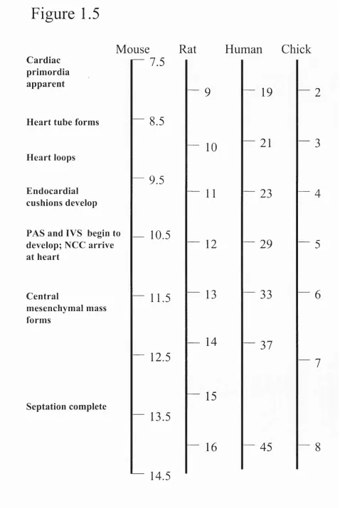
and stress-mediated apoptosis 45 Figure 1.5 Temporal comparison of events in cardiac development
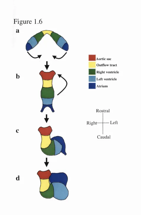
in different species 53 Figure 1.6 The paired cardiac primordia and looping 54 Figure 1.7 Early chamber-specific gene expression 58 Figure 1.8 Realignment of the outflow tract, ventricles and atria 61 Figure 1.9 Atrial septation 63 Figure 1.10 Development of the interventricular septum 66 Figure 1.11 Possible mechanisms for the development of the
interventricular septum 67 Figure 1.12 The central mesenchymal mass 70 Figure 1.13 Outflow tract terminology 72 Figure 1.14 Outflow tract septation I 74 Figure 1.15 Outflow tract septation II 79 Figure 2.1 Developmental staging 102 Figure 2.2 PCR genotyping of litters 106 Figure 2.3 Transverse sectioning of embryos 109 Figure 2.4 Dissection for whole embryo culture 119
Figure 2.5 Templates 125
Figure 2.6 Morphological comparison between sections
made for templating and TUNEL-assayed sections 126 Figure 2.7 Summation of data by templating 128 Figure 2.8 Assessment of cell number 129 Figure 3.1 Controls for TUNEL and anti-cleaved caspase-3 140 Figure 3.2 Templates showing PCD in the atrioventricular cushions 143 Figure 3.3 PCD in the atrioventricular cushions 144
' Figure omitted following viva.
Figure 3.4 PCD illustrated by templating in the outflow tract cushions 146 Figure 3.5 The development of high intensity foci o f PCD in the
outflow tract cushions 148 Figure 3.6 Transmission electron microscopy of E l 2.5 outflow tract 151
cushions
Figure 3.7 Anti-cleaved caspase-3 labelling in E l 2.5 endocardial cushions 152 Figure 3.8 Template data of low intensity foci in the caudal part of the atria 154 Figure 3.9 Template data of PCD in the septum spurium 156 Figure 3.10 Template data of PCD in the sinus septum and sinus horns 157 Figure 3.11 Templates showing the rightward expansion of the
atrioventricular junction 159 Figure 3.12 Templates showing PCD in the mesenchymal cap of the
primary atrial septum 160 Figure 3.13 Templates of PCD at the right ventricular/outflow tract junction 162 Figure 3.14 Templates of PCD at the left ventricular/outflow tract junction 163 Figure 3.15 Visualisation of PCD foci at the atrioventricular
junction by templating 165 Figure 3.16 PCD in the interventricular septum 166 Figure 3.17 Templates showing PCD foci in the ventral ventricles
and the interventricular septum 167 Figure 3.18 PCD in the great vessels and aortic sac shown by templating 169 Figure 3.19 Templates showing PCD foci in the outflow tract wall 171 Figure 4.1 Comparison of cell numbers and PCD index through
time in atrial structures 195 Figure 4.2 Comparison of cell numbers through time
in ventricular structures 196 Figure 4.3 Comparison of cell numbers through time in outflow tract
structures and the atrioventricular cushions 197 Figure 4.4 Comparison of PCD indices in atrial structures 199 Figure 4.5 Comparison of PCD indices in ventricular structures 200 Figure 4.6 Comparison of PCD indices in the outflow tract 201 Figure 4.7 Comparison of PCD indices in endocardial cushion tissues 202 Figure 4.8 Comparison of PCD indices in non-trabeculated ventricular
myocardium and the outflow tract wall 203 Figure 4.9 Schematic of myocardialisation in the outflow tract 218
Figure 5.1 Controls for TUNEL and immunohistochemistry 225 Figure 5.2 Image analysis following double labelling for
TUNEL and skeletal slow myosin 227 Figure 5.3 Image analysis following double labelling for TUNEL
and a-smooth muscle actin 228 Figure 5.4 TUNEL and anti-skeletal slow myosin double labelling of
outflow tract walls at the 54 and 57 somite stages, and at E l 3.5 230 Figure 5.5 TUNEL and anti-skeletal slow myosin double-labelling
in the outflow tract of 49 and 54 somite embryos 232 Figure 5.6 TUNEL and anti-skeletal slow myosin double labelling
in the outflow tract of 57 somite embryos 233 Figure 5.7 TUNEL and anti-skeletal slow myosin double-labelling
of the outflow tract of E13.5 embryos 234 Figure 5.8 Reduction in size of the outflow tract cushions in
the pulmonary trunk between E l 2.5 and E l 3.5 236 Figure 5.9 TUNEL and anti-a-smooth muscle actin double labelling 237 Figure 5.10 Transmission electron microscopy of E l2.5 outflow tract
septum showing ultrastructure of the septation complex 240 Figure 5.11 Comparison of morphology and PCD between CD 1, +/+
and Sp^^/Sp^^ embryo atrioventricular cushions 241 Figure 5.12 Atrioventricular morphology and PCD in CD 1, +/+
and Sp^^/Sp^^ embryos at E13.5 243
Figure 5.13 Outflow tract morphology in CD 1, +/+ and Sp^^/Sp^^
embryos at E l2.5 244
Figure 5.14 Outflow tract PCD in CD 1, +/+ and Sp^^/Sp^^ embryos
at E l 2.5 245
Figure 5.15 Outflow tract morphology and PCD in CD 1, +/+ and Sp^^/Sp^^
embryos at E13.5 247
Figure 5.16 F4/80 immunohistochemistry 249 Figure 5.17 F4/80 immunohistochemistry and TUNEL 250 Figure 5.18 Transmission electron microscopy of E l 2.5 outflow
tract cushions showing engulfed apoptotic body 251
Figure 6.1 Controls for immunohistochemistry using antibodies
to human FADD, Fas and Fas ligand 272 Figure 6.2 Expression of FADD dominant negative construct in
transgenic mice shown by immunohistochemistry for
human FADD 274
Figure 6.3 Cardiac morphology in FADD dominant negative wild
type and transgenic embryos at E l2.5 275 Figure 6.4 Cardiac morphology in FADD dominant negative wild
type and transgenic embryos at E l 3.5 276 Figure 6.5 PCD in FADD dominant negative wild type and transgenic
embryos at E l2.5 278
Figure 6.6 Templating of PCD in FADD dominant negative wild type
and transgenic mice 280 Figure 6.7 Comparison of cell numbers in the interventricular septum of
CD l, and FADD dominant negative wild type and
transgenic mice 282
Figure 6.8 Comparison of PCD indices in the hearts of FADD dominant
negative wild type and transgenic mice 283 Figure 6.9 Expression of Fas in El 0.5 and El 3.5 hearts 284 Figure 6.10 Expression of Fas ligand at E12.5 286 Figure 7.1 Anti-WT-1 immunohistochemistry in the interventricular septum 301 Figure 7.2 Changes in length and cell number of the interventricular septum
from 31 to 48 somites 303 Figure 7.3 Histological comparison of the interventricular septum
as shown by myosin/TUNEL double -labelling and the
expression of lacZ in the endocardium 304 Figure 7.4 TUNEL and anti-skeletal slow myosin double-labelling of the
interventricular septum from E 10.5- E l l . 5 embryos 306 Figure 7.5 Morphological comparison of cultured and uncultured embryos 309 Figure 7.6 Cardiac morphology in cultured and uncultured embryos 312 Figure 7.7 Apoptosis in cultured and uncultured embryos 314
Figure 7.8 Morphology of the interventricular septum in cultured and
uncultured embryos 316
Figure 7.9 Graphs showing analysis of interventricular septal development
following embryo culture 317 Figure 8.1 Effects of high intensity PCD in the proximal outflow
tract cushions 331
Figure 8.2 Internalisation of the pericardium and development
of the great veins 335
CHAPTER 1
G
e n e r a l
I
n t r o d u c t i o n
Cardiac septation is the process by which the linear heart tube is divided into four chambers and two outflow vessels. Perturbations in this process are the
commonest cause of congenital cardiac defects (Clark, 1987), which are the most common life-threatening congenital malformations, occurring in up to one percent of live births (Larsen, 1993; Edmonds and James, 1993; Hoffman, 1995; Tynan and Anderson, 1996). Currently, although we have a good understanding of the changes in morphology and histology of the heart during this crucial period, our knowledge of the nature of the mechanisms that underlie these changes is relatively poor. An improved appreciation of these mechanisms is essential in order to understand normal and abnormal cardiac development.
Programmed cell death is an important mechanism that has been shown to sculpt tissues and act to control cell numbers, and could therefore regulate many of the
changes that are observed during cardiac septation. The last major study of programmed cell death in the heart was in 1975 (Pexieder). In the last ten years, however, our understanding of the molecular pathways regulating programmed cell death and techniques for its in situ identification have increased dramatically.
The aim of this thesis is to assess the timing and location of programmed cell death during mouse cardiac septation. In addition, recent advances in our knowledge of the molecular mechanisms and function of programmed cell death will be used to try to understand how it is controlled and what its functions are during cardiac septation.
The General Introduction is divided into three parts. The first part concerns the process of programmed cell death and the second concerns cardiac development, particularly focusing on events during the process of septation. Finally, these two parts are brought together in an analysis of the existing literature on the role of programmed cell death during cardiac development.
1.1
Programmed cell death
Programmed cell death (PCD) is the process by which cells die in a highly organised and enzymatically-mediated fashion (Huppertz et a l, 1999). Essentially, it is a feature of multicellular organisms, including plants (Greenberg, 1996a), and is a mechanism by which cells can be efficiently removed when, and if, necessary. The various cues that prompt this cell death may trigger different initial molecular pathways, but the final results are the same. The cell undergoes a series of changes that
irreversibly disable it, including DNA cleavage and cytoskeletal breakdown. While maintaining membrane integrity, the dying cell detaches from neighbouring cells and the extracellular matrix, and is phagocytosed by neighbouring cells or macrophages. Since, unlike in necrosis, cell membrane integrity is maintained, cellular contents do not spill into the extracellular compartment and potentially harmful inflammation does not ensue. The term apoptosis refers to the morphological appearance of cells undergoing this process (Kerr et a l, 1972).
Originally PCD was so named because it was thought that specific cells were fated, or programmed, to die (Lockshin and Williams, 1965a and b). Now that it is evident that neighbouring cells or environmental conditions can regulate PCD, the “programme” is considered to refer to the highly conserved, intracellular cascade of molecules that leads to apoptosis, rather than to the specification of which cells die (Jacobson et a l, 1997). Although the term PCD originally referred to, and technically still refers to, the whole process involved in this type of cell death, and apoptosis to its morphological features, the expressions are frequently used interchangeably.
In this section, an overview will be given of PCD, with particular emphasis on aspects that are relevant to the current study, such as death receptor-mediated apoptosis and its roles in development. The section will commence with a look at the history of investigation into PCD. This is followed by a sequential look at the literature
concerning the common executive mechanisms and features of PCD, and then the molecular changes that elicit it. Finally, the role of PCD in physiological and pathological situations will be briefly described.
1.1.1 Overview and history of programmed cell death
Despite the discovery of cells in the latter half of the seventeenth century following Malphigi’s invention of the microscope (Hooke, 1665, from Vaux and
Korsmeyer, 1999), it seems that the first report of cells dying in physiological situations did not appear until 1842 (Vogt, from Vaux and Korsmeyer, 1999), describing cell death that occurs during amphibian morphogenesis. Over the following seventy years, cell death was largely ignored, until a study in 1914 by Graper (from. Rich et a i, 1999). In this study, which looked at carcinogenesis, particularly in the breast, Graper made the observation that under normal circumstances, proliferation is counterbalanced by the death o f cells. However, until the early fifties, the importance of this work was missed (Rich et a l, 1999), perhaps because during this time cellular and biochemical science largely focused on understanding how cells proliferate and pass on hereditary
information.
The first major work on cell death in non-pathological conditions after 1914 was published by Glucksmann, in 1951. The study made a detailed survey of cell death found during normal vertebrate development. Initially, no clear distinction was made between the appearance of this cell death and that which occurs during pathology (Glucksmann, 1951; Rich et a l, 1999). Classically, cell death, or necrosis, is
considered to involve a generalised loss of cellular homeostasis, rapidly followed by cellular swelling and lysis (Kerr et a l, 1972). In 1965, non-pathological cell death that occurs in a regular, predictable fashion was observed during insect development by Lockshin and Williams, and so was termed programmed cell death.
A notable difference between PCD and necrosis was observed in 1969, when Tata showed that the PCD required to remove the tail of tadpoles could be blocked by cyclohexamide and so, unlike necrosis, was dependent on protein synthesis.
Furthermore, during a study on cytotoxicity in the liver, some dying cells were observed to have a different appearance to necrotic cells (Kerr et a l, 1972). These cells
demonstrated a distinct cellular and nuclear morphology, in which there was cell shrinkage, detachment from neighbouring cells and chromatin condensation (Kerr et a l,
1972). This morphological appearance was termed “apoptosis”, from the Ancient Greek for “falling-off’, due to the metaphorical similarity of the physiological necessity for this cell death in animals to the necessity of leaf fall in deciduous trees (Kerr et a l,
1972; Rich graZ., 1999).
During the 1970s, PCD was also being analysed during animal development, for example in the developing interdigital webs and in the developing nervous system (Schweichel and Merker, 1973; Hamburger; 1975; Oppenheim et a l, 1978). It was during this time that Pexieder’s work on cell death in heart development was published (1975), which provided the first evidence that inappropriate cell death may cause cardiac malformations. This research focused on when and where PCD occurred, and on its possible functions, as knowledge of its molecular and genetic controls was very limited.
In the early 1980s, genetic analysis of PCD was carried out in the nematode
Caenorhabditis elegans (C. elegans). During its development, 131 of the somatic cells of C.elegans are fated to die by apoptosis, and mutations in a certain group of their genes were found to alter this otherwise invariant number of cell deaths (Horvitz et a l,
1982; Ellis and Horvitz, 1986). These genes were named the cell death {Ced) genes. Some of these genes were shown to promote cell death {Ced-3 and -4) and one (Ced-9)
Interest in the intracellular mechanisms of PCD in mammals was accelerated by the observation, in 1988, that the B-cell lymphoma-causing oncogene, Bcl-2, encodes an anti-apoptotic molecule (Vaux et al). Subsequently, Vaux et a l (1992) showed that Bcl-2 is a highly conserved mammalian homologue of Ced-9 and can inhibit apoptosis in C.elegans (Vaux et a l, 1992). This marked the beginning o f the understanding that cellular mechanisms induce apoptosis when proliferation becomes uncontrolled, and that these mechanisms are invariably faulty in cancers. It might be expected, then, that if neoplastic cells could be caused to apoptose this would provide a theoretical cure for cancer. With a prize of such potential clinical importance (and potential financial reward), interest in the molecular aspects of PCD increased dramatically. In the mid nineties, it became apparent that all apoptosis, irrespective of the context in which it occurs, relies on a common group of enzymes, that when activated, cleave proteins at specific aspartate residues (Yuan et a l, 1993; Heusel et a l, 1994; Greenberg, 1996b; Rich et a l, 1999). In vertebrates, these are known as the caspases (cysteine aspartases, for review of nomenclature, see Alnemri et a l, 1996). These enzymes form a cascade, with the “upstream” caspases causing the early changes in PCD and initiating the cleavage of “downstream” caspases, which execute the later stages of the process (reviewed in Huppertz et a l, 1999).
Further insight into the molecular biology of PCD was derived in part from the development o f mouse knockouts of specific apoptotic genes (reviewed by Vaux and Korsmeyer, 1999, and Wang and Leonardo, 2000). Many o f these null mutants develop abnormally, and some die in utero. Predictably, several of these mutants display defects in tissues and systems previously shown to exhibit apoptosis. Examples include the
capsase-3 and -9 null mutants (Kuida et a l, 1996; Kuida et a l, 1998), which both exhibit an overgrowth of neural tissue, and the caspase-8, FADD and FLIP knockouts.
which develop heart defects (Varfolomeev et a l, 1998; Yeh et a l, 1998; Yeh et a l,
2000).
1.1.2 Characteristic features of PCD and their molecular basis
A summary of the major molecular pathways leading to PCD and their effects is shown in Figure 1.1. As described above, apoptosis was first shown to be distinct from necrosis on the basis of its characteristic morphology under the electron microscope (Kerr et a l, 1972), During apoptosis, cells first shrink and their cellular membranes develop a characteristic “blebbed appearance” (Kerr et a l, 1972). These features appear to be mediated by the cleavage of cytoskeletal proteins such as actin and vimentin (Kayalar et a l, 1996; van Engeland et a l, 1997). Concurrently there is an
externalisation of some cell membrane molecules such as phosphatidylserine, following cleavage of the translocase/fiippase enzyme (Martin et a l, 1995). Phosphatidylserine, and other similar externalised molecules, are then believed to act as signals instructing either neighbouring cells or macrophages to phagocytose the apoptotic cell (Fadok et a l, 1992). This is the basis for the annexin V assay for PCD, in which annexin V binds to the externalised phosphatidylserine molecule (Koopman et a l, 1994; Martin et a l,
1995). Phagocytosis of apoptotic cells is discussed in more detail in Section 1.1.6. These early events in apoptosis are thought to be directly due to enzymatic cleavage by upstream/initiator caspases such as caspase-8 and -9 (Greidinger et a l, 1996; Kayalar et a l, 1996; van Engeland et a l, 1997). Activation of caspase-8 and -9 is described in more detail in Section 1.1.3. The upstream/initiator caspases also cause the cleavage and activation of downstream/executor caspases such as caspase-3, -6 and -7 (Green and Kroemer, 1998; Huppertz et a l, 1999; Kruidering and Evan, 2000).
Following these early events, the chromatin within the nucleus of the dying cells becomes condensed, as does the nucleus itself. The cells also become detached from
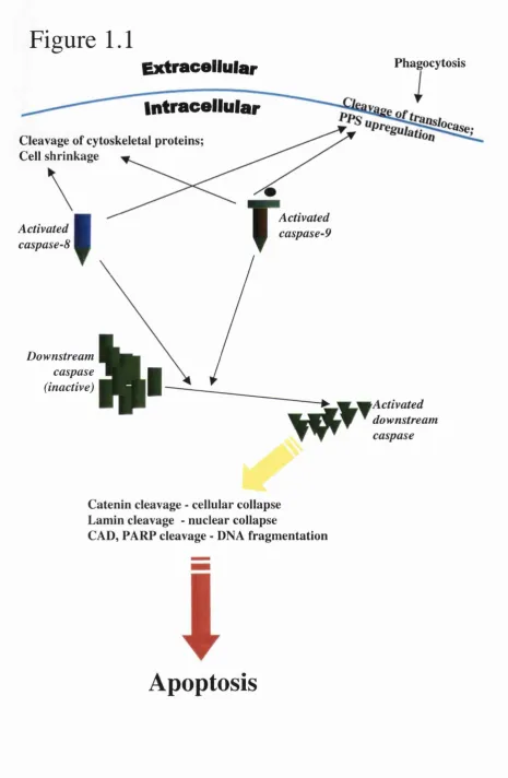
Figure 1.1 Overview of the molecular and cellular aspects of PCD
Overview o f molecular pathways and cellular events that follow activation of upstream caspases (8 or 9) and downstream, executor caspases.
Activated caspases-8 and -9 cleave cytoskeletal components and cause cell surface upregulation o f phosphatidyl serine, which in turn leads to phagocytosis. The activated upstream caspases also activate downstream effector caspases, which irreversibly commits a cell to an apoptotic fate by causing cellular and nuclear collapse and DNA fragmentation.
Figure 1.1
Extracellular
PhagocytosisActivated caspase-8
Intracellular
Cleavage of cytoskeletal proteins; Cell shrinkage
Activated caspase-9
I
Downstreamcaspase (inactive)
Activated downstream caspase
Catenin cleavage - cellular collapse Lamin cleavage - nuclear collapse
CAD, FA R ? cleavage - DNA fragm entation
neighbouring cells and the extracellular matrix, and are phagocytosed. Chromatin margination against the nuclear envelope then occurs, giving it an annular shape. This is followed by fragmentation of the nucleus to give small “apoptotic bodies” (Kerr et a l, 1972). These terminal events in apoptosis result from the activation of the executor caspases-3, -6 and -7 (Huppertz et a l, 1999). Immunochemical labelling of cleaved- caspase-3 can therefore be used as an effective assay for PCD (Black et a l, 1998; Srinivasan, 1998). These caspases cleave, and thereby activate, one another, although the hierarchy of these interactions remains unclear (Cohen, 1997; Hirata et a l, 1998; Grossmann et a l, 1998; Lincz, 1998; Slee et a l, 1999). Cleaved-caspase-3 can also cleave the upstream caspase-8 and, by a positive feedback loop, accelerate its own cleavage, and so apoptosis (Kruidering and Evan, 2000). In addition, the downstream caspases directly cleave other substrates such as a - and p-catenin (Brancolini et a l,
1997). These proteins regulate the anchoring of cells to neighbouring cells, so their cleavage leads to cellular collapse (but not loss of membrane integrity). Lamins, which form structural supports for the nucleus, are also cleaved specifically by caspase-6, leading to nuclear collapse (Orth et a l, 1996; Takashi et a l, 1996). Simultaneously, the downstream, executor caspases cleave and thus activate other, non-caspase, enzymes, such as cytoplasmic transglutaminase. This enzyme, once activated, moves to the inner aspect o f the cell membrane and, by cross-linking cytoskeletal components in this region, prevents loss of cell membrane integrity (Cummings 1996; Fesus et a l, 1996).
The executor caspases also cleave enzymes required for DNA maintenance such as poly-ADP ribose polymerase (PARP), and caspase-activated DNAse (CAD), which normally inhibit DNA kinases (Tewari et a l, 1995; Enari et a l, 1998). As a
consequence, DNA cleavage occurs. This is the basis for the detection of PCD by the laddering seen when DNA from apoptotic cells is run on an agarose gel, and also underlies the terminal deoxynucleotidyl transferase (TdT)-mediated dUTP nick
labelling (TUNEL) assay. In this latter assay, the TdT enzyme attaches labelled nucleic acid molecules to the terminal ends of DNA fragments, and since there are millions more terminal ends following apoptotic DNA cleavage than in either live cells or necrotic cells, the effect is to specifically label apoptotic cells (Gavrieli et a l, 1992). If phagocytosis does not occur, then the apoptotic cell finally loses membrane integrity and ruptures in what is known as secondary necrosis (Kerr et a l, 1972).
1.1.3 Molecular pathways leading to PCD
As described in the previous section, the execution o f PCD can occur following two molecular pathways. One is frequently termed the stress-mediated pathway and is largely dependent on caspase-9, and the other, the death receptor-mediated pathway, involves caspase-8. For a time, these pathways were thought to be relatively separate. Recent evidence suggests, however, that there is cross talk and co-operation between them. Nonetheless, for the sake of descriptive clarity, the two pathways will be considered independently.
1.1.3.1 PCD following cellular stress
Cells in multicellular organisms are constantly exposed to stress of one type or another, from deprivation of nutrients to genotoxic irradiation. Broadly speaking, exposed cells can either succumb to this stress or recover from it. However, each of these responses can have potentially disastrous consequences for the organism. For example, in the case of a cell that is exposed to genotoxic radiation, if the cell is irreparably damaged and simply lyses, spilling its intracellular contents into the extracellular compartment, inflammation will ensue. This is a highly energy- demanding process, which may also result in the loss of neighbouring cells, and, furthermore, poor wound healing often follows the inflammatory response, which may
be a reason why embryonic wound healing, which does not involve inflammation, can occur without scar formation (Mast et a l, 1992; Nodder and Martin, 1997). On the other hand, the cell may survive but with DNA damage. In this case, there is always the chance, however minimal, that the cell has undergone a potentially carcinogenic
mutation, and could therefore threaten the whole organism. Stress-mediated apoptosis is an alternative to these two scenarios. In outline, cellular stress appears to upregulate pro-apoptotic molecules in the cytoplasm (p53 and certain members of the Bcl-2 family) or in the mitochondrial outer membrane (members of the Bcl-2 family), which cause apoptosis largely by allowing the release of cytochrome c from the mitochondria, which activates caspase-9. The molecular interactions involved in stress-mediated apoptosis are summarised in Figure 1.2.
The mitochondria are central to stress-mediated apoptosis. Following cellular stress, release of the haem-containing form of cytochrome c from the mitochondria allows the apoptotic protease-activating factor 1 (Apaf-1) to activate cytoplasmic
caspase-9 (Li et a l, 1997; Zou et a l, 1997), although exactly how this occurs is unclear. Recent evidence suggests that Apaf-1 and caspase-9 form a holoenzyme, which then performs the various enzymatic cleavages, described in Section 1.1.2, necessary to bring about apoptosis (Rodriguez and Lazebnik, 1999). Biochemical evidence suggests that caspase-3 is a major downstream target of caspase-9 (Li et a l, 1997; Zou et a l, 1997). This is supported by evidence from knockout studies, which show very similar
abnormal phenotypes in caspase-9, Apaf-1 and caspase-3 null mutants, including neuronal overgrowth (Section 1.1.9). Furthermore, since death receptor-mediated PCD is not affected in these null mutants, this suggests that caspase-3 is not vital for the action of caspase-8 (Section 1.1.4). Recently, it has been shown that at the same time as cytochrome-c is released from the mitochondria, so is the second mitochondria-derived activator of caspases (Smac; Du et a l, 2000). This protein, upon release, inhibits by
Figure 1.2 Stress-mediated apoptosis
A summary of molecular pathways leading to apoptosis that occur following various forms o f cellular-stress. The stress either directly or indirectly (via p53) increases the amount of pro-apoptotic Bcl-2 family members in the outer mitochondrial membrane. This allows the release o f cytochrome-c into the cytoplasm, where it allows Apaf-1 to activate caspase-9, thus stimulating apoptosis (Figure 1.1). Under normal
circumstances, the inhibitor of apoptosis proteins help prevent autocatalysis of caspases, but when the apoptotic programme is triggered, Smac is released from the mitochondria inhibiting them and facilitating apoptosis.
Note that p53 is normally maintained at a constant level because of a negative feedback loop with Mdm2.
Figure 1.2
Extracellular
Intracellular
MDM2\
Cyto. c
Smac
Inactive
caspase-9 Activated
caspase-9 lAPs
Downstream caspase (inactive)
Stress
Proapoptotic Bcl-2 family member (inactive)
Proapoptotic Bcl-2family member (active)
Antiapoptotic Bcl-2 ''amily member
w
Activated downstream
caspase \ %
&
direct interaction the inhibitor of apoptosis proteins (lAPs), which normally themselves inhibit caspases from spontaneously activating and initiating apoptosis (Deveraux and Reed, 1999; Shi, 2001), and so Smac acts to facilitate apoptosis.
In the mechanism described above for caspase-9-mediated PCD, the factor deciding whether or not apoptosis occurs is the release of cytochrome c from the mitochondria. This appears to be controlled largely by the Bcl-2 family of proteins. These molecules comprise various combinations of the four Bcl-2 homology (BH) domains (Adams and Cory, 1998; Kelekar and Thompson, 1998; Gross et a l, 1999), which allow the different members of the family to form homo- or heterodimers.
Certain members of the family are pro-apoptotic whereas others are anti-apoptotic. The pro-apoptotic members, which all have the BH3 domain, are sub-divided into the Bax subfamily (including Bax and Bak) and the BH3 subfamily (including Bad and Bid) (Knudson and Korsmeyer, 1997; Adams and Cory, 1998; Kelekar and Thompson,
1998). It seems that the relative proportions of activated anti-apoptotic and pro
apoptotic Bcl-2 family molecules in the outer mitochondrial membrane, and whether, or how they dimerise, determines whether cytochrome c is released, causing apoptosis (Knudson and Korsmeyer, 1997; Adams and Cory, 1998; Kelekar and Thompson, 1998; Gross et a l, 1999; Vlaeminck-Guillem et a l, 2001). The exact manner, however, in which these proteins allow the release of cytochrome c into the cytoplasm, in the case of the pro-apoptotic molecules, or inhibit its release, in the case of the anti-apoptotic
molecules, remains elusive (Adams and Cory, 1998; Gross et a l, 1999; Shi et a l, 2001) A variety of cellular stresses can result in a relative increase in the pro-apoptotic members o f the Bcl-2 family. For example, deprivation of the survival factor
interleukin-3 (IL-3), which normally keeps cytoplasmic Bad phosphorylated, results in its dephosphorylation and relocation to the outer mitochondrial membrane, where it has a pro-apoptotic effect (Gross et a l, 1999). In some types of cellular stress, there can
also be a transcriptional upregulation of pro-apoptotic Bcl-2 family members. For example, some genotoxic stimuli can result in an upregulation of Bax transcription (Evan and Littlewood, 1998; Vousden, 2000). This particular upregulation is caused by the tumour suppressor protein, p53. The importance of this protein can be gauged by the fact that 50% of all human cancers carry a mutation in the p52 gene (Raff, 1998; Steller, 2000).
Similarly to the Bcl-2 family, p53 acts as a sensor for cell stresses, including genotoxicity. Following DNA damage, for example, intracellular levels of p53 increase. Exactly how this occurs is uncertain, but it seems likely to be linked to the DNA binding properties of p53 (Liu and Kulesz-Martin, 2001). The relatively high levels of p53 in a cell then appear to increase transcription of pro-apoptotic proteins, such as Bax (see above) or death receptors that transduce pro-apoptotic signals, such as Fas, rendering a cell susceptible to apoptosis (Bennett et a l, 1998; Muller et a l, 1998; Hill et a l, 1999; Vousden, 2000; Vlaeminck-Guillem et a l, 2001). Simultaneously, p53 causes arrest of the cell cycle, for example by causing upregulation of the p2jWAF/ciPi cyclin-dependent kinase inhibitor, which binds and thereby inhibits 01 cyclin-dependant kinases (Norbury and Hickson, 2001). It seems that normally, levels of p53 are maintained in a “non-apoptotic” range by a negative feedback loop in which p53 causes the transcription of the ligase Mdm2, which itself causes the proteosomic degradation of p53 (by an as yet undetermined mechanism) (Prives, 1998; Vousden, 2000; Evan and Vousden, 2001). Mdm2 is also negatively regulated by p i 9 ^ ^ , which binds and inactivates it. This interaction allows mitogenic factors, such as Myc and Ras, a method by which they can predispose a cell to apoptosis since they upregulate pjçARF same time as stimulating proliferation (Vousden, 2000). This coupling of the proliferative and apoptotic pathways, making proliferating cells particularly
vulnerable to PCD, may sound paradoxical, but has been suggested to be important as mutations may occur following mitosis.
This section has given a brief overview of stress-mediated apoptosis, in an attempt to outline some of its key components and principles, particularly the importance o f the balance existing between pro- and anti-apoptotic signals in
determining whether or not a cell undergoes PCD. Attention is now turned to what has been described as “death receptor-mediated apoptosis”.
1.1.3.2 Death receptor-mediated PCD
Apoptosis can be triggered by activation of a trans-membrane death receptor, which, via an adaptor molecule, results in the cleavage of pro-caspase-8 into activated caspase-8, which is then believed to cleave downstream caspases by the mechanisms described in Section 1.1.2.
Trauth et al. (1989) and Yonehara et al. (1989) both identified a cell surface receptor responsible for lymphocytic induction of apoptosis. This was named Fas, or APO-1, although it is the former term that is now generally used. The Fas/Fas ligand interaction, and the subsequent molecular pathways it regulates, forms the archetypal death receptor/ligand apoptotic signal, and it is this that will be described first. A summary of death receptor-mediated PCD is shown in Figure 1.3.
Fas ligand is a cytokine belonging to the tumour necrosis factor (TNF) family of cytokines. It is synthesised as a type II membrane protein, having an extracellular carboxy-terminus and an intracellular amino-terminus (Suda et al. 1993; Nagata, 1997). Although it may be cleaved from the cytoplasmic membrane, in mouse models, its potency as an endocrine agent is much reduced compared to when it is membrane bound, and so it generally activates Fas in an autocrine or paracrine manner. Indeed, cleavage of Fas ligand from the cell membrane may serve to inactivate it (Nagata,
Figure 1.3 Death receptor-mediated apoptosis
A summary o f molecular pathways following activation of death receptors. Upon trimérisation, death receptors either directly, or indirectly via TRADD, activate FADD. This results in the oligomerisation o f caspase-8 (unless there is sufficient competition from FLIP). Caspase-8 then cleaves downstream caspases to cause apoptosis.
Sometimes, the trimérisation of Fas may activate Daxx. The exact functions of Daxx are unknown, but it is believed that it can both enhance and attenuate apoptotic
induction, depending on the circumstances. TRADD may also activate RIP, which can activate NF-kB and so inhibit apoptosis. However, RIP cleaved by caspase-8 may actually have a pro-apoptotic function.
DD = death domain
Figure 1.3
Fas ligandDaxx
TNF-a
Extracellular
Intracellular
I
Inactive caspase-8 ADDTRADD FLIP
FADD
Active caspase-8 Inactive
downstream caspase
Active caspase-8
t
I Ap^tosis!;
Activated downstream caspase
1 DD
1997). Fas, itself, is a member of the TNF receptor (TNFR) family, which are all type I membrane proteins and have a characteristic intracellular, carboxy-terminal death domain (DD) consisting of 65-80 amino acids (Itoh et a l, 1991; Oehm et a l, 1992; Nagata, 1997; Kruidering and Evan, 2000). On binding of Fas ligand to Fas, there is a trimérisation of the receptor via the death domain of Fas, which is thereby activated (Banner et a l, 1993; Nagata, 1997; Ashkenazi and Dixit, 1998; Kruidering and Evan,
2000).
In 1995, two independent yeast two-hybrid studies found that activated Fas associates with a cytoplasmic molecule, which was called the Fas-associated death domain (FADD) (Boldin et a l, 1995; Chinnaiyan et a l, 1995). FADD has a carboxy- terminal death domain comparable to that of Fas, and also an amino-terminal domain called the death effector domain (DED). Upon aggregation of Fas receptors, FADD cross-links to their death domains, via its own, in a homotypic manner. This renders the death effector domain of FADD active (Kischkel et a l, 1995; Muzio et a l, 1996; Nagata, 1997; Yeh et a l, 1998). In 1996, another yeast two-hybrid study, again by Boldin et a l, identified a cytoplasmic molecule that was named the FADD-like IL-ip converting enzyme (FLICE) or caspase-8, which was found to have its own death effector domain in its amino terminus. The active death effector domain on FADD recruits, again via homotypic interactions, the death effector domain of caspase-8. Recruitment of multiple caspase-8 molecules appears to result in their oligomerisation, which in turn causes the caspase-8 molecules to auto-cleave and consequently become active (Yang et a l, 1998; Kruidering and Evan, 2000). This structure, consisting of Fas, FADD and capase-8 is known as the death-inducing signalling complex (DISC) (Kischkel et a l, 1995). The DISC then releases activated caspase-8 into the cytoplasm. Caspase-8 can then cleave downstream caspases, resulting in apoptosis, as described in Section 1.1.2.
Another important group of death ligands and death receptors are TNF-a and -p, and their receptors, TNFR-1 and -2. The cascade from TNF-a and -p-induced
trimérisation of TNFR-1 and -2, leading to activation o f caspase-8, is similar to that following Fas trimérisation, with the exception that the activated receptor complex, rather than recruiting FADD directly, recruits the TNFR-associated death domain (TRADD) (Hsu et a l, 1995; Nagata, 1997). TRADD does not have a death effector domain of its own, but has two death domains through which it recruits and activates FADD (Hsu et a l, 1996; Nagata, 1997). Other less well investigated death receptors, such as death receptor 3, 4 and 5 (DR-3, -4 and -5) also act either via the FADD- caspase-8 pathway (Ashkenazi and Dixit, 1998; Kuang et a l, 2000).
Confusingly, in some situations, activation of death receptors - particularly TNFRl - can have a pro-survival effect (Nagata, 1997). TRADD, in addition to
recruiting FADD, may also recruit the receptor interacting protein (RIP) (Ashkenazi and Dixit, 1998). RIP can then activate nuclear factor k B (N F-kB), which resides in an inactive form in the cytoplasm, by activating the I-kB kinase (IKK) which
phosphorylates, and so inactivates, the inhibitor of N F-kB (I-kB) (Ashkenazi and Dixit, 1998). Once active, N F -kB translocates to the nucleus, where it transcribes
cytoprotective and pro-inflammatory proteins (Ashkenazi and Dixit, 1998). Although in some circumstances, it appears that RIP may have an anti-apoptotic role. For example, cleavage of RIP by caspase-8 in cell culture, and overexpression of RIP can induce apoptosis (Nagata, 1997; Lin et a l, 1999). Death receptor activation can also be inhibited by the presence of FLICE (caspase-8) inhibitory protein (FLIP). FLIP has two death effector domains, which mimic that of caspase-8, and therefore FLIP behaves as a competitive inhibitor of caspase-8 (Thome et a l, 1997; limier et a l, 1997; Shu et al, 1997; Yeh et a l, 2000). In overexpression systems, however, FLIP can actually accelerate cell death by an as yet unknown mechanism (Irmler et a l, 1997; Shu et
al., 1997; Yeh et a l, 2000). In addition, it has recently been shown that activated Fas receptor can activate Daxx (Yang et a l, 1997; Michaelson et a l, 1999). Little is known about this promiscuous 120 kD protein, although it appears that it may have both pro- and anti-apoptotic effects (Michaelson et a l, 1999). It seems that these may be mediated by the c-Jun amino-terminal kinase (JNK) pathway, which causes activation o f c-Jun (Yang et a l, 1997; Michaelson et a l, 1999; Jochum et a l, 2001). c-Jun, in turn, forms part of the activating protein-1 (AP-1) transcription factor (Jochum et a l,
2001; Mechta-Grigoriou, et a l, 2001). AP-1 is a heterodimer and can be made up of various combinations of members of the Jun and Fos protein families. AP-1 has multiple functions and may be both pro- and anti- apoptotic (Jochum et a l, 2001). The circumstances that pre-dispose Daxx and AP-1 to being pro- or anti-apoptotic are as yet unclear.
1.1.3.3 Cross-talk between stress-mediated and death receptor-mediated apoptosis
Stress-mediated and death receptor-mediated PCD are not completely
independent of one another. p53, which is upregulated following genotoxic stress, can upregulate Fas, and thus accelerate PCD by the death receptor-mediated pathway. The practical effect of this is shown by a study in which skin cancer, following UV light exposure (i.e. genotoxic stress), was shown to be very frequent in Fas ligand deficient mice (70% of mice examined), whereas in control, wild type mice, the rate was only 5% (Hill et a l, 1999). Furthermore, PCD following detachment o f cells from their
neighbours or the extracellular matrix (anoikis) can be inhibited via dominant-negative FADD and Bcl-2 expression, suggesting that both pathways are activated (Rytomaa et a l, 1999; Kruidering and Evan, 2000). Also, recent evidence has shown that the cytosolic Bcl-2 protein Bid can be cleaved by activated caspase-8 (Luo et a l, 1998). Cleaved Bid then translocates to the outer mitochondrial membrane where it has a
apoptotic effect by facilitating the release of cytochrome-c into the cytoplasm. It has also been shown that downstream caspases can potentially activate upstream caspases (Kruidering and Evan, 2000), so it is theoretically possible for activated caspase-9 to activate downstream caspases, which could then, in turn, activate caspase-8. Some of the main components involved in the interaction between the stress- and death receptor- mediated pathways are shown in Figure 1.4.
1.1.4 Clearance of apoptotic cells
As stated in Section 1.1.2, if apoptotic cells are not rapidly cleared, they undergo secondary necrosis, with all the features of inflammation that would normally follow necrosis. To prevent this, upregulation of cell surface molecules on the apoptotic cell, such as phosphatidylserine, stimulate other cells to phagocytose them (Fadok et a l,
1992). This process is very rapid, and as a result, by the time apoptotic morphology is visible, the apoptotic cell has usually been phagocytosed by another cell in in vivo
situations (Camp and Martin, 1996; Jacobson et a l, 1997). Cleared apoptotic cells are then rapidly digested by the phagocyte.
Although this phagocytosis can be performed by neighbouring cells, in tissues where the intensity of PCD is high, it is often specialised cells, that is to say
macrophages, that perform phagocytosis (Hopkinson-Woolley et a l, 1994; Camp and Martin, 1996; Pamaik et a l, 2000). This occurs, in the developing kidney and also in the developing limbs, where apoptosis removes the interdigital webs (Hopkinson- Woolley et a l, 1994; Camp and Martin, 1996). Interestingly, in mouse mutants in which no macrophages develop, clearance of apoptosis in the interdigital webs still occurs, but by neighbouring cells (Wood et a l, 2000). The neighbouring cells are not as efficient as macrophages in clearing the apoptosis, taking approximately three times as long to do so. Correspondingly, the regression of the interdigital webs takes three
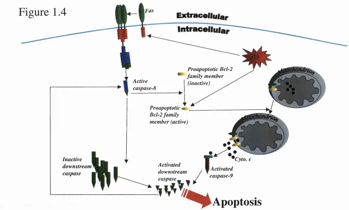
Figure 1.4 Cross-talk between death receptor-m ediated apoptosis and stress- mediated apoptosis
A summary o f some molecular pathways that link death-receptor- and stress-mediated apoptosis. Death receptor-mediated apoptosis may result in the cleavage of cytosolic members of the Bcl-2 family of molecules that then relocate to the mitochondria, facilitating cell death by the stress-mediated pathway.
Also, activation o f stress-mediated mechanisms for apoptosis can cause an upregulation of Fas via p53, facilitating cell death by the receptor-mediated pathway.
Figure 1.4
Extracellular
Active caspase-8
Proapoptotic Bcl-2 family member
(inactive)
Inactive downstream caspase
Proapoptotic Bcl-2 family member (active)
Activated downstream caspase^
Cyto. c
Activated caspase-9
times longer. In some developmental situations macrophages have also been shown to actually induce apoptosis, for example in the development of the lens, which requires removal of the hyaline vessels by apoptosis (Lang and Bishop, 1993).
1.1.5 Necrosis and apoptosis
Apoptosis was originally defined by the morphological features that distinguish it from necrosis. Recently, however, it has been observed that there are circumstances in which apoptosis and necrosis can occur at the same time, and furthermore, individual cells can be seen to be simultaneously showing signs of apoptosis and necrosis. This occurs in the heart wall following myocardial infarction, or in the regressing vestigial tail during human development (Sapunar, Vilovic and England, 2001; Kajstura et a l,
1996; Saraste and Pulkki, 2000). This is, perhaps, unsurprising. PCD is often a response to cellular stress, as is necrosis, albeit markedly different. It is, then,
conceivable that apoptosis and necrosis may “compete” with one another to kill a cell exposed to some cellular stress. In this case, if one or other mechanism is inhibited, the other one may kill the cell. For example, inhibition with the pan-apoptotic inhibitor benyloxycarbonyl-valine-alanine-aspartate fluoromethylketone (zVAD-fmk) prevents apoptosis in the mouse interdigital webs (Chautan et a l, 1999). zVAD-fmk is a peptide that mimics the target sequences of the caspases, binds to them, and thereby inhibits their function (McCarthy et a l, 1997; Garcia-Calvo et a l, 1998; Sun et a l, 1999). However, despite this, “programmed” cell death may still proceed by necrosis independently of caspases (Chautan et a l, 1999).
1.1.6 Programmed cell death in development
Ever since the seminal study by Glucksmann (1951), it has been evident that apoptosis is an important part of development. More recently, new methods for
studying apoptosis, and the development of animals with mutations in apoptotic genes, has led to an enhanced understanding of the role of apoptosis during development which include regulating cell numbers and sculpting of structures.
In the developing nervous system, there is strong evidence for a role for apoptosis in controlling cell numbers (reviewed by Burek and Oppenheim, 1996, and Pettmann and Henderson, 1998). It is estimated that in the vertebrate nervous system, up to twice as many neurons develop as are finally required. There is evidence to suggest that some of this neuronal cell death follows competition for growth factors, such as members of the neurotrophin family. For example, amputation of an embryonic chick limb during development leads to a vastly increased loss of the neurons that would normal innervate it, but this increased loss can be reduced by grafting another limb bud onto the amputation site, or by supplementation with muscle extract (Caldero
et a l, 1998; Pettmann and Henderson, 1998). In support of the importance of apoptosis in regulating cell numbers in the nervous system, and potentially providing the
molecular mechanism by which at least some developmental neuronal PCD may occur, the caspase-9, caspase-3 and Apaf-1 null mutants all display a developmental
overgrowth of central nervous tissue (Kuida et a l, 1996; Kuida et a l, 1996; Yoshida et a l, 1998). lnterestingly,a recent study has provided evidence that neurones from the
caspase-9 and caspase-3 knockout mice may still undergo cell death, but that this cell death is delayed (Oppenheim et a l, 2001).
Cell death can also play an important part in sculpting the embryo, by removing vestigial structures. A relatively well-understood example of this is in the interdigital webs, which are removed by apoptosis under the influence of the bone morphogenetic proteins (BMPs; Zou and Niswander, 1996). This cell death is signifcantly reduced in the Hammertoe mouse mutant, in which, correspondingly, the interdigital webs remain (Zakeri et a l, 1994; Chautan et al, 1999).