Western University Western University
Scholarship@Western
Scholarship@Western
Electronic Thesis and Dissertation Repository
8-28-2012 12:00 AM
Reliability of flow-mediated dilation measures in the popliteal
Reliability of flow-mediated dilation measures in the popliteal
artery and implications for use in clinical and research practices
artery and implications for use in clinical and research practices
Kaitlin M. McLay
The University of Western Ontario
Supervisor
Dr. Donald H Paterson
The University of Western Ontario Graduate Program in Kinesiology
A thesis submitted in partial fulfillment of the requirements for the degree in Master of Science © Kaitlin M. McLay 2012
Follow this and additional works at: https://ir.lib.uwo.ca/etd
Part of the Systems and Integrative Physiology Commons
Recommended Citation Recommended Citation
McLay, Kaitlin M., "Reliability of flow-mediated dilation measures in the popliteal artery and implications for use in clinical and research practices" (2012). Electronic Thesis and Dissertation Repository. 831. https://ir.lib.uwo.ca/etd/831
This Dissertation/Thesis is brought to you for free and open access by Scholarship@Western. It has been accepted for inclusion in Electronic Thesis and Dissertation Repository by an authorized administrator of
i
Reliability of flow-mediated dilation measures in the popliteal artery and
implications for use in clinical and research practices
(Spine Title: Reliability of popliteal artery flow-mediated dilation)
(Thesis Format: Integrated Article)
By
Kaitlin M. McLay
Graduate Program in Kinesiology
A thesis submitted in partial fulfillment
of the requirements for the degree of
Master of Science
School of Graduate and Postdoctoral Studies
The University of Western Ontario
London, Ontario, Canada
© Kaitlin M. McLay
ii
THE UNIVERSITY OF WESTERN ONTARIO
SCHOOL OF GRADUATE AND POSTDOCTORAL STUDIES
CERTIFICATE OF EXAMINATION
Supervisor
______________________________ Dr. Donald Paterson
Supervisory Committee
______________________________ Dr. John Kowalchuk
Examiners
______________________________ Dr. John Kowalchuk
______________________________ Dr. Peter Lemon
______________________________ Dr. Jan Polgar
The thesis by
Kaitlin Marlisa McLay
entitled:
Reliability of flow-mediated dilation measures in the popliteal artery and
implications for use in clinical and research practices
is accepted in partial fulfillment of the requirements for the degree of
Master of Science
Date__________________________ _______________________________
iii ABSTRACT
PURPOSE: The aim of the present study was to assess the test-retest and stability reliability of
flow-mediated dilation (FMD) in the popliteal artery and to investigate the effect of occlusion
pressure on the FMD response. METHODS: A series of FMD tests were performed on ten
healthy young adult males to assess reliability. Ultrasound-derived artery diameter of the
popliteal was measured and FMD was calculated as the percent change in diameter from
baseline. RESULTS: FMD measurements for intra- and interday comparisons demonstrated
poor reliability (Repeatability 5.62 and 4.82%, Intraclass correlation coefficient [ICC] 0.36 and
0.25, respectively). Repeatability values were as large as the FMD measures themselves for both
intra- and interday reliability. CONCLUSION: Popliteal artery FMD has poor reliability for
test-retest and stability reliability. Interpretation of individual or group changes using this
technique should be interpreted with caution.
iv
ACKNOWLEDGEMENTS
There are several people who I will always be appreciative to for making these past two
years at Western so enjoyable. I would like to start by thanking my thesis supervisor, Dr. Donald
Paterson. Don, I cannot believe how lucky I was to have you as my supervisor. No matter
where you were, you provided feedback and support for everything I did. I look forward to our
next chapter!
Another individual who deserves more thanks that I can provide is Dr. Juan Murias. Not
only did I have a great supervisor, I had an amazing “unofficial co-supervisor”. Juan, I can’t
imagine these past two years without having you around to encourage me. You were always
available to provide support for everything I tried (research-related or not) and most importantly,
to BBQ.
Thank you to Dr. John Kowalchuk, who was always available if I needed anything, and
to my labmates. Thank you for all the assistance in the lab but more importantly for the unique
work environment you all provide. Our lab truly is one of a kind!
I would also like to thank my friends who have provided me with incredible memories
and helped guide me through this process. My experience here at Western wouldn’t have been
nearly as enjoyable without you guys.
Finally, to my family: Mom, Dad and Jas. I owe you all more than I could ever hope to
repay. The unrelenting love and support you always provide is incomprehensible, especially
with everything we have experienced in the past two years. Thank you for all the opportunities
you have provided to me and your continued encouragement is appreciated more than you know.
I know it is a cliché to say “I couldn’t have done it without you”, so to everyone I
v
TABLE OF CONTENTS
CERTIFICATE OF EXAMINATION ii
ABSTRACT iii
ACKNOWLEDGEMENTS iv
TABLE OF CONTENTS v
LIST OF TABLES vii
LIST OF FIGURES viii
LIST OF APPENDICES ix
LIST OF ABBREVIATIONS x
CHAPTER 1: REVIEW OF LITERATURE 1
1.1 Introduction 2
1.2 Physiology of FMD 3
1.3 Methodological Considerations 5
1.4 Rationale 7
CHAPTER 2: RELIABILITY OF FLOW-MEDIATED DILATION MEASURES IN THE POPLITEAL ARTERY AND IMPLICATIONS FOR USE IN CLINICAL AND
RESEARCH PRACTICES 10
2.1 Introduction 11
2.2 Methods 14
2.2.1 Participants 14
2.2.2 Study Design 14
2.2.3 Protocol 15
2.2.4 Popliteal Artery Diameter Analysis 16
2.2.5 Statistical Analysis 16
2.3 Results 20
2.3.1 Subject Characteristics 20
2.3.2 Internal Consistency Reliability 20
2.3.3 Day-to-day Reliability 20
2.3.4 Different Occlusion Pressure 21
2.3.5 Time Course of Popliteal FMD 21
2.4 Discussion 30
vi
APPENDIX A 45
vii
LIST OF TABLES
Table Description Page
2.1 Participant characteristics 22
2.2 Test-retest reliability statistics for 23
FMD properties
2.3 Day-to-day reliability statistics for FMD 25
properties
2.4 Statistics for FMD properties associated with 27
viii LIST OF FIGURES
Figure Description Page
1.1 Schematic representation of FMD pathway 9
2.1 Study design outlining the series of FMD 18 tests
2.2 Schematic representation of FMD timeline 18
2.3 Sample calculation of baseline and peak 19 diameter and FMD
2.4 Variability of measures between three FMD 24 tests performed on the same day
2.5 Day-to-day variability of measures 26 between five FMD tests performed on
separate days
2.6 Variability of measures between five FMD 28 tests performed at five different occlusion
pressures
2.7 Time course of popliteal artery dilation 29
following reactive hyperemia
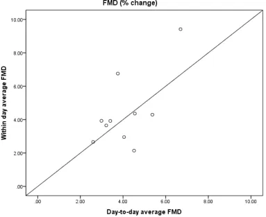
2.8 Relationship between the average of three FMD 37
ix
LIST OF APPENDICIES
Appendix Description Page
x
LIST OF ABBREVIATIONS
CV – Coefficient of variation
CVD – Cardiovascular Disease
Ca++ – Calcium
ECG - Electrocardiography
EDHF – Endothelial-derived relaxing factor
eNOS – Endothelial nitric oxide synthase
FMD – Flow-mediated dilation
ICC – Intraclass correlation coefficient
L-NMMA – NG-monomethyl-L-arginine
MBV – Mean blood velocity
NO – Nitric oxide
PGs - Prostaglandins
SD – Standard deviation
1
Chapter 1:
2 1.1 Introduction
When blood flow increases through a blood vessel, a resultant frictional force parallel to
the vessel, termed shear stress, is applied. An increase in shear stress on the vascular
endothelium causes endothelium-dependent vasodilation (flow-mediated vasodilation [FMD]).
In 1992, Celermajer et al. (Celermajer et al., 1992) developed a technique that took advantage of
this physiological response and is currently a widely used, noninvasive technique to provide
insight into vascular health.
The reactive hyperemia endothelial function test, commonly referred to as an FMD test,
uses ultrasound assessment of FMD in response to cuff release following an occlusion period.
This technique generates a shear stress stimulus, resulting in a dilation of downstream resistance
vessels following the occlusion of blood flow to the limb with a pressure cuff. Upon release of
the occlusion, inflow through the conduit artery is transiently increased (reactive hyperemia) and
acts as the stimulus for FMD.
In humans FMD is typically assessed in the large peripheral conduit arteries and is
considered representative of the response in more clinically relevant coronary circulation
(Anderson et al., 1995; Takase et al., 1998). As a result of the close relationship to the coronary
circulation, the FMD test has become widely used as a measure of endothelial dysfunction in
both clinical and asymptomatic patients. Experimental and clinical studies suggest that
endothelial dysfunction is an important feature of vascular disease and is strongly associated
with several cardiovascular conditions, including atherosclerosis (Ross, 1999), hypertension
(Taddei et al., 1993; Perticone et al., 2001; Modena et al., 2002), and coronary and peripheral
3
Brevetti et al., 2003), and that endothelial dysfunction predicts cardiovascular events in these
groups (Gokce et al., 2002; Widlansky et al., 2003).
1.2 Physiology of FMD
The endothelium is a single layer of cells lining all of the blood vessels in the body, also
known as the tunica intima, and has been identified as playing a major role in smooth muscle
dilation. Animal studies established that FMD in arteries was dependent on the presence of an
intact endothelial lining (Smiesko et al., 1985; Pohl et al., 1986). Rubanyi et al. (Rubanyi et al.,
1986) indicated that, in response to shear stress, the endothelium released a substance that
possessed the characteristics of Furchott’s endothelium-derived relaxing factor, now known as
nitric oxide (NO) (Moncada et al., 1988).
Reductions in FMD are widely assumed to reflect diminished NO production as several
pivotal human studies have concluded that brachial and radial artery FMD are dependent on an
NO pathway (Joannides et al., 1995; Mullen et al., 2001; Doshi et al., 2001; Jiang et al., 2011).
Studies involving the administration of NO blockades, such as NG-monomethyl-L-arginine
(L-NMMA), have confirmed the major role that NO plays in vasoregulation. Joannides et al.
(Joannides et al., 1995) found that radial artery dilation following 3 minutes of ischemia was
abolished in the presence of L-NMMA. Similarly, Mullen et al. (Mullen et al., 2001) found that
NO blockade decreased the radial artery FMD response to 5 minutes of ischemia from 5.3% to
0.7% dilation, with no difference in hyperemic stimulus, concluding that it was unlikely that
stimulus magnitude was responsible for the abolished FMD response.
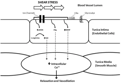
Figure 1 outlines the pathway of endothelial-dependent vasodilation in response to
increases in fluid shear stress. Although several vasodilators are released by the endothelium,
4
an FMD test, the resultant increase in blood flow creates a shear stress stimulus causing
deformation of mechanosensitive structures on the endothelial cell membrane, such as membrane
proteins (glycocalyx), primary cilia and mechanosensitive ion channels (Pyke & Tschakovsky,
2005; Davies, 2009). The acute response to the shear stress stimulus is the opening of calcium
(Ca++)-activated potassium channels, causing hyperpolarization of the endothelial cell (Olesen et
al., 1988; Cooke et al., 1991; Miura et al., 2001). This results in an increased driving force for
Ca++ entry into the cell. The Ca++ activates endothelial nitric oxide synthase (eNOS) which in
turn increases the conversion of L-arginine to NO (Pohl et al., 1986; Joannides et al., 1995).
Over prolonged periods of shear stress stimulus (minutes), the mechanosensitive structures signal
increases G-protein expression and resultant phosphorylation of eNOS (Corson et al., 1996;
Dimmeler et al., 1999). The increase in eNOS activity increases the production of NO, even at
low concentrations of calcium. Vasodilators diffuse from the endothelial cell into the tunica
media (composed of smooth muscle), trigger a signalling cascade which ultimately reduces
intracellular [Ca++] and induces relaxation of the smooth muscle and subsequent vasodilation.
As mentioned previously, there is some redundancy in the mechanisms controlling
vasodilation. In addition to multiple mechanosensitive structures on the endothelial cell surface,
there are also multiple vasodilatory pathways within the endothelial cell which may facilitate
FMD (Sun et al., 1999; Mullen et al., 2001; Doshi et al., 2001; Pyke et al., 2009; Parker et al.,
2011). A study using blood vessels from mice genetically engineered to lack eNOS still
responded to shear stress by dilating (Sun et al., 1999). Other animal models have confirmed
prostaglandins (PGs) and endothelial-derived hyperpolarizing factor (EDHF) also contribute to
endothelium dependent vasorelaxation (Huang et al., 1998; Pak et al., 2002; Scotland et al.,
5
with a large dose of L-NMMA and concluded that there may be heterogeneous vasodilator
phenotypes which affect the contribution of NO to FMD.
1.3 Methodological Considerations
FMD has emerged as a popular technique in both clinical and physiological studies to
examine the mechanisms that impact endothelial and vascular function. It is clear, that minor
changes in the methodological approach can significantly alter the nature and magnitude of the
FMD response. As a result of this, a series of reviews and tutorials have been published in an
attempt to standardize the protocol of this widespread technique (Corretti et al., 2002; Pyke &
Tschakovsky, 2005; Harris et al., 2010; Thijssen et al., 2011a). Most recently, Thijssen et al.
(Thijssen et al., 2011a) published methodological and physiological guidelines for several key
aspects of the assessment of FMD in humans. These guidelines included recommendations
regarding subject preparation, test protocol, Doppler ultrasound technique and data analysis.
FMD is typically assessed in peripheral conduit arteries, such as the brachial, radial and
superficial femoral, although the popliteal artery has recently emerged as an alternative site for
assessing endothelial function in the lower limbs. During an FMD test, baseline
ultrasound-derived artery diameter and Doppler mean blood velocity (MBV) is established prior to the
occlusion period. Once baseline measures have been taken a pressure cuff is inflated to occlude
blood flow to the forearm or lower leg, depending on the location of the FMD test. Cuff position
(distal or proximal to ultrasound measurements) has been shown to alter the FMD response by
altering the contribution of vasoactive substances (Uehata et al., 1997; Mannion et al., 1998;
Vogel et al., 2000). Distal cuff occlusion on the brachial artery was associated with a 7% FMD,
which was abolished by administration of NO blockade (Doshi et al., 2001). Alternatively,
6
reduced (to 7.5%) with the administration of an NO blockade. These data suggest that cuff
placement may affect the nature of the FMD response, by influencing the heterogeneity of
vasodilatory pathways in the endothelium. Distal cuff occlusion is considered to be greatly
NO-mediated, whereas additional factors are contributing to the dilation associated with proximal
cuff placement. Consequently, distal cuff placement is acknowledged as the standard practice to
elicit an NO-dependent response.
In addition to cuff position, occlusion duration can also affect the FMD response. The
change in brachial FMD increases as the duration of cuff inflation increases from 30 seconds to 5
minutes (Corretti et al., 2002). Although it has been suggested that there is no change in dilation
following 5 or 10 minutes of occlusion (Corretti et al., 2002), this remains unclear. Some studies
have found that a 10 minute occlusion period results in a greater FMD response that is a result of
contributions from non-NO, ischemic-induced vasodilators (Kooijman et al., 2008; Harris et al.,
2009). As a result, a 5 minute occlusion period is the accepted duration to mediate an
NO-dependent FMD response.
Following the 5 minutes of distal cuff occlusion, diameter and MBV are monitored for at
least 3 minutes in upper limb arteries and 5 minutes in lower limb arteries. Studies have found
that peak diameters in the brachial artery, and similarly in the radial, typically occur within the
first 120 seconds following release (Black et al., 2008; Irace et al., 2008; Padilla et al., 2009;
Liuni et al., 2010). Arteries in the legs demonstrate a significantly later peak than those in the
arms (Thijssen et al., 2008). Therefore, arteries such as the superficial femoral and popliteal
should be monitored for 5 minutes following cuff release to ensure adequate detection of peak
7 1.4 Rationale
The magnitude of FMD of the conduit arteries is a widely used test of endothelial
function. The FMD test has been documented to correlate with invasively assessed endothelial
function in the coronary arteries (Anderson et al., 1995), and is now commonly used as an index
of endothelial function and is often applied as a surrogate marker of cardiovascular disease
(CVD). As a diagnostic tool, it is important to understand the repeatability of this measure and
the test-retest reliability, or the ability to reproduce similar results from consecutive tests within a
single day. It is critical to comprehend the limitations, if there are any, to taking a single
measure compared to an average of multiple FMD tests. Additionally, due to the technique’s
application in tracking changes in pre-post interventions, it is important to understand the
accuracy of repeating measurements on separate days, and what percentage of changes is due to
measurement error compared to a physiological adaptation.
Studies in the brachial (Welsch et al., 2002; Peretz et al., 2007; Magda et al., 2012) and
radial arteries (Brook et al., 2005) have found conflicting results concerning the reliability of the
FMD test. Some studies have found that the FMD technique is a stable and reproducible marker
of vascular function in the brachial artery (Welsch et al., 2002). Alternatively, other studies have
shown that brachial FMD may only be satisfactory (Magda et al., 2012), or in the case of the
radial artery, a very poor (Brook et al., 2005) indicator of vascular function due to high
variability between repeated measures. We are unaware of any studies detailing acceptable
values for day-to-day variability and test-to-test repeatability within the ultrasound measure of
FMD of the popliteal artery. Due to the increasing use of this noninvasive technique in clinical
settings as a prediction of CVD risk, and in physiological research as an assessment of
8
to examine the test-retest reliability (variability between repeated trials within a day) and
stability reliability (day-to-day variability in measurements) of the FMD measurement in the
popliteal artery.
Between-laboratory comparisons of the magnitude of FMD are often made difficult due
to the use of different experimental protocols that may affect the physiological response. The
use of different occlusion pressures is a major aspect of FMD protocol that is inconsistent
between laboratories, and the effect on the physiological response of the endothelium is unclear.
Theoretically any pressure above resting systolic pressure (suprasystolic) should be sufficient to
occlude blood flow however little research has been done to look at the effects of different
pressures used on the FMD response. The present study will permit insight regarding the effect
of different occlusion pressures on the FMD response.
Overall, the main objectives of the study were 1) to assess the test-to-test and day-to-day
reliability of flow-mediated dilation in the popliteal artery; and 2) to examine the effects of five
different occlusion pressures on the flow-mediated dilation response in the popliteal artery. We
hypothesized that 1) there will be good test-retest and stability reliability of flow-mediated
dilation between tests repeated within the same day and on separate days; and 2) there will be no
9
Fig. 1.1 Schematic representation of the pathways involved in flow-mediated dilation (FMD)
10
Chapter 2:
11 2.1 INTRODUCTION
Flow-mediated dilation (FMD) describes the vasodilation of a conduit artery following an
increase in shear stress. This is typically induced by a five minute period of ischemia generated
by an inflated cuff placed distal to the artery of interest (Corretti et al., 2002). The FMD
response is largely nitric oxide (NO)-mediated (Joannides et al., 1995; Mullen et al., 2001) and
provides information about the integrity of the endothelium (Vita & Keaney, 2002). Studies
have shown that impaired endothelial vasodilation is an important feature of vascular disease and
is strongly associated with several chronic cardiovascular conditions (Neunteufl et al., 2000;
Perticone et al., 2001; Kuvin et al., 2001; Gokce et al., 2002; Modena et al., 2002; Widlansky et
al., 2003). The reactive hyperemia endothelial function test, commonly referred to as an FMD
test, is presently a widely used, noninvasive technique to provide insight into peripheral conduit
artery vasoreactivity.
The FMD technique has increasingly been applied in both clinical and physiological
studies to examine the mechanisms that impact endothelial and vascular function. FMD is
typically assessed in the peripheral conduit arteries, such as the brachial, radial and superficial
femoral. More recently, the popliteal artery has emerged as an alternative location for assessing
endothelial function in the lower limbs. The magnitude of FMD in the conduit arteries is a
widely used test of endothelial function. The FMD test has been documented to correlate with
invasively assessed endothelial function in the coronary arteries (Anderson et al., 1995), and is
now commonly used as an index of endothelial function and a surrogate marker of
cardiovascular disease (CVD). As a diagnostic tool, it is important to understand the test-retest
reliability, or the ability to reproduce similar results from consecutive tests within a single day, to
12
important to understand the accuracy of repeating measurements on separate days, and what
percentage of changes is due to measurement error instead of physiological adaptation.
Studies in the brachial (Welsch et al., 2002; Peretz et al., 2007; Magda et al., 2012) and
radial arteries (Brook et al., 2005) have found conflicting results concerning the reliability of the
FMD test. Some studies have found that the FMD technique is a stable and reproducible marker
of vascular function (Welsch et al., 2002). Alternatively, other studies have shown that FMD
may only be satisfactory (Magda et al., 2012), or a very poor (Brook et al., 2005) indicator of
vascular function due to high variability between repeated measures. We are unaware of any
studies detailing acceptable values for day-to-day variability and test-to-test repeatability within
the ultrasound measure of FMD in the popliteal artery. With the increasing use of this
noninvasive technique in clinical settings to predict risk of CVD, and in physiological research
as an assessment of endothelial function, there is a strong need for stability of this measure. The
present study aims to examine the test-retest reliability (variability between repeated tests within
a single day) and stability reliability (day-to-day variability in measurements) of the FMD
measurement in the popliteal artery.
Additionally, between-laboratory comparisons of the magnitude of FMD are often
difficult to make because of different experimental protocols which may affect the physiological
response. The use of different occlusion pressures is a major aspect of FMD protocol that
remains inconsistent between laboratories, and the effect on the physiological response is
unclear. Theoretically any occlusion pressure above resting systolic pressure (suprasystolic)
should be sufficient to occlude blood flow however little research has been done to look at the
effects of different pressures used on the FMD response. The present study will permit insight
13
Overall, the main objectives of the study were 1) to investigate the day-to-day and
test-to-test reliability of flow-mediated dilation in the popliteal artery; and 2) to examine the effects of
five different occlusion pressures on flow-mediated dilation responses in the popliteal artery.
We hypothesized that 1) there will be good test-retest and stability reliability of FMD between
tests repeated within the same day and on separate days; and 2) there will be no difference in
14 2.2 METHODS
2.2.1 Participants
Ten healthy young men (27 ± 6 yr; mean ± SD; Table 2.1) volunteered and gave written
consent to participate in the study. All procedures were approved by The University of Western
Ontario Research Ethics Board for Health Sciences Research Involving Human Subjects. All
participants were recreationally active (regularly exercising to maintain fitness) and
non-smokers. Additionally, all subjects were normotensive and no subjects were taking medications
that would affect hemodynamic responses.
2.2.2 Study Design
A series of FMD tests were performed on each participant over five consecutive days
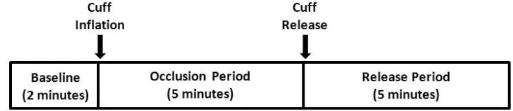
(Figure 2.1). Tests were performed at the same time each day to minimize diurnal effects. Two
FMD tests were performed on each of four days, with a fifth day involving three FMD tests.
Each FMD test was separated by a 30 minute rest period to allow blood flow and arterial dilation
to return to resting conditions (Harris et al., 2006). The first FMD test of each day was
performed with a different occlusion pressure found throughout the literature: 175; 200; 225;
250; and 300 mmHg. The succession of the first FMD test occlusion pressure was randomized
between subjects, such that one subject may have an occlusion pressure of 175 mmHg for their
first test on day 1, while another subject may have an occlusion pressure of 250 mmHg. The
second FMD test of each day was performed with an occlusion pressure of 250 mmHg, resulting
in five tests performed in five consecutive days at the same pressure – allowing analysis
15
occlusion pressure of 250 mmHg, a third test at that pressure was performed allowing
determination of the test-retest reliability for three tests within the same day.
2.2.3 Protocol
FMD of the popliteal artery was assessed in accordance with previously published
guidelines for the current standardized methodology (Corretti et al., 2002; Thijssen et al.,
2011a). All participants were instructed to refrain from caffeine, alcohol and exercise for 12
hours prior to their scheduled appointments. Following at least 10 minutes of supine rest,
participants were instructed to lie prone and a small pillow was placed under their ankle. The
left popliteal artery was measured immediately proximal to the bifurcation (usually at or slightly
above the popliteal fossa), and a pneumatic cuff (Flexiport; Welch Allyn Inc., Skaneateles Falls,
NY, USA) was placed around the calf (approximately 2-3 inches distal to the popliteal fossa).
Heart rate was continuously monitored with a three-lead ECG to allow for consistent and
accurate selection of arterial diameter measurements during the cardiac cycle.
The popliteal artery was imaged with a 10-MHz multifrequency linear-array transducer
attached to a Doppler ultrasound machine (VingMed System FiVe, GE Medical Systems,
Horten, Norway). All scans were performed by an experienced investigator with training
obtained through pilot studies, other research projects and comparisons to a phantom artery to
insure accurate assessment of arterial diameters. All scans were made under similar conditions
and all images were recorded on an external video camera (HDD Everio; JVC, Canada) for later
offline analysis. Baseline diameter was recorded prior to manual inflation of the pneumatic cuff.
The cuff was then inflated to the predetermined occlusion pressure, according to which test in the
series was being performed. The cuff was inflated for 5 minutes during which diameter was not
16
5 minutes the pneumatic cuff was released and arterial diameter was continuously monitored for
5 minutes post-release (Figure 2.2).
2.2.4 Popliteal Artery Diameter Analysis
All videos were uploaded and then analyzed using a software program (VirtualDub
1.6.19.0 by Avery Lee) which allows for frame by frame analysis to ensure that diameter
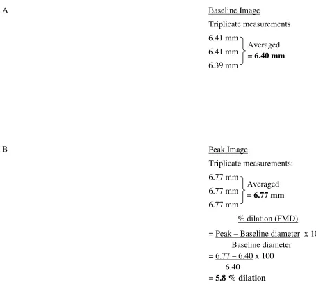
measurements were always taken at end diastole (determined by ECG gating). Triplicate
measurements of diameter were taken for each of five baseline images and averaged to determine
the baseline diameter of the artery. Similarly, triplicate measurements of diameter were
averaged for a single image taken every 15 seconds following cuff release. Diameter
measurements were defined as the distance between the media and intima interface of the near
wall and far wall (see Figure 2.3). Peak diameter was determined as the post-occlusion image
with the largest diameter and FMD was then calculated as the percent change in diameter from
resting baseline. Delta was calculated as the difference between peak and baseline diameters
[Delta (mm) = Peak diameter – Baseline diameter].
2.2.5 Statistical Analysis
All statistical analyses were performed using SPSS software, version 19 (SPSS Inc.,
Chicago, Ill., USA) and Microsoft Excel 2010 (Microsoft, Seattle, Wash., USA).
Group mean, standard deviation (SD) and coefficient of variation (SD/mean) were
calculated for four variables (%FMD, Baseline diameter, Peak diameter and Delta) for each test.
A one-way repeated measures analysis of variance (ANOVA) was used to determine if there
were significant differences within the four variables for three comparisons: the FMD tests
performed at different pressures; the five FMD tests performed over consecutive days; and the
17
comparisons was calculated by multiplying the within-subject standard deviation (Sw) by 2.77
[or (1.96 × √2) × Sw] (Bland & Altman, 1996). The difference between two or more
measurements for the same subject is less than 2.77 × measurement error for 95% of
observations (Bland & Altman, 1996). P < 0.05 was considered significant. Reliability of three
FMD tests repeated in a single day and the five tests performed over consecutive days were
assessed using the intraclass correlation coefficient (ICC (1,1)), which was based on the repeated
18
Figure 2.1 Study design outlining the series of FMD tests. The succession of the occlusion pressure for test one was randomized between subjects.
19
Figure 2.3 Example calculation of baseline diameter (A) and peak diameter and FMD (B) for a representative subject.
Baseline Image
Triplicate measurements
6.41 mm
6.41 mm
6.39 mm
Peak Image
Triplicate measurements:
6.77 mm
6.77 mm
6.77 mm
% dilation (FMD)
= Peak – Baseline diameter x 100 Baseline diameter
= 6.77 – 6.40 x 100 6.40
= 5.8 % dilation Averaged = 6.77 mm A
B
20 2.3 RESULTS
2.3.1 Subject Characteristics
Subject characteristics are listed in Table 2.1.
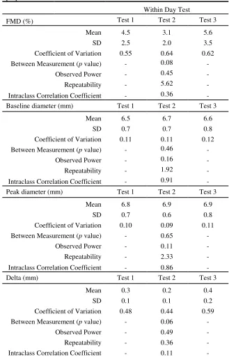
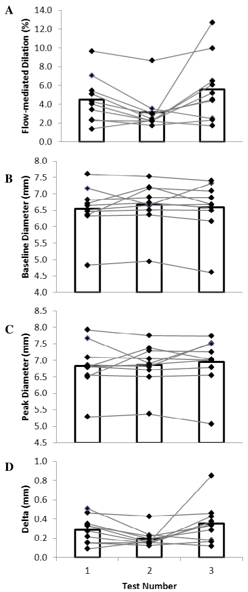
2.3.2 Test-retest Reliability
The means, SDs and coefficient of variation for FMD, baseline diameter, peak diameter
and delta of the three tests performed are listed in Table 2.2. There was no significant difference
between the three repeats within a day at 250 mmHg occlusion pressure for any of the four
variables (p > 0.05). The repeatability of the three FMD tests is also listed in Table 2.2. The
repeatability represents the critical value at which a measurable change is observed in a given
participant between tests. The repeatability of FMD was 5.62%, which means that a percent
change in arterial diameter would have to be greater than 5.62 to be considered a physiological
adaptation rather than measurement error. The repeatability of baseline diameter, peak diameter
and delta were 1.92 mm, 2.33 mm and 0.36 mm respectively. ICCs (also listed in Table 2.2) for
FMD, baseline diameter, peak diameter and delta were 0.36, 0.91, 0.86 and 0.11 respectively.
The high ICCs for baseline and peak diameter indicates that there is a good reliability for those
measures between tests. Alternatively the low ICCs for FMD and delta indicates that those
measures have a very poor reliability over the three tests. This is also illustrated in Figure 2.4;
the baseline (B) and peak diameter (C) show more consistent measurements over the three FMD
tests performed whereas FMD (A) and delta (D) show much more variability between tests.
2.3.3 Day-to-day reliability
The means, SDs and coefficient of variation for FMD, baseline diameter, peak diameter
21
pressure) are listed in Table 2.3. There was no significant difference between the five tests for
any of the four variables (p > 0.05). The repeatability of the five tests is also listed in Table 2.3.
The repeatability of FMD, baseline diameter, peak diameter and delta were 4.82%, 1.00 mm,
1.32 mm and 0.33 mm respectively. ICCs (also listed in Table 2.3) for FMD, baseline diameter,
peak diameter and delta were 0.25, 0.62, 0.52 and 0.11 respectively. Similar to the trends
observed with the test-retest reliability, the day-to-day reliability of baseline and peak diameter
measurements were stronger than that of FMD and delta. Again this is illustrated in Figure 2.5
depicting greater variability between subjects with FMD (A) and delta (D) compared to baseline
(B) and peak (C).
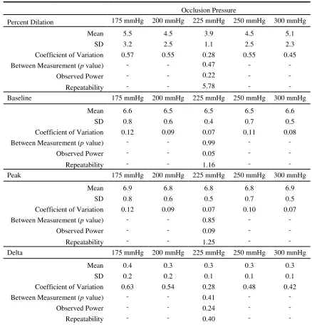
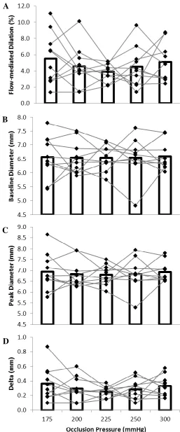
2.3.4 Different Occlusion Pressures
The means, SDs and coefficient of variation for FMD, baseline diameter, peak diameter
and delta of the five FMD tests performed at different occlusion pressures are listed in Table 2.4.
There was no significant difference between the FMD tests performed at different pressures for
any of the four variables (p>0.05). The repeatability of the five FMD tests for each of the
variables is also listed in Table 2.4. The repeatability of the five FMD tests for FMD, baseline
diameter, peak diameter and delta were 5.78, 1.16, 1.25 and 0.40 respectively. Figure 2.6
illustrates the variability of subjects across the different occlusion pressures. Similar to the
test-retest and day-to-day reliability, there appears to be less variation with baseline (B) and peak
diameter (C) measurements compared to FMD (A) and delta (D).
2.3.5 Time Course of Popliteal FMD
Pooling all the popliteal FMD tests together (a total of 110 tests), we were able to
describe the time course of the average FMD response of the popliteal artery over 15 second
22 Table 2.1 Subject Characteristics (n=10)
Age (yrs) Mass (kg) Height (cm)
Resting Blood Pressure (mmHg)
Systolic Diastolic
Mean 27 81 179 120 65
23
Table 2.2 Test-retest reliability statistics for flow-mediated dilation properties.
Within Day Test
FMD (%) Test 1 Test 2 Test 3
Mean 4.5 3.1 5.6
SD 2.5 2.0 3.5
Coefficient of Variation 0.55 0.64 0.62 Between Measurement (p value) - 0.08 -
Observed Power - 0.45 -
Repeatability - 5.62 -
Intraclass Correlation Coefficient - 0.36 - Baseline diameter (mm) Test 1 Test 2 Test 3
Mean 6.5 6.7 6.6
SD 0.7 0.7 0.8
Coefficient of Variation 0.11 0.11 0.12 Between Measurement (p value) - 0.46 -
Observed Power - 0.16 -
Repeatability - 1.92 -
Intraclass Correlation Coefficient - 0.91 - Peak diameter (mm) Test 1 Test 2 Test 3
Mean 6.8 6.9 6.9
SD 0.7 0.6 0.8
Coefficient of Variation 0.10 0.09 0.11 Between Measurement (p value) - 0.65 -
Observed Power - 0.11 -
Repeatability - 2.33 -
Intraclass Correlation Coefficient - 0.86 -
Delta (mm) Test 1 Test 2 Test 3
Mean 0.3 0.2 0.4
SD 0.1 0.1 0.2
Coefficient of Variation 0.48 0.44 0.59 Between Measurement (p value) - 0.06 -
Observed Power - 0.49 -
Repeatability - 0.36 -
Intraclass Correlation Coefficient - 0.11 -
24
Figure 2.4 Variability of measures between three FMD tests performed on the same day for 10 subjects. Group mean for each test is represented by the bar graph.
A
B
25
Table 2.3 Day-to-day reliability statistics for flow-mediated dilation properties.
Day-to-Day Tests
FMD (%) Test 1 Test 2 Test 3 Test 4 Test 5
Mean 3.3 3.5 4.8 4.5 4.5
SD 2.1 1.6 1.8 2.3 2.0
Coefficient of Variation 0.64 0.45 0.37 0.52 0.45
Between Measurement
(p value) - - 0.28 - -
Observed Power - - 0.28 - -
Repeatability - - 4.82 - -
Intraclass Correlation
Coefficient - - 0.25 - -
Baseline diameter (mm) Test 1 Test 2 Test 3 Test 4 Test 5
Mean 6.7 6.7 6.7 6.5 6.7
SD 0.7 0.5 0.6 0.5 0.5
Coefficient of Variation 0.11 0.08 0.09 0.08 0.07
Between Measurement
(p value) - - 0.79 - -
Observed Power - - 0.10 - -
Repeatability - - 1.00 - -
Intraclass Correlation
Coefficient - - 0.62 - -
Peak diameter (mm) Test 1 Test 2 Test 3 Test 4 Test 5
Mean 6.9 6.9 7.0 6.8 7.0
SD 0.6 0.6 0.6 0.5 0.4
Coefficient of Variation 0.09 0.08 0.08 0.08 0.06
Between Measurement
(p value) - - 0.73 - -
Observed Power - - 0.11 - -
Repeatability - - 1.32 - -
Intraclass Correlation
Coefficient - - 0.52 - -
Delta (mm) Test 1 Test 2 Test 3 Test 4 Test 5
Mean 0.2 0.2 0.3 0.3 0.3
SD 0.1 0.1 0.1 0.2 0.1
Coefficient of Variation 0.44 0.52 0.22 0.62 0.42
Between Measurement
(p value) - - 0.28 - -
Observed Power - - 0.25 - -
Repeatability - - 0.33 - -
Intraclass Correlation
Coefficient - - 0.11 - -
26
Figure 2.5 Day-to-day variability of measures between five FMD tests performed on separate days for 10 subjects. Group mean for each test is represented by the bar graph.
A
27
Table 2.4 Statistics for flow-mediated dilation properties associated with five different occlusion pressures.
Occlusion Pressure
Percent Dilation 175 mmHg 200 mmHg 225 mmHg 250 mmHg 300 mmHg
Mean 5.5 4.5 3.9 4.5 5.1
SD 3.2 2.5 1.1 2.5 2.3
Coefficient of Variation 0.57 0.55 0.28 0.55 0.45
Between Measurement (p value) - - 0.47 - -
Observed Power - - 0.22 - -
Repeatability - - 5.78 - -
Baseline 175 mmHg 200 mmHg 225 mmHg 250 mmHg 300 mmHg
Mean 6.6 6.5 6.5 6.5 6.6
SD 0.8 0.6 0.4 0.7 0.5
Coefficient of Variation 0.12 0.09 0.07 0.11 0.08
Between Measurement (p value) - - 0.99 - -
Observed Power - - 0.05 - -
Repeatability - - 1.16 - -
Peak 175 mmHg 200 mmHg 225 mmHg 250 mmHg 300 mmHg
Mean 6.9 6.8 6.8 6.8 6.9
SD 0.8 0.6 0.5 0.7 0.5
Coefficient of Variation 0.12 0.09 0.07 0.10 0.07
Between Measurement (p value) - - 0.85 - -
Observed Power - - 0.09 - -
Repeatability - - 1.25 - -
Delta 175 mmHg 200 mmHg 225 mmHg 250 mmHg 300 mmHg
Mean 0.4 0.3 0.3 0.3 0.3
SD 0.2 0.2 0.1 0.1 0.1
Coefficient of Variation 0.63 0.54 0.28 0.48 0.42
Between Measurement (p value) - - 0.41 - -
Observed Power - - 0.24 - -
Repeatability - - 0.40 - -
28
Figure 2.6 Variability of measures between five FMD tests performed at different occlusion pressures for 10 subjects. Group mean for each test is represented by the bar graph.
A
B
C
29
Figure. 2.7 Time course of popliteal artery dilation following reactive hyperemia. Time course in 15-second intervals of popliteal artery FMD expressed as percent difference from baseline after distal cuff occlusion. Each point on the graph represents an average of all the data available at that time-point.
6.4 6.45 6.5 6.55 6.6 6.65 6.7 6.75 6.8
30 60 90 120 150 180 210 240 270 300
30 2.4 DISCUSSION
The main goals of this study were to investigate the test-to-test (intraday) and day-to-day
(interday) reliability of FMD in the popliteal artery, and to examine the effects of five different
occlusion pressures on the dilatory response in the popliteal artery. The main findings were as
follows: 1) repeatability values for these tests were large indicating a high systematic error of the
technique; 2) reliability of FMD tests was poor both within and between testing days (low ICCs);
3) there was no significant difference between FMD tests performed in the same day, across five
days or tests performed at five different occlusion pressures.
Repeatability is used to examine the influence of measurement errors on data analysis
and is an indicator of absolute reliability, such that the difference between repeated
measurements for the same subject is expected to be less than 2.77 × within subject SD (Sw) for
95% of observations [where 2.77 is derived as (1.96 × √2)] (Bland & Altman, 1996). In the
present study, large values of repeatability indicated poor reliability of the technique.
Repeatability values were high for intra- and interday FMD, such that a difference of 5.62% and
4.82% respectively would be needed to observe a change in FMD that would not be associated
with systemic error. These values for repeatability are as large as the mean FMD measures
themselves. This is particularly important for pre- versus post-intervention study designs that
use FMD tests as an indicator of endothelial function. With high values of repeatability, the
likelihood of detecting a difference is small. The repeatability between days was no worse than
the within day measures, suggesting it is measurement error rather than variability in day-to-day
physiological responses. Studies in the brachial and radial arteries have also shown poor
reliability as estimated by repeatability. Hardie et al. (Hardie et al., 1997) demonstrated that
31
for two FMD tests separated by an average of 90 days. Repeatability calculated from reported
values for Sw indicated FMD in the brachial artery would need to be approximately 19% to be
able to detect changes that could not be attributed to systemic error. This study however, did not
use the “present-day” standardized technique, consider age or sex differences in participants and
did not control for diurnal variation, recent exercise or caffeine intake. More recently, Brook et
al. (Brook et al., 2005) assessed intra- and interday reliability for two FMD tests performed in
the same day and two tests performed approximately 7 days apart. The repeatability calculated
from reported values for Sw were high for both intraday (10.75%) and interday (10.72%). The
authors concluded that radial artery FMD was highly variable. Although this group did control
for diurnal variation and dietary intake, they also did not utilize the “present day” standardized
technique and did not control for sex differences in participants. Unlike the previous studies, the
present study utilized the current standardized technique for FMD tests and controlled for
multiple factors.
This study also reported ICC, which provides a measure of relative reliability. Unlike
measures of absolute reliability, correlation coefficients are influenced by the range of values
measured and give no indication of actual measurement values or systemic variability within the
measure itself (Hopkins, 2000). As suggested by Portney and Watkins (Portney & Watkins,
2000), ICC values >0.75 are considered to be reliable. In the present study, ICC values for
intraday (0.36) and interday (0.25) reliability of FMD indicated very poor reliability of the
technique. These ICC values are similar to those reported by Brook et al (Brook et al., 2005) for
intraday (0.38) and interday (0.23) measures analyzed by the same reader. Alternatively,
Welsch et al. (Welsch et al., 2002) reported high reliability of two FMD measures in the brachial
32
previous studies where only two measures were compared, the present study was the first to
systematically compare five measures for the interday reliability and three measures for intraday
reliability.
Although reliability of FMD was poor for both intra- and interday comparisons, it is
interesting to note that in both cases the reliability of baseline and peak diameters (which are
used to calculate the change in diameter and then FMD) were much better than that of delta
diameter and FMD (expressed as a % change from the baseline value). The repeatability for
intraday comparisons was much smaller for baseline (1.92 mm ie. ~30% of the baseline
diameter) and peak diameter (2.33 mm ie. ~35% of the peak diameter) compared with delta
diameter (0.36 mm ie. greater than 100% of the delta diameter) and FMD (5.62% ie. greater than
100% of the FMD). Additionally, repeatability for interday comparisons was much smaller for
baseline (1.00 mm i.e. ~ 15% of the baseline value) and peak diameter (1.32 mm, i.e. ~ 20% of
the peak value) compared to delta diameter (0.33 mm, i.e. greater than 100% of the delta) and
FMD (4.82% ie. greater than 100% of the FMD). Similarly, ICC values for intra- and interday
reliability were much higher for baseline and peak diameter indicating a higher reliability. In the
case of intraday reliability, ICC values indicated a good reliability of baseline and peak diameter
(0.91 and 0.86, respectively). In agreement with these results, West et al. (West et al., 2004)
found baseline diameter to have a small coefficient of variation (CV range of 1-13%), whereas
the maximal FMD was associated with a much larger variability (CV range 1-84%). It appears
that although baseline and peak diameters can be measured with adequate reliability, the delta
diameter is so small that when adjusted to ratios of % change in diameter or FMD, the error is
33
The FMD values reported in the present study are comparable to others in literature
(Thijssen et al., 2008, 2011b). Thijssen et al. (Thijssen et al., 2008) reported similar values for
the popliteal artery of baseline diameter (6.2 ± 1 mm), peak diameter (6.6 ± 1 mm) and FMD
(6.1 ± 3.3%). Additionally, the time course of popliteal artery FMD depicted in Figure 2.7 is
comparable to the time to peak diameter reported by the same group (181 ± 85 seconds)
(Thijssen et al., 2008).
In the present study, there were no significant differences between FMD tests performed
on the same day or on different days. Nevertheless it is acknowledged that the study was
underpowered to detect these differences and thus there is the possibility of a type II error
(stating that there are no differences when in fact there actually are); in fact, for the three
within-day tests a p-value of 0.08 approaches significance (further demonstrating variability of the
measure). Although we acknowledge this is a limitation in the present study, we were interested
in measuring reliability in this sample size as the majority of research focused studies use small
sample sizes (often 10 or less subjects) to test physiological adaptations to various exercise
protocols, or to examine differences between sexes and clinical populations (Joannides et al.,
1995; Mullen et al., 2001; Betik et al., 2004; Green et al., 2006, 2010; Parker et al., 2006, 2011;
Pyke & Tschakovsky, 2007; Black et al., 2009; Pyke et al., 2009; Tinken et al., 2010). For this
reason we wanted to determine the reliability of the FMD technique in these smaller sample sizes
and understand the limitations when applied to these samples. That being said, using
computational modelling of day-to-day FMD measures we generated data for an increased
sample size, maintaining the same variability in the data. By doubling the sample size (n=20) we
doubled the statistical power (0.57), approached significance for differences between tests (p =
34
size was increased to 40, statistical power above 0.8 was achieved and there was a significant
difference between tests performed on different days (p = 0.002). Interestingly, repeatability
remained relatively unchanged (4.63% vs 4.82%) compared to the original sample size of 10.
Thus, although the present study was not powered to state that there were no statistically
significant differences in measures within or between days, it is likely that the repeatability and
ICC values (which were the main focus of the study) would not be greatly altered with an
increased sample size. The measurement error of the FMD technique with 10 subjects is likely
to remain consistent in larger samples.
Another important aspect of this study was to assess the intraday reliability, to determine
if a single test was repeatable, and therefore an accurate representation of vasoreactivity in the
popliteal artery. By averaging two tests (on each day there was an FMD test at 250 mmHg and
one at another occlusion pressure), we reduced measurement error (decreased repeatability from
4.82% to 3.90%) suggesting that multiple measures averaged may provide a more accurate
assessment of changes in FMD. Sorensen et al. (Sorensen et al., 1995) noted that the
repeatability of the measure could be reduced from 5.2% to 2.6% when the number of FMD tests
is increased from one to four for both pre- and post-measures for a given intervention. In the
present data we were able to examine the effect of averaging by comparing the average of the
three tests within a day and the five tests over five days (at 250 mmHg occlusion pressure). As
shown in Figure 2.8, the correlation of the averaged values for FMD was not high (r = 0.641).
Although significant (p = 0.046) there is not a good reliability and there is considerable scatter
about the line of identity Therefore, although averaging tests may reduce the repeatability
slightly, the reliability of the measure is not greatly improved in this sample. Further analysis is
35
The standardization of the FMD technique is critical for the comparison of FMD values
obtained between different clinicians and research centres. An aspect of the FMD protocol that
remains variable is the occlusion pressure used for the five minute cuff occlusion period. As
hypothesized, there were no significant differences between FMD tests performed at five
different suprasystolic occlusion pressures. In that regard, however, a larger sample size is
needed to definitively conclude that there is no effect of occlusion pressure on FMD response in
the popliteal artery.
Shear rate (calculated as blood velocity/vessel diameter) is commonly used as a proxy
measure of shear stress when viscosity measures cannot be obtained. The shear stress stimulus
has been identified as the major contributor to the magnitude of FMD (Pyke & Tschakovsky,
2007). In some studies of FMD the shear stress is reported in order to estimate the dilation per
shear rate (Pyke & Tschakovsky, 2005). In the present study, although we were confident in our
vessel diameter measures we were not confident in the blood velocity measures which showed
large variability. Therefore, it is plausible that the variability in shear stress may affect
variability and repeatability of FMD, and understanding this interplay requires further study.
In addition to limitations mentioned above, the present study did not control for dietary
intake in the hours leading up to FMD tests. No blood samples were taken and assessed for
hematological variables known to influence arterial vasodilation such as; lipids (Vogel et al.,
1996; Steinberg et al., 1997); homocysteine (Tawakol et al., 1997; Chambers et al., 1999);
fibrinogen (Allen et al., 2000); and blood glucose (Title et al., 2000). Although dietary intake
should not account for the poor reliability of FMD tests within a day, it is possible that some of
the variability in FMD measures between days could be attributed to changes in hematological
36
All ultrasound imaging and analysis were performed by the same investigator. To ensure no
bias, analysis should have been completed by a blinded observer. Furthermore, this study was
completed with manual analysis of all FMD tests. This restricted the frequency at which images
could be selected for diameter measurements. Manually-derived diameter measurements were
taken every 15 seconds in the present study. Current edge-detection software programs allow for
diameter measurements to be obtained as frequently as each cardiac cycle during the time period
of interest. This may provide a more representative time course of vasodilation in the artery
being imaged.
In conclusion, this study demonstrated the popliteal artery FMD measure has poor
reliability for both test-retest and day-to-day reliability. The lack of reliability using this
technique suggests that interpretation of individual or group changes should be made with
caution, particularly when the FMD values are used for clinical diagnosis. Baseline and peak
diameter measures have stronger reliability; however the change in diameter is so small that the
variation in these measures may be magnified when converted to a delta diameter and the FMD
ratio, ultimately accounting for the poor reliability exhibited in those measures. Studies
involving larger sample sizes are required to confirm this. Even with the ever increasing number
of studies addressing reliability of the FMD tests, there remains little consensus. Due to the
noninvasive nature of this technique it is likely that it will continue to be a popular research and
diagnostic tool for assessing endothelial function. As such, known factors affecting vascular
reactivity should be strictly controlled for and studies should adhere to standardized protocol and
37
38 3.0 REFERENCES
Allen JD, Wilson JB, Tulley RT, Lefevre M & Welsch MA (2000). Influence of age and normal plasma fibrinogen levels on flow-mediated dilation in healthy adults. The American Journal
of Cardiology86, 703–5, A9.
Anderson TJ, Uehata A, Gerhard MD, Meredith IT, Knab S, Delagrange D, Lieberman EH, Ganz P, Creager MA, Yeung AC & Selwyn AP (1995). Close relation of endothelial function in the human coronary and peripheral circulations. Journal of the American
College of Cardiology26, 1235–1241.
Betik AC, Luckham VB & Hughson RL (2004). Flow-mediated dilation in human brachial artery after different circulatory occlusion conditions. American Journal of Physiology Heart and
Circulatory Physiology286, H442–8.
Black MA, Cable NT, Thijssen DHJ & Green DJ (2008). Importance of measuring the time course of flow-mediated dilatation in humans. Hypertension51, 203–210.
Black MA, Cable NT, Thijssen DHJ & Green DJ (2009). Impact of age, sex, and exercise on brachial artery flow-mediated dilatation. American Journal of Physiology Heart and
Circulatory Physiology297, H1109–16.
Bland JM & Altman DG (1996). Measurement error. British Medical Journal Clinical Research
Edition313, 744.
Brevetti G, Silvestro A, Schiano V & Chiariello M (2003). Endothelial dysfunction and cardiovascular risk prediction in peripheral arterial disease: additive value of flow-mediated dilation to ankle-brachial pressure index. Circulation108, 2093–2098.
Brook R, Grau M, Kehrer C, Dellegrottaglie S, Khan B & Rajagopalan S (2005). Intrasubject variability of radial artery flow-mediated dilatation in healthy subjects and implications for use in prospective clinical trials. The American Journal of Cardiology96, 1345–1348.
Celermajer DS, Sorensen KE, Gooch VM, Spiegelhalter DJ, Miller OI, Sullivan ID, Lloyd JK & Deanfield JE (1992). Non-invasive detection of endothelial dysfunction in children and adults at risk of atherosclerosis. Lancet340, 1111–1115.
Chambers JC, McGregor A, Jean-Marie J, Obeid OA & Kooner JS (1999). Demonstration of rapid onset vascular endothelial dysfunction after hyperhomocysteinemia: an effect reversible with vitamin C therapy. Circulation99, 1156–1160.
Cooke JP, Rossitch E, Andon NA, Loscalzo J & Dzau VJ (1991). Flow activates an endothelial potassium channel to release an endogenous nitrovasodilator. The Journal of Clinical
39
Corretti MC, Anderson TJ, Benjamin EJ, Celermajer D, Charbonneau F, Creager MA, Deanfield J, Drexler H, Gerhard-Herman M, Herrington D, Vallance P, Vita J & Vogel R (2002). Guidelines for the ultrasound assessment of endothelial-dependent flow-mediated vasodilation of the brachial artery: a report of the International Brachial Artery Reactivity Task Force. Journal of the American College of Cardiology39, 257–265.
Corson MA, James NL, Latta SE, Nerem RM, Berk BC & Harrison DG (1996). Phosphorylation of Endothelial Nitric Oxide Synthase in Response to Fluid Shear Stress. Circulation
Research79, 984–991.
Davies PF (2009). Hemodynamic shear stress and the endothelium in cardiovascular pathophysiology. Nature Clinical Practice Cardiovascular Medicine6, 16–26.
Dimmeler S, Fleming I, Fisslthaler B, Hermann C, Busse R & Zeiher AM (1999). Activation of nitric oxide synthase in endothelial cells by Akt-dependent phosphorylation. Nature 399, 601–605.
Doshi SN, Naka KK, Payne N, Jones CJ, Ashton M, Lewis MJ & Goodfellow J (2001). Flow-mediated dilatation following wrist and upper arm occlusion in humans: the contribution of nitric oxide. Clinical Science (London, England : 1979)101, 629–635.
Gokce N, Keaney JF, Hunter LM, Watkins MT, Menzoian JO & Vita JA (2002). Risk stratification for postoperative cardiovascular events via noninvasive assessment of endothelial function: a prospective study. Circulation105, 1567–1572.
Green DJ, Maiorana AJ, Tschakovsky ME, Pyke KE, Weisbrod CJ & O’Driscoll G (2006). Relationship between changes in brachial artery flow-mediated dilation and basal release of nitric oxide in subjects with Type 2 diabetes. American Journal of Physiology Heart and
Circulatory Physiology291, H1193–9.
Green DJ, Swart A, Exterkate A, Naylor LH, Black MA, Cable NT & Thijssen DHJ (2010). Impact of age, sex and exercise on brachial and popliteal artery remodelling in humans.
Atherosclerosis210, 525–530.
Hardie KL, Kinlay S, Hardy DB, Wlodarczyk J, Silberberg JS & Fletcher PJ (1997). Reproducibility of brachial ultrasonography and flow-mediated dilatation (FMD) for assessing endothelial function. Australian and New Zealand Journal of Medicine 27, 649– 652.
Harris R a, Nishiyama SK, Wray DW & Richardson RS (2010). Ultrasound assessment of flow-mediated dilation. Hypertension55, 1075–1085.
40
Harris RA, Nishiyama SK, Wray DW, Tedjasaputra V, Bailey DM & Richardson RS (2009). The effect of oral antioxidants on brachial artery flow-mediated dilation following 5 and 10 min of ischemia. European Journal of Applied Physiology107, 445–453.
Hopkins WG (2000). Measures of reliability in sports medicine and science. Sports Medicine
(Auckland, NZ)30, 1–15.
Huang A, Sun D, Koller A & Kaley G (1998). Gender difference in flow-induced dilation and regulation of shear stress: role of estrogen and nitric oxide. The American Journal of
Physiology275, R1571–7.
Irace C, Tschakovsky ME, Carallo C, Cortese C & Gnasso A (2008). Endothelial dysfunction or dysfunctions? Identification of three different FMD responses in males with type 2 diabetes.
Atherosclerosis200, 439–445.
Jiang B, Seddon M, Fok H, Donald A & Chowienczyk P (2011). Flow-mediated dilation of the radial artery is offset by flow-induced reduction in transmural pressure. Hypertension 57, 1145–1150.
Joannides R, Haefeli WE, Linder L, Richard V, Bakkali EH, Thuillez C & Lüscher TF (1995). Nitric oxide is responsible for flow-dependent dilatation of human peripheral conduit arteries in vivo. Circulation91, 1314–1319.
Kooijman M, Thijssen DHJ, de Groot PCE, Bleeker MWP, van Kuppevelt HJM, Green DJ, Rongen GA, Smits P & Hopman MTE (2008). Flow-mediated dilatation in the superficial femoral artery is nitric oxide mediated in humans. The Journal of Physiology 586, 1137– 1145.
Kuvin JT, Patel AR, Sliney KA, Pandian NG, Rand WM, Udelson JE & Karas RH (2001). Peripheral vascular endothelial function testing as a noninvasive indicator of coronary artery disease. Journal of the American College of Cardiology38, 1843–1849.
Liuni A, Luca MC, Lisi M, Dragoni S, di Stolfo G, Mariani JA, Uxa A, Gori T & Parker JD (2010). Observations of time-based measures of flow-mediated dilation of forearm conduit arteries: implications for the accurate assessment of endothelial function. American Journal
of Physiology Heart and Circulatory Physiology299, H939–45.
Magda SL, Ciobanu AO, Florescu M & Vinereanu D (2012). Comparative reproducibility of the noninvasive ultrasound methods for the assessment of vascular function. Heart and Vessels; DOI: 10.1007/s00380-011-0225-2.