Efficient Retinal Image Segmentation Using Wavelets and
Neural Networks
Mohd Mazhar Hussain Prof. Prakash J.Patil
Dr.B.R.Vikram
M.Tech Scholar Head of the Department(ECE) Professor and Principal Vijay Rural Engineering College Vijay Rural Engineering College Vijay Rural Engineering College
ABSTRACT:-
Retinopathy has turned into a commonly spread
disease in the world and it causes many
complications. One of the common vision threatening
debilitating complexities of Diabetic Retinopathy. It
occurs when blood vessels in the patient's retina
begin to leak into the macula region of eye. The
purpose of this paper is to extricate features from
retina digital images based on a further analysis of
high frequency components (HH) obtained with the
Discrete Wavelet Transform (DWT).
In particular,
the DWT is applied to the retina photograph to obtain
its high-high (HH) image sub band using
db1,symlet,biorthogonal wavelet transform. Then, a
further decomposition by DWT is applied to the HH
image subband of the previous step to obtain HH*.
Finally, statistical features are computed from HH*
Discrete Wavelet Transform (DWT) based features
and Adaptive Neural Inference System is reported.
The computational results show that present
stage(i.e., normal or abnormal) and gives overall
accuracy and sensitivity, specificity.
Keywords:- Discrete wavelet transform, Neural
network , Diabetic retina, Hard exudates, fundus.
I-INTRODUCTION
Computer-aided diagnosis (CAD) has been the
subject of a considerable measure of research as a
tool to help health professionals in medical decision
making. Subsequently, numerous, many CAD
systems integrate image processing, computer vision,
and intelligent and statistical machine learning
methods to aid radiologists in the interpretation of
medical images and ultimately help improve
diagnostic accuracy. The typical process begin with a
segmentation stage to recognise one or more regions
of interest (ROI) in the image of interest. Then, the
ROI(s) is processed for image enhancement and/or
feature extraction before classification. Because the
segmentation step requires prior knowledge of
discriminate image features and its implementation
typically calls for numerous parameter settings,
recent works have attempted to eliminate it. These
methodologies acknowledge feature space reduction
by applying one or more transforms to the whole
image and extracting the feature vector to classify
from one or more of the obtained components.
Diabetic Retinopathy is caused because of the
increase in intraocular pressure of the eye. The
intraocular pressure increases due to malfunction or
malformation of the drainage system of the eye. The
increased intraocular pressure within the eye
damages the optic nerve through which retina sends
light to the brain where they are perceived as images
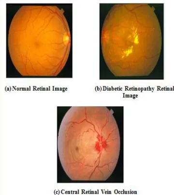
and makes vision possible[1]. The objective of this
paper is to develop an algorithm which automatically
eye images and diseased Diabetic Retinopathy eye
images. The two central issues to automatic
recognition feature extraction from the retinal images
and classification based on the chosen feature
extracted.Several pathologies affecting the retinal
vascular structures due to diabetic retinopathy can be
found in retinal images.
Figure 1: Retinal Images
Ophthalmologists use digital fundus cameras to
non-intrusively view the optic nerve, fovea, surrounding
vessels and the retinal layer .Since retinal imaging
is non-invasive, there is a rapid increase in the
number of images which are being collected.
Diagnosing these large volumes of images is
expensive, tedious and may be prone to human
error. To aid the doctors with this diagnostic task, a
computer-aided diagnosis scheme could offer an
objective, secondary opinion of the images.
II-RELATED PROBLEMS
Dynamic Thresholding
In this method we have applied median filtering onto
the input image directly if it is in grayscale, otherwise
we have to convert the input image into grayscale
before applying median filtering. It corresponds to
the boundary between two regions or a set of points
in the image where luminous intensity changes very
sharply.The presence of an edge within a grayscale
image indicates that there is a change in the grayscale
from one region to another. This approach is
subtraction of median filtered image from input
image (in case of the input image is in grayscale
form) or subtraction of median filtered image from
grayscale form on input image (in case of the input
image is in RGB form). Image subtraction is used to
find changes between two images of same scene. The
algorithm uses the information about color and size
of hemorrhages as a tool for classifying hemorrhages
from other dark lesions present in the retinal images.
The algorithm uses the concepts of contrast
enhancement, background estimation and intensity
variation at edges that is gradient magnitude
information supported by some morphological
operations. The algorithm follows a simple approach
of step by step removal of unwanted features from
targeted images using concepts of thresholding and
morphology without compromising with accuracy
and time of execution. The experimental results
indicate that hemorrhages are detected with good
accuracy in the retinal images.
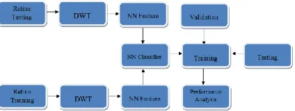
III-PROPOSED METHOD
Multi-Level Discrete Wavelet Transform
Discrete Wavelet transform (DWT) is a mathematical
tool for hierarchically decomposing an image. The
DWT decomposes an input image into four
first letter corresponds to applying either a low pass
frequency operation or high pass frequency operation
to the rows, and the second letter refers to the filter
applied to the columns. The lowest resolution level
LL consists of the approximation part of the original
image. The remaining three resolution levels consist
of the detail parts and give the vertical high (LH),
horizontal high (HL) and high (HH) frequencies.
Many famous coders have been proposed to
effectively compress images or frames processed via
DWT.
Figure 2: Three-Level Wavelet Decomposition of an
Image
Figure 3: Wavelet-based texture analysis on retina
Images
Wavelet-based texture analysis gives a multi
determination analytical platform which enable us to
characterize a signal (an image) in multiple
spatial/frequency spaces. The multi-scale
characteristics of wavelet can be extremely useful.
The 2D wavelet transform has been broadly applied
in image processing applications. There exists two
wavelet structure; (1) Pyramid-structured wavelet
transform which decomposes a signal into a set of
frequency channels with narrower bandwidths in
lower frequency channels, useful for signals which
their important information lies in low frequency
components [8], (2) Tree-structured wavelet analysis
which provides low, middle and high frequency
decomposition which is done by decomposing both
approximate and detail coefficients. In dermoscopy
image analysis, the lower frequency components
reveal information about the general properties
(shape) of the lesion, which is clinically important,
and the higher frequency decomposition provides
information about the textural detail and internal
patterns of the retina which is also significant in the
diagnosis. Thus the decomposition of all frequency
channels are useful in this application. Therefore, the
tree-structured wavelet analysis can be more
informative for classification of retina funds.
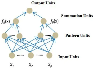
Neural Networks
Neural networks have been used to solve the image
segmentation problem. Generally, the method
involves mapping the problem into a neural network
by means of an energy function, and enabling the
network to converge so as to minimize the engery
function. The network classifies input vector into a
specific class because that class has the maximum
probability to be correct. In this paper, the PNN has
three layers: the Input Layer, Radial Basis Layer and
the Competitive layer. Radial Basis Layer evaluates
vector distances between input vector and row weight
vectors in weight matrix. These distances are scaled
by Radial Basis Function nonlinearly. Competitive
thus finds the training pattern closest to the input
pattern based on their distance.
Figure 4: Neural network analysis on retina
.RESULT Analysis:-
Accuracy:- Accuracy is also used as a statistical
measure of how well a binary classification test
correctly identifies or excludes a condition. among
the total number of cases examined. To make the
context clear by the semantics, it is often referred to
as the "rand accuracy. It is a parameter of the test.it
shows in the command window..
Acc=(Tp+Tn)/(Tp+Tn+Fp+Fn)
Sensitivity:-In medical diagnosis, test sensitivity is
the ability of a test to correctly identify those with the
disease (true positive rate).
Sensitivity =Tp/(Tp+Fn).
Specificity:-Whereas testspecificity is the ability of
the test to correctly identify those without the disease
(true negative rate).
Specificity =Tn/(Tn+Fp).
Output image:-
CONCLUSION:-
This project implemented an retina fund effected on
image classification using texture features and it will
be classified effectively based on neural network.
Here, probabilistic neural network was used for
classification based on unsupervised learning using
wavelet and curve let statistical features and target
vectors. The clustering was estimated from
smoothing details of images accurately for effective
retina disease affected part on segmentation.
REFERENCES:-
[1]. R. Varma et al., “Disease progression and the
need for neuroprotection in glaucoma management,”
Am. J. Manage Care, vol. 14, pp. S15–S19, 2008.
[2]. R. George, R. S. Ve, and L. Vijaya, “Glaucoma in India: Estimated burden of disease,” J. Glaucoma,
vol. 19, pp. 391–397, Aug. 2010.
[3]. K. R. Sung et al., “Imaging of the retinal nerve
fiber layer with spectral domain optical coherence
tomography for glaucoma diagnosis,” Br. J.
[4]. J. M. Miquel-Jimenez et al., “Glaucoma
detection by wavelet-based anal-ysis of the global
flash multifocal electroretinogram,” Med. Eng. Phys.,
vol. 32, pp. 617–622, 2010.
[5]. B. Brown, “Structural and functional imaging of the retina: New ways to diagnose and assess retinal
disease,” Clin. Exp. Optometry, vol. 91, pp. 504–514,
2008.
[6]. S. Weiss, C. A. Kulikowski, and A. Safir,
“Glaucoma consultation by computer,” Comp. Biol.
Med., vol. 8, pp. 24–40, 1978.
[7]. S. Weiss et al., “A model-based method for
computer-aided medical decision-making,” Artif.
Intell., vol. 11, pp. 145–172, 1978.
[8]. R. O. Duncan et al., “Retinotopic organization of
primary visual cortex in glaucoma: A method for
comparing cortical function with damage to the optic
disk,” Invest. Ophthalmol. Vis. Sci., vol. 48, pp. 733–
744, Feb. 2007.
[9]. M. Balasubramanian et al., “Clinical evaluation
of the proper orthagonal decomposition framework
for detecting glaucomatous changes in human
subjects,” Invest. Ophthalmol. Vis. Sci., vol. 51, pp.
264–271, 2010.
[10]. U. R. Acharya, S. Dua, X. Du, V. S. Sree, and
C. K. Chua, “Automated diagnosis of glaucoma using texture and higher order spectra features,” IEEE
Trans. Inf. Technol. Biomed., vol. 15, no. 3, pp. 449–
455, May 2011.
[11]. S. Dua, U. R. Acharya, and E. Y. K. Ng,
Computational Analysis of the Human Eye With
Applications. World Scientific Press, 2011.
[12]. E. A. Essock, Y. Zheng, and P. Gunvant,
“Analysis of GDx-VCC polarime-try data by
wavelet-Fourier analysis across glaucoma stages,”
Invest. Ophthalmol. Vis. Sci., vol. 46, pp. 2838–
2847, Aug. 2005.
[13]. K. Huang and S. Aviyente, “Wavelet feature
selection for image classifi-cation,” IEEE Trans.
Image Process., vol. 17, no. 9, pp. 1709–1720, Sep.
2008.
[14]. A. Arivazhagan and L. Ganesan, “Texture classification using wavelet transform,” Pattern
Recog. Lett., vol. 24, pp. 1513–1521, 2003.
[15]. I. Daubechies, Ten Lectures on Wavelets.
Philadelphia, PA: Society for Industrial and Applied
Mathematics, 1992.
[16]. R. C. Gonzalez and R. E. Woods, Digital Image
Processing. NJ: Prentice Hall, 2001.
[17]. M. H. Dunham, Data Mining Introductory and
Advance Topics. NJ: Pren-tice Hall, 2002.
[18]. H. Liu and R. Setiono, “Chi2: Feature selection and discretization of numeric attributes,” in Proc.
IEEE 7th Int. Conf. Tools WithArtif. Intell., 1995,
pp. 338–391.
[19]. J. R. Quinlan, “Induction of desion trees,”
Mach. Learning, vol. 1, pp. 81– 106, 1986.
[20]. J. R. Quinlan, C4.5 Programs for Machine
Learning. San Mateo: Morgan Kaufmann, 1993.
[21]. K. Kira and L. A. Rendell, “A practical approach to feature selection,” in Proc. 9th Int.
Workshop Mach. Learning, San Francisco, CA, 1992,