ISSN(Online): 2320-9801
ISSN (Print): 2320-9798
I
nternational
J
ournal of
I
nnovative
R
esearch in
C
omputer
and
C
ommunication
E
ngineering
(A High Impact Factor, Monthly, Peer Reviewed Journal)
Website: www.ijircce.com
Vol. 5, Issue 10, October 2017
A Survey on Brain Tumor Detection and
Classification Techniques
Prof.Nitesh Dangre1, Akshata Suravse2, Bhagyashree Kamble3, Shital Baraskar 4
Dept. of Computer Engineering, Keystone School of Engineering, Pune, India1,2,3,4
ABSTRACT: Till date we have seen that many imaging techniques have been used in health care techniques for
determining the brain related diseases such as CT scan or MRI Scan etc. Since the use of unwanted devices such as mobile phones etc. have increased rapidly in last decade, so the increase in the victims of brain tumor has been observed tremendously. Depending upon the abnormalities depicted by the brain tumor it can be either considered as primary or secondary. The theoretical study to be done on the MRI image is a very lengthy process and can be prone to errors and inefficiency. The major concern in determining the brain tumor is proper technique of analysis of MRI image where human intervention is deeply required. This type of difficulty can be recovered by the Computer-aided diagnosis (CAD) scheme have capability of improve the diagnosis and less time required for accurate result The main objective of this review paper is to define the various previously proposed detection techniques for the brain MRI images.
KEYWORDS: Medical imaging, Brain Tumor, CAD, Segmentation, Feature extraction, Classification.
I.INTRODUCTION
AllNow days the biomedical imaging is very important for many applications for radiologist to diagnose the patient treatment related problems. At present imaging technology is must for patient diagnosis. The various medical images like MRI ,Ultrasound, CT, X-ray etc play an important role in the field of process of disease, diagnosing and treating [36, 45,35]. The recent revolution in medical imaging results from techniques such as CT and (MRI) can provide detailed information about disease. and can identify many pathologic conditions giving an accurate diagnosis.
Furthermore, the new techniques are helping to advance fundamental biomedical research. Medical imaging is one of the most common techniques used to improving the diagnoses, understanding and treatment of a large variety of diseases. [41]
The brain imaging analysis is main objective in the field of medical image analysis. Magnetic resonance (MR) imaging have many benefits over the medical imaging modalities such as a useful non invasive technique for assisting in clinical diagnoses, the high level of contrast resolution, multispectral characteristics[46, 9] and ability to provide rich information about human soft tissue[46, 15]. MRI provides useful information in the field of surgery, radiotherapy treatment planning, stereotactic neurosurgery [46, 5] Computer Aided Diagnosis system has been developed for Automatic Detection of Brain Tumor through MRI. Improving the ability to identify early stage tumors is an important goal for physicians, because early detection of class of disease is a key factor in producing successful treatments.
There are different type of detection techniques which is use to develop the CAD. To create a CAD system, the integration of various image processing techniques such as segmentation, feature extraction and classification are essential.[41, 42, 13, 11].
II. LITERATURESURVEY
In recent years, various methods have been proposed for image segmentation, classification and detection techniques for brain tumors.
ISSN(Online): 2320-9801
ISSN (Print): 2320-9798
I
nternational
J
ournal of
I
nnovative
R
esearch in
C
omputer
and
C
ommunication
E
ngineering
(A High Impact Factor, Monthly, Peer Reviewed Journal)
Website: www.ijircce.com
Vol. 5, Issue 10, October 2017
enhancement and skull stripping is performed to improve the speed and accuracy. Segmentation was done by Fuzzy C-Mean (FCM) clustering. Grey level run length matrix (GLRLM) is used for extraction of feature from the brain image, after which SVM technique is applied to classify the brain MRI images, which provide accurate and more effective result for classification of brain MRI images.
Hatice Cinar Akakin and Metin [2], propped the system for multi image queries. Feature is extracted in two part; For Color extraction they have used the color spaces are CIELab (Lab) and hue–saturation–value (HSV) color spaces additional to RGB. The total 26 color and gray-scale features are extracted using three different color spaces for a given image. Texture feature is extracted using Co-occurrence histograms. They have used the two separate classifier (SVM and k-NN) for the classifications of images. They have achieved about 93% and 86% average classification accuracy.
R. Guruvasuki and A. Josephine Pushpa [3], have designed the method using multi support vector machine classifier. The image is preprocessed with median filter. The Gray Level Cooccurrence Matrix is used for feature extraction. Multi Support Vector Machine (M-SVM) classifier is used for classification of three types of image. System performance is improved by the multiple image queries than single image query.
Mohanapriya.S and Vadivel.M [4], propose a robust retrieval using a supervised classifier which concentrates on extracted features. Gray level co-occurrence matrix algorithm is implemented to extract the texture features from images. The feature optimization is done on the extracted features to select best features out of it to train the classifier. The classification is performed on the dataset and it is classified into three categories such as normal, benign and malignant. They have used the SVM (Support Vector machine) classifier followed by KNN (K-nearest neighbor).
B.Ramasubramanian, G.Prabhakar and S.Murugeswari [5], designed the Multitier system for microscopic images having more than one disease. The features based on colour and texture is extracted. In the first tier, the images are classified by recursive SVM classifier with the help of extracted features. In the next tier, the similar images are retrieved using Decision tree algorithm. They have achieved the accuracy 96% (for FL) and 98% (for NB).
Yudong Zhang, Zhengchao Dong, Lenan Wua, Shuihua Wanga [6], have developed a novel hybrid classifier to distinguish normal and abnormal brain MRIs. In this paper, they present a neural network (NN) based method to classify a given MR brain image as normal or abnormal. This method first employs wavelet transform to extract features from images, and then applies the technique of principle component analysis (PCA) to reduce the dimensions of features. The reduced features are sent to a back propagation (BP) NN, with which scaled conjugate gradient (SCG) is adopted to find the optimal weights of the NN.
Hashem Kalbkhania, Mahrokh G. Shayesteha, Behrooz Zali-Vargahan [7], have proposed method which can classifies MRI into normal or one of the seven different diseases. The coefficients of two-level 2D DWT of brain MRI are computed. The calculated coefficients of detail sub-bands are modeled by GARCH. After feature vector normalization, principal component analysis (PCA) and linear discriminant analysis (LDA) are used to extract the proper features and remove the redundancy from the primary feature vector. Finally, the extracted features are applied to the K-nearest neighbor (KNN) and support vector machine (SVM) classifiers separately to determine the normal image or disease type.
Sandeep Chaplot, L.M. Patnaik, N.R. Jagannathan [8], propose a novel method using wavelets as input to neural network self-organizing maps and support vector machine for classification of magnetic resonance (MR) images. In this paper, they have used the wavelets as input to support vector machine and neural network. Classification accuracy of more than 94% was achieved using the neural network self-organizing maps (SOM) and 98% from support vector machine.
Zafer Iscan, Zümray Dokur, Tamer Ölmez [9], proposed method for the detection of tumor in magnetic resonance (MR) brain images. First 2D continuous wavelet transform (CWT) and then each MR image is segmented into seven classes (six head tissues and the background) by using the incremental supervised neural network (ISNN). Symmetry axis of the head is determined by using moment properties. Asymmetry is analyzed using the Zernike moments of each of six tissues. The two vectors are individually formed for the left and right hand sides of the symmetry axis. The two vectors are used to determine the asymmetry and tissue with the tumor.
ISSN(Online): 2320-9801
ISSN (Print): 2320-9798
I
nternational
J
ournal of
I
nnovative
R
esearch in
C
omputer
and
C
ommunication
E
ngineering
(A High Impact Factor, Monthly, Peer Reviewed Journal)
Website: www.ijircce.com
Vol. 5, Issue 10, October 2017
easily realizes a solution and requires fewer parameters than SOINN; using some smoothing techniques, ESOINN is also more stable than SOINN.
Monika Jain, Shivanky Jaiswal, Sandeep Maurya, Mayank Yadav [11], have proposed strategy for detection of tumor with the help of segmentation techniques in MATLAB; which incorporates preprocessing stages of noise removal, image enhancement and edge detection. Processing stages includes segmentation. Tumor region is extracted using over global thresholding method. Post proposing stage include histogram clustering, morphological operations. In this step the shape of tumor is determine and also area is calculated.
R. S. RajKumar and G. Niranjana [12], proposes segmentation using cellular automata and classification of tumors using Gray level Co-occurrence matrix features and artificial neural network. Seed pixel selection is done by using the GLCM and after selection by calculating the run length it is checked that the seed pixel is belong to abnormal region or not. The segmentation using cellular automata done and then classification done using Radial basis function which is the type of ANN. The approach is limited by the fact that it necessitates fresh training each time whenever there is a change in image database.
Ketan Machhale, HariBabu Nandpuru, Vivek Kapur and Laxmi Kosta [13], presented an intellectual classification system to recognize normal and abnormal MRI brain images. For preprocessing Median filter and morphological operations are used. In feature extraction phase, gray scale, symmetrical and texture features are extracted. They have used the three classifier; Support Vector Machine (SVM), K- Nearest Neighbor (KNN) and Hybrid Classifier (KNN). They used these classifiers to classify 50 images. The result observation shows that the Hybrid classifier SVM-KNN demonstrated the highest classification accuracy rate of 98% among others.
Padma Nanda Gopal & R.Sukanesh [14], in their paper they presented a combination of wavelet statistical features (WST) and wavelet co-occurrence texture feature (WCT) obtained from two level discrete wavelet transform (DWT). is used for the classification of abnormal brain tissues in to benign and malignant. The proposed system consists of four phases: segmentation of region of interest, discrete wavelet decomposition, feature extraction and feature selection and classification and evaluation. The support vector machine is employed to segment the shape of tumor information. A combination of both WST and WCT texture features is extracted from tumor region of two-level discrete wavelet transformed images. Genetic algorithm (GA) is used to select the optimal texture features from the set of extracted features. The probabilistic neural network classifier (PNN) is built to classify the abnormal brain tissues into benign, malignant tumor images. Comparing the classifications results of PNN, LVQ, BPN classifiers for the texture analysis methods, the results shows that best performence3 is achieved by PNN. The 97.5% accuracy is achieved.
Kailash Sinha and G.R.Sinha [15], presented a comparative study of three segmentation methods implemented for extraction of tumor in the MRI images. The methods include kmeans clustering with watershed segmentation algorithm, optimized k-means clustering with genetic algorithm and optimized c- means clustering with genetic algorithm. Using all three methods exact position and the shape are determined. Results shows that genetic c-means algorithm provide fast and efficient clustering results and also eliminate the over segmentation problem.
Pranita Balaji, Kanade and P.P. Gumaste [16], proposed brain tumor detection method for MRI images. In this paper, the brain tumor is detected & classified stages of the tumor by using testing & training the database. Proposed methodology consists of following main stages: image preprocessing, de noising, SWT & segmentation, feature extraction an classification. In the first step, median based filters and SWT technique are used for de-noising the image. Then spatial FCM technique is used for segmentation and Stationary wavelet transform (SWT) technique is used for feature extraction, as SWT coefficients will not change even if the signal is shifted. In the last step, using Probabilistic neural networks (PNN) images are classified with the help of extracted features.
BRAINTUMORCLASSIFICATIONTECHNIQUE
1. “Pre-processing and Enhancement for MR image”
ISSN(Online): 2320-9801
ISSN (Print): 2320-9798
I
nternational
J
ournal of
I
nnovative
R
esearch in
C
omputer
and
C
ommunication
E
ngineering
(A High Impact Factor, Monthly, Peer Reviewed Journal)
Website: www.ijircce.com
Vol. 5, Issue 10, October 2017
Image pre-processing and enhancement is the starting stage which is important for the further stages. This gives the higher accuracy of CAD system. Pre-processing and enhancement used to improve the detect the regions in MRI. This stage is used for reducing image noise, highlighting edges, or displaying digital images. The removal of unwanted parts from the brain MR image, finding edge position for removing labels and smoothing the image will be to be much superior to other techniques processed by using innovatively new pre-processing methods. The enhancement stage used some techniques for MRI images and gives the noise free, film artifact images. [2, 22]
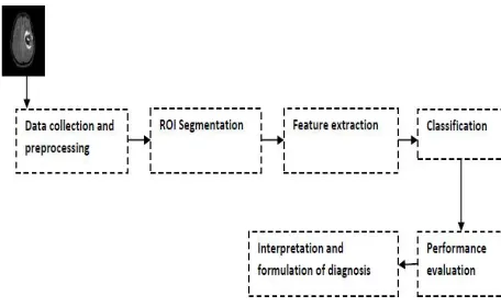
Fig 1. Methodology of CAD System
2. “Segmentation Techniques For MR Image”
Radiologist first to analysis the condition and nature of the brain image abnormality, and then plan for treatment. In case of MR image abnormalities, the abnormal growth may be in increasing or decreasing form which indicates the positive effect and sometimes indicates the negative effect. In any condition the volumetric analysis is very important for analysis the brain tumor growth in MR image. Segmentation methods has ability to detect or identify the abnormal portion from the image which is useful for analyzing the size, volume, location, texture and shape of the extracted image. Several researchers are currently working on this medical image segmentation area. The most famous detection methods are: (i) fuzzy based methods (ii). Thresholding based methods (iii) region-growing based methods and (iv) Clustering based methods used for tumor segmentation.[46]
Table 1 shows the summary of segmentation techniques and Table 3 compares the different segmentation methods. Clearly, advantages and disadvantages of the most famous detection techniques used for medical imaging analysis are summarized briefly.
ISSN(Online): 2320-9801
ISSN (Print): 2320-9798
I
nternational
J
ournal of
I
nnovative
R
esearch in
C
omputer
and
C
ommunication
E
ngineering
(A High Impact Factor, Monthly, Peer Reviewed Journal)
Website: www.ijircce.com
Vol. 5, Issue 10, October 2017
3. “Feature Extraction for MR Image”
In The feature extraction techniques represent the real biomedical/non medical image database in an alternate way by measuring the most popular properties or to extract the features of the image. The different type of feature like shape, texture and boundary used for biomedical images.
4. “Classification for MR Image”
The biomedical image classification is very important stage for automated CAD system. In this step define the different normal or abnormal cases for disease with the help of the calculated feature set. Classification is the best approaches for identification of images like any kind of medical imaging. All classification algorithms are based on the prediction of image, where one or more features and that each of these features belongs to one of several classes. Table 2 shows the summary of classification and feature extraction techniques and Table 3 compares the different classification methods. Clearly, advantages and disadvantages of the most famous classification techniques used for medical image classification are summarized briefly.
III. CONCLUSION
Thus In this paper we have accomplished a comprehensive and deep survey of various brain tumor classification and detection techniques for MRI brain image. A comparative study is made for various techniques. There are various methods which can detect the tumor efficiently and accurately. This work will be extended for the development of new algorithm for brain tumor classification and detection, which will provide more efficient result than the existing methods in near future. Accuracy, reliability and computational time are the most importance to be considered to compare this technique efficiently, as the diagnosis of brain tumor is a complicated and sensitive task.
ACKNOWLEDGMENT
The All faith and honor to our HOD for his grace and inspiration. I would like to thank all my Friends and Family members they were always been there to support us. We sincerely thanks to our Department Head, Project coordinator, our project guide and all other staff members to give us the guidelines for this paper.
REFERENCES
[1] Parveen and Amritpalsingh, “Detection of Brain Tumor in MRI Images, using Combination of Fuzzy C-Means and SVM,” 2nd International Conference on Signal Processing and Integrated Networks (SPIN),pp. 98-102, 2015.
[2] Hatice CinarAkakin and Metin N. Gurcan,“Content-based microscopic image retrieval system for multi-image queries”, IEEE Transaction on Information Technology in Biomedicine, Vol. 16, No. 4, pp 758-768, 2012.
[3] Guruvasuki, A. Josephine Pushpa Arasi, “MRI brain image retrieval using multisupport vector machine classifier”, International Journal of Advanced Information Science and Technology, Vol. 10, No 10, pp 29-36, 2013
[4] Mohanpriya S., Vadivel M, “Automatic Retrieval of MRI Brain Image using Multiqueries System”, IEEE Conference, pp 1099- 1103, 2013. [5] B.Ramasubramanian, G. Praphakar, S. Murugeswari, “ A Novel Approach for Content Based Microscopic Image Retrieval system Using
Decision Tee Algorithm”, International journal of scientific& engineering research, Vol. 4, No 6, pp 584-588, 2013.
[6] Yudong Zhang, Zhengchao Dong, LenanWua, ShuihuaWanga, “A hybrid method for MRI brain image classification”, Elsevier journal Expert system and Application, Vol. 20, No 2, pp 10049- 10053 ,2011.
[7] Hashem Kalbkhani, Mahrokh G Shayesteh, Behrooz Zalivargahan “Robust algorithm for Brain Magnetic Resonance Image Classification based on GARCH variances Series”, ELSEVIER Biomedical Signal Processing and Control 8(2013) 909-919
[8] Sandeep Chaplot , L.M. Patnaik , N.R. Jagannathan, “Classification of magnetic resonance brain images using wavelets as input to support vector machine and neural Network”, Elsevier Journal on Biomedical Signal Processing and Control, Vo.1, No 1,pp 86 -92 ,2006.
[9] Z. Iscan, Z. DokurandT. Olmez, “Tumor detection by using Zernike moments on segmented magnetic resonance brain images”, Elsevier Journal of Expert system and Application, Vol. 37, No 3, pp 2540-2549, 2010.
[10] ShenFurao, Tomotaka Ogura, Osamu Hasegawa, “An Enhanced Self Organizing Incremental Neural network For Online Unsupervised learning”, Elsevier Journal on Neural Network, Vol. 20, No 8, pp 893-903, 2007.
[11] Monika Jain, Shivanky Jaiswal, Sandeep Maurya, Mayank Yadav “ A Novel Approach for the Detection & Analysis of Brain Tumor,” International Journal of Emerging Technology and Advanced Engineering, vol. 5, Issue 4, pp. 54–59, 2015.
ISSN(Online): 2320-9801
ISSN (Print): 2320-9798
I
nternational
J
ournal of
I
nnovative
R
esearch in
C
omputer
and
C
ommunication
E
ngineering
(A High Impact Factor, Monthly, Peer Reviewed Journal)
Website: www.ijircce.com
Vol. 5, Issue 10, October 2017
[13] KetanMachhale, HariBabuNandpuru , VivekKapur and LaxmiKosta, “MRI Brain Cancer Classification Using Hybrid Classifier (SVM-KNN),” International Conference on Industrial Instrumentation and Control (ICIC), pp.60-65, 2015.
[14] Padma Nanda Gopal & R.Sukanesh,” wavelet stat ist ical feature based segmentat ion and classificat ion of brain computed tomography images”IET Image P rosess Vol7 pp 25 -32 2013
[15] KailashSinha and G. R. Sinha, “Efficient Segmentation Methods for Tumor Detection in MRI Images,” IEEE Student’s Conference on Electrical, Electronics and Computer Science, pp.1-6, 2014.