The Structure, Function and
Regulation of Mycobacterial
Porin-Encoding Genes
Richard Alan Speight
A thesis submitted in partial fulfilment of the requirements of University College London for the
degree of Doctor of Philosophy
Division of Mycobacterial Research
National Institute for Medical Research
Mill Hill
London
ProQuest Number: U642571
All rights reserved
INFORMATION TO ALL USERS
The quality of this reproduction is dependent upon the quality of the copy submitted.
In the unlikely event that the author did not send a complete manuscript and there are missing pages, these will be noted. Also, if material had to be removed,
a note will indicate the deletion.
uest.
ProQuest U642571
Published by ProQuest LLC(2015). Copyright of the Dissertation is held by the Author.
All rights reserved.
This work is protected against unauthorized copying under Title 17, United States Code. Microform Edition © ProQuest LLC.
ProQuest LLC
789 East Eisenhower Parkway P.O. Box 1346
Index of Contents
INDEX OF FIGURES...V INDEX OF TABLES______________________________________________________________ VII ABBREVIATIONS... VIII ABSTRACT... X
1. INTRODUCTION... 1
1.1 Th e My c o b a c t e r ia... 1
1.2 Ta x o n o m yo ft h em y c o b a c t e r ia...2
1.3 Th e St a in in g Pr o p e r t ie so f My c o b a c t e r ia... 5
1.4 Ho s tr e s p o n s et ot u b e r c u l o s is... 5
1.5 Th e My c o b a c t e r ia l Ce l l En v e l o p e... 7
1.5.1 T h e P lasm a M e m b ra n e ...8
1.5.2 T h e C ell W a ll...8
1.5.3 T h e C apsular la y e r ... 10
1.6 Pu b l ic a t io no ft h e My c o b a c t e r ia lg e n o m e... 13
1.7 B ACTERIAL PERMEAB ILITY...13
1.7.1 P erm eability in G ram positive b a c te ria... 13
1.7.2 P erm eab ility in G ram negative b a c te ria ...14
1.7.3 P erm eab ility in th e M y c o b a c te ria ...16
1.8 Me t h o d so ft r a n s p o r ta c r o sst h eb a c t e r ia lo u t e re n v e l o p e... 17
1.8.1 S pecific D iffusion C h a n n e ls...17
1.8.2 A ctive E fflux P u m p s... 19
1.8.3 N on-S pecific C hannels, T he P o rin s ...20
1.9 Po r in sf r o m Gr a m Po s it iv e Ba c t e r ia... 23
1.10 Po r in sf r o mt h e My c o b a c t e r ia... 24
1.10.1 P orins in the cell w all o f Mycobacterium tuberculosis...25
1.11 Vo lta g e Gatingo f Pordm Ch a n n e l s... 27
1.12 Dy n a m ica ltera tio no fb a c t e r ia lm e m b r a n ep e r m e a b il it y...28
1.13 M o d u l a t i o n o f P o r i n l e v e l s in Es c h e r ic h iac o u - a b a c t e r i a l t w o - c o m p o n e n t REGULATORY SYSTEM...29
1.14 Ot h e rt w o-c o m p o n e n ts y st e m sinb a c t e r ia...32
1.15 T w o - c o m p o n e n t s y s t e m s in My c o b a c t e r iu mt u b e r c u l o s is...32
1.16 Dis r u pt io no fg e n ee x p r e s s io nint h em y c o b a c t e r ia...34
1.16.1 M obile genetic e le m e n ts... 34
1.16.2 U se o f antisense R N A ... 37
AIMS OF THE PROJECT... 41
2. M A T E R IA L S A N D M E T H O D S ... 42
2.1 Ba c t e r ia ls t r a in sa n dg r o w t h CONDITIONS u s e d...42
2.2 P G R AMPLIFICATION OF D N A ... 43
2.3 Ag a r o s eg e le l e c t r o p h o r e siso f D N A ... 43
2.4 Ex c is io no f D N A b a n d sf r o ma g a r o s eg e l s...43
2.5 Lig a tio no fp l a s m ida n din se r t D N A ...44
2.6 P r e p a r a t i o n o f c o m p e t e n t Es c h e r ic h iac o u... 44
2.7 T r a n s f o r m a t i o n o f Esc h e r ic h iac o u b y h e a t s h o c k ... 45
2.8 E x t r a c t i o n o f p la s m id s f r o m Esc h e r ic h iac o u... 45
2.9 Prepa ra tio no fe l e c t r o c o m p e t e n tm y c o b a c t e r ia...45
2 .10 El e c t r o po r a t io no fm y c o b a c t e r ia... 46
2.11 Au t o m a t e ds e q u e n c in go fp l a s m id s... 46
2.12 P G R PROBING OF M y c o b a c t e r i a l s p e c ie s f o r om pA Tb...47
2.13 We s t e r nb l o ta n a ly siso fm y c o b a c t e r ia lc e l ll y s a t e s...47
2.13.1 P rep aratio n o f m ycobacterial cell ly sa te s... 47
2.13.2 D eterm ination o f p rotein c o n c e n tra tio n ... 48
2.13.3 P olyacrylam ide gel electro p h o resis...48
2.13.4 W estern transfer o f p rotein g e l s ...49
2.13.5 A ntibody b ased detection o f O m pA Tb in W estern b lotted cell ly s a te s ... 49
2.14 Id e n t if ic a t io no faputativereg u la to r ys y s t e m...50
2.14.1 G ene R v 0903c is transcribed in m y c o b ac teria ...50
2.15 Ex p r e s s io na n dp u r ific a t io no f Rv0903 p r o t e in...50
2.15.1 C onstruction o f plasm id p R A S l ... 50
2.15.2 E xp ressio n o f recom binant R v0903 p ro te in ...51
2.15.3 P u rification o f recom binant R v0903 p ro tein ...51
2.16 Ge l Re t a r d a tio na s s a y s... 52
2.17 Ph o s p h o r y l a t io no f Rv0903 p r o t e inp r io rtog e lr eta rd a tio na s s a y...53
2.18 E x p r e s s io n o f OmpATb in Myc o b a c t e r iu ms m e g m a t is...55
2.18.1 O verexpression o f the po rin g e n e ... 55
2 .18.2 H igh-level expression o f th e porin g e n e ...55
2.18.3 E x p ressio n o f the porin gene and the p u tative regulatory com plex u n d er a native p ro m o te r ... 55
2.18.4 E x p re ssio n o f the porin gene under a native p ro m o te r...57
2.19 C o n s t r u c t i o n o f a s u ic id e v e c t o r f o r o m pA Tb k n o c k o u t b y h o m o l o g o u s RECOMBINATION... 57
2.19.1 C onstruction o f plasm id p R A S 4 ... 57
2.19.2 C onstruction o f plasm id p R A S 5 ... 58
2.2 0 Co n s t r u c t io no fas u ic id ev e c t o rf o r Rv0 9 0 3ck n o c k o u tb yh o m o l o g o u s
RECOMBINATION...60
2.20.1 C on stru ctio n o f p la sm id pR A S 1 4 ... 60
2.20.2 C o n stru ctio n o f p la sm id pR A S 1 7 ... 62
2.20.3 C on stru ctio n o f p la sm id pR A S 1 8 ... 64
2.21 C o n s t r u c t i o n o f l a cZ r e p o r t e r v e c t o r s ...65
2.22 p-GALACTOSIDASE ASSAYS FOR PROMOTER ACTIVITY... 66
2.23 Ex t r a c t io no f My c o b a c t e r ia l Nu c l e ic Ac i d s...68
2.23.1 D N A ex tractio n fro m M yco b a cteriu m tuberculosis grow n o n 7 H 1 1 agar p la te s ... 68
2.23.2 D N A extraction usin g Instag en e M a tr i x ...68
2.23.3 D N A E x tractio n fro m M ycobacterial bro th c u ltu re s... 69
2.23.4 Iso latio n o f M ycobacterial R N A fro m B C G ...69
2.23.5 Iso latio n o f R N A fro m M yco b a cteriu m tu b e rc u lo sis...70
2.24 Re m o v a lo f D N A co n t a m in a t io nf r o mm y c o b a c t e r ia l R N A ...70
2.25 Sy n t h e s iso fcD N A b yr e v e r s et r a n s c r ip t io n...70
2.26 N a C l s h o c k o f B C G c u l t u r e s t o i n v e s t i g a t e o m pA Tb r e g u l a t i o n ...71
2.27 Re a l-t im e P C R t od e m o n st r a t er e g u l a t io no ft h ep o r ing e n e...71
2.28 So u t h e r n b l o t t in go fm y c o b a c t e r ia l D N A ...72
2.29 A n a l y s i s o f t h e g r o w t h o f a p o r i n - d e f i c i e n t s t r a i n o f My c o b a c t e r iu mt u b e r c u l o sis . 74 2 .3 0 Mic r o a r r a yh y b r id is a t io n...74
2.30.1 L ab e llin g o f genom ic D N A ... 74
2.30.2 L ab ellin g o f R N A ... 75
2.30.3 H y b rid isatio n o f s lid e s ...75
3. RESULTS... 77
3.1 P C R PROBING OF M y c o b a c t e r i a l s p e c ie s f o r om pA Tb... 77
3.2 We s t e r nb l o ta n a ly siso fm y c o b a c t e r ia lc e l ll y s a t e s...81
3.3 Id e n t if ic a t io no fapu ta tiv er eg u la to r ys y s t e m...81
3.4 Ex p r e s s io na n dp u r ific a t io no f Rv0903 p r o t e in...90
3.5 Ge l Re t a r d a t io na s s a y s... 90
3.6 E x p r e s s io n o f OmpATb in My c o b a c t e r iu ms m e g m a t is... 91
3.7 A n a l y s i s o f My c o b a c t e r iu mt u b e r c u l o sis t r a n s f o r m e d w i t h pR A S5 o r pO m pA -aph-r p s L t b f o -aph-r o m pA Tb k n o c k o u t ... 100
3 .7.1 M yc o b a cte riu m tuberculosis transform ed w ith p R A S 5 ... 100
3.7.2 M yc o b a cte riu m tuberculosis transform ed w ith pO m p A -a p h -rp sL t b ...100
3.8 A n a l y s i s o f My c o b a c t e r iu mt u b e r c u l o sis t r a n s f o r m e d w i t h pRAS 18 f o r R v 0 9 0 3 c KNOCKOUT...101
3.8.1 A ttem p t at second cro sso v er o f pR A S 18 transform ants in liquid b r o t h ...104
3.8.2 A ttem p t at second cro sso v er on solid m e d ia ...104
3.10 Re a l-t im e P C R t od e m o n st r a t er e g u la tio no ft h ep o r ing e n e... 108
3.10.1 R eal tim e R T -PC R o f B C G ...108
3.10.2 R eal tim e R T -PC R o f Mycobacterium tuberculosis...113
3.11 Mic r o ARRAY h y b r id isa t io n...113
3.11.1 C om parison o f genom ic D N A from Mycobacterium tuberculosis 1424 and AompATb m utant using a m icroarray... 113
3.11.2 C om parison o f R N A from Mycobacterium tuberculosis 1424 and AompATbm utant using a m ic ro a rra y ... 114
3.11.3 C om parison o f Mycobacterium tuberculosisH 3 7 R v w ith N aC l shocked bacteria using a m icroarray ... 114
3.12 A n a l y s i s o f g r o w t h o f My c o b a c t e r iu mt u b e r c u l o s is w i t h o u t t h e p o r i n e n c o d i n g g e n e . ... 115
4. DISCUSSION...122
Ov e r v ie w...122
4.1 PCR-BASED SCREENING OF MYCOBACTERIAL CULTURES FOR THE OMPATB GENE AND THE RV0903C GENE... 124
4.2 P-GALACTOSIDASE ASSAYS FOR PROMOTER ACTIVITY... 125
4.3 E x p r e s s io n o f t h e pordm dm My c o b a c t e r iu ms m e g m a t i s... 126
4.4 Reg u la tio no f t h epordmass h o w nb yr e a l-t im e R T -P C R ...129
4.5 Ge lr eta rd a tio na ssa ysu s in gr e c o m b in a n t Rv0903 p r o t e in...131
4.5.1 P hosphorylation o f R v0903 protein enhances bin d in g affin ity ...133
4 .6 G e n e k n o c k o u t o f o m pA Tb u s in g pRAS5 a n d p O m p A -a p h -rp sL t b ... 134
4.7 Ge n ek n o c k o u to f Rv09 0 3cu sin gpR A S 1 8 ... 137
4.8 M i c r o ARRAY ANALYSIS OP My c o b a c t e r iu mt u b e r c u l o s is... 139
5. GENERAL CONCLUSION AND FUTURE PERSPECTIVES... 141
ACKNOWLEDGEMENTS... 143
Index of Figures
Figure 1 Schematic diagram of the mycobacterial cell envelope... 12
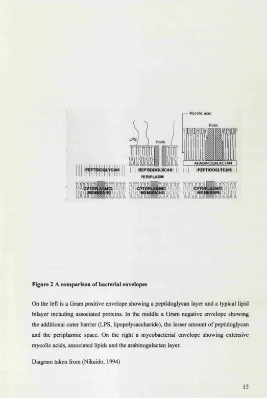
Figure 2 A comparison of bacterial envelopes... 15
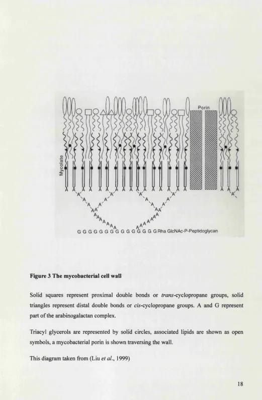
Figure 3 The mycobacterial cell w all... 18
Figure 4 Regulation of porin levels in Escherichia coli... 30
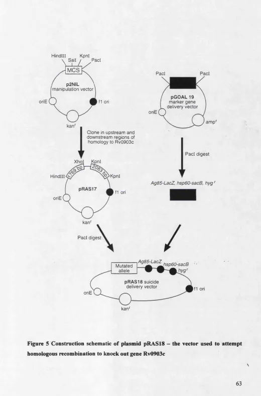
Figure 5 Construction schematic of plasmid pR A SlS - the vector used to attempt homologous recombination to knock out gene R v0903c... 63
Figure 6 PCR based detection of ompATb gene amongst different mycobacterial species ... 79
Figure 7 PCR based detection of Rv0903c gene amongst different mycobacterial species 80 Figure 8 Western blot analysis o f Mycobacterium microti with anti-OmpATb antibodies 83 Figure 9 Result of a BLAST search of the Mycobacterium tuberculosis genome with the Escherichia coli OmpR sequence... 84
Figure 10 Rv0903 gene is transcribed in Mycobacterium bovis B C G ... 85
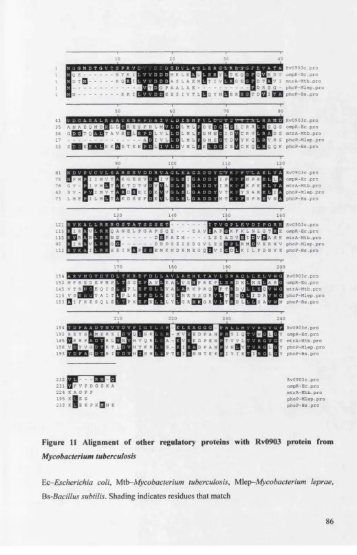
Figure 11 Alignment of other regulatory proteins with Rv0903 protein from Mycobacterium tuberculosis... 86
Figure 12 Alignment of Mycobacterium tuberculosis Rv0902 protein m d Escherichia coli E nvZ ...87
Figure 13 Position of coding sequences on the Mycobacterium tuberculosis genome 88 Figure 14 Purification of the recombinant Rv0903 p ro tein ...89
Figure 15 Sample results of a band-shift assay on regions of the Mycobacterial genome using recombinant Rv0903 protein ... 93
Figure 16 Schematic representation of fragments used in gel shift assay...94
Figure 17 Phosphorylation of the recombinant protein enhances binding to D N A 95 Figure 18 Expression of the OmpATb protein in Mycobacterium smegmatis assayed by western b lo t... 96
Figure 19 Growth of Mycobacterium smegmatis expressing the porin from a single copy of the hsp60 promoter per cell (transformed with plasmid p R A S ll) ... 97
Figure 21 Expression of the porin (pRAS21) or the porin and associated regulatory
machinery (pRAS20) in Mycobacterium smegmatis with 200 mM NaCl in the culture
m edium ...99
Figure 22 PCR-based analysis of pOmpA-aph-rpsL tb transformants (8 isolates) 102
Figure 23 Southern blot analysis of Mycobacterium tuberculosis IsompATb knockout
stra in s... 103 Figure 24 Results of PCR-based screen of potential Rv0903c knockouts...106
Figure 25 p-galactosidase activity of Mycobacterium smegmatis cell lysates after
transformation with pRAS3, pRAS6, pRAST orpR A SS ... 107 Figure 26 Sample amplification of a cDNA target in the real-time PCR machine, the cycle threshold (Ct) can be seen as the point where the curve crosses a predefined fluorescence value... 110
Figure 27 Molarity of culture medium affects levels of ompATb transcript...I l l
Figure 28 Time after addition of N a C l shock (400 mM) affects levels of ompATb
transcript... 112 Figure 29 Sample results from microarray hybridisation using Cy3 and Cy5 labelled nucleotides...116 Figure 30 Scatter plot of microarray hybridisation from cDNA prepared from RNA
harvested 6 hours after 0.2 M NaCl shock of Mycobacterium tuberculosis cu ltu res 117
Figure 31 Growth of Mycobacterium tuberculosis 1424 vs AompATb in Dubos broth ...118
Figure 32 Growth of Mycobacterium tuberculosis 1424 vs AompATb in Dubos broth with
0.2 M N aC l...119
Figure 33 Growth of Mycobacterium tuberculosis 1424 vs AompATb in Dubos broth with
0.1 M raffin o se... 120
Figure 34 Growth of Mycobacterium tuberculosis 1424 vs AompATb in Dubos broth with
0.2 M sucrose... 121
NOTE ADDED AFTER EXAMINATION
A revised and additional list o f figure legends can now be found as Supplementary Appendix 1.
A list o f plasmids used can also be found as Supplementary Appendix 2.
Index of Tables
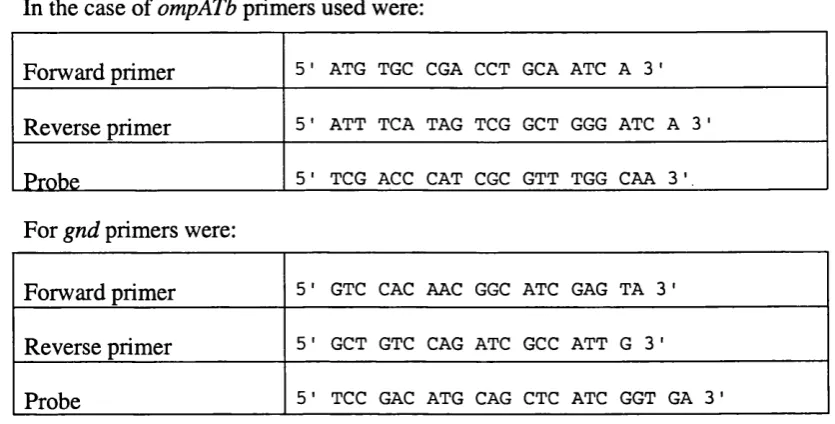
Table 1 Primers used to generate PCR products for gel-shift experim ents...54 Table 2 Details of primers and probes used for TaqMan experiment... 72 Table 3 Results of a PCR-based screen for the porin gene and the regulator gene Rv0903c
... 78 Table 4 Transformation efficiency of suicide delivery vector is enhanced by pre-treatment of plasmid DNA with ultraviolet light... 101
Table 5 Level of ompATb transcript varies with time after addition of 400 mM N a C l.. 108
Table 6 Level of ompATb transcript varies with molarity 1 h after NaCl addition 108
Table 7 Result of addition of 200 mM sucrose to growth media of B C G ...109
Table 8 Effect of addition of 200 mM NaCl on transcript levels of ompATb in
Abbreviations
APS Ammonium persulphate
BCA Bicinchoninic acid
BCG Bacille Calmette-Guérin
BLAST Basic local alignment search tool
bp Base pairs
BSA Bovine serum albumin
cDNA complementary single-stranded DNA
GIF Calf intestinal phosphatase
DEPC Diethyl pyrocarbonate
DMSO Dimethylsulphoxide
DNA Deoxyribonucleic acid
dNTP 2’-Deoxynucleoside 5'-triphosphate
DTT Dithiothreitol
dUTP Deoxyuracil triphosphate
EDTA Ethylenediaminetetraacetic acid
ELISA Enzyme linked immunosorbent assay
IPTG Isopropyl-p-D-thiogalactoside
kb Kilobase pairs
NIMR National Institute for Medical Research
ONPG o-Nitrophenyl P-D-galactopyranoside
PBS Phosphate buffered saline
PCR Polymerase Chain Reaction
PM SF Phenylmethylsulphonylfluoride
PVDF Polyvinylidene fluoride
r.p.m. Revolutions per Minute
RNA Ribonucleic acid
RT-PCR Reverse transcription polymerase chain reaction
SDS Sodium dodecyl sulphate
SRS Sequence retrieval service
SSC Saline-sodium citrate buffer
TAE Tris(hydroxymethyl)aminomethane-acetic
acid-Ethylenediamine-tetraacetic acid buffer
TEE Tris(hydroxymethyl)aminomethane-boric acid-Ethylenediamine-tetraacetic
acid buffer
TBS Tris(hydroxymethyl)aminomethane-buffered saline
TE Tris(hydroxymethyl)aminomethane-ethylenediaminetetraacetic acid buffer
TEMED N, N, N', N' tetramethylene-ethylene-diamine
Tris Tris(hydroxymethyl)aminomethane
TTBS Tris(hydroxymethyl)aminomethane-buffered saline with 0.05 % Tween20
U Unit(s)
Abstract
Two years ago Senaratne et a l, (1998) published research into a gene, ompATb, which
encodes an outer membrane protein in the genus Mycobacterium. It was shown that the
protein, OmpATb, possesses a pore-forming ability in liposomes and can be incorporated into lipid bilayers in which it exhibits voltage-gated pore characteristics.
This study examines the distribution of this gene, and the protein product OmpATb, amongst different mycobacterial species using a PCR based method, and demonstrates immunoblotting using an antibody previously raised against the protein.
Regulation of the porin genes in Escherichia coli by a two-component sensor-regulator
pair is discussed and parallels drawn to a putative method of regulation in the mycobacteria. Regulation of the mycobacterial porin is demonstrated by real-time
RT-PCR of Mycobacterium bovis BCG RNA and the addition of osmotic stress to the
bacterial culture is shown to have a dramatic effect on the levels of ompATb transcript.
Two genes, Rv0902c and Rv0903c, are proposed to have a mode of action in controlling
the expression of the porin gene in Mycobacterium tuberculosis. These were identified by
homology to their Escherichia coli counterparts by searching the published genome of
Mycobacterium tuberculosis (Cole et al., 1998). The regulatory component, Rv0903c, is cloned into an expression vector and the recombinant Rv0903 protein overexpressed in
Escherichia coli. This protein product is purified using immobilised metal affinity chromatography and is used in gel retardation assays to show that two regions around the
ompATb gene bind the protein. Phosphorylation of the protein in vitro is also shown to enhance the binding affinity.
The porin-encoding gene is expressed in a non-native organism, Mycobacterium
smegmatis, both from its native promoter and from the mycobacterial hsp60 promoter, the effect of overexpression of this gene and the protein product is investigated. The protein
is also cloned into Mycobacterium smegmatis in the presence and the absence of the
putative regulatory machinery.
An attempt to characterise the promoter region of the ompATb gene is made using the
in Mycobacterium smegmatis. It is shown that the ompATb promoter functions in this organism and that the length of upstream sequence included affects the promoter activity.
A suicide vector for knockout of the ompATb gene is constructed and transformed into
Mycobacterium tuberculosis H37Rv in an attempt to disrupt the function of the gene and a AompATb strain of Mycobacterium tuberculosis 1424 is characterised by Southern blot and PCR based methods. This strain is investigated by DNA and cDNA microarray analysis and by growth in media providing a variety of environmental stresses.
Gene disruption by homologous recombination is also attempted on the regulatory gene Rv0903c, employing a different technique for suicide vector construction that uses 3 counterselectable markers. This technique has been previously applied with some success in the mycobacteria (Parish and Stoker, 2000; Parish and Stoker, 2000). A number of colonies exhibiting the correct phenotype for gene knockouts are examined using a PCR based method.
NOTE ADDED AFTER EXAMINATION
A revised and additional list o f figure legends can now be found as Supplementary Appendix 1.
A list o f plasmids used can also be found as Supplementary Appendix 2.
1. Introduction
1.1
The Mycobacteria
In 1882 Robert Koch isolated and characterised the organism that causes the disease
tuberculosis, Mycobacterium tuberculosis. Since that time much research has been
devoted to understanding this causative agent and how it may be controlled.
Significant progress has been made since the days when 'consumption' caused 20-25 % of
all deaths in European cities (Daniel et al., 1994). However to this day tuberculosis
remains an extremely significant cause of human mortality and morbidity world-wide, with the latest World Health Organisation report estimating that 3 % of deaths globally per annum are due to tuberculosis (WHO, 2000), a total of some 1.7 million people. This makes tuberculosis the leading cause of microbial mortality. It is also estimated that around 1 in 3 of the world’s population are carriers of the disease in a latent form (Preface
to Bloom, (1994)) and of these around 10 % will go on to develop clinical symptoms later
in life (Bloom and Murray, 1992).
Furthermore there is a continued threat from tuberculosis as the rise in immunocompromised individuals continues due to the A.l.D.S. pandemic and increasing use of chemotherapy and radiotherapy. Loss of immune function increases susceptibility to the disease and is leading to a global re-emergence of tuberculosis, in 1993 the W.H.O announced that tuberculosis was a 'global emergency' (Colston, 1997). Also of concern is
the rise in so called 'multidrug resistant' forms of Mycobacterium tuberculosis which are
able to withstand the (85 % effective) combination chemotherapy of isoniazid, rifampicin and other first line antituberculous drugs (Petrini and Hoffner, 1999). Such resistant forms arise as a result of patients not continuing therapy for the required term or by providing a selection pressure towards drug resistance due to the amount of antibiotics already in use. This is often the case with H.l.V infected patients who are surrounded by other tuberculosis sufferers and by those at risk of developing tuberculosis infection (Morse, 1994).
antibiotics, many of which are unable to easily permeate the cell and can be broken down by mycobacterial enzymes such as the 6-lactamases. Antibiotics which are effective, such as the lipid soluble rifampicin, are often ineffective against a small sub-population of 'persistent' mycobacteria which seem to have entered a dormant state. In this state they are not actively growing and hence are not susceptible to the actions of the drug. It is the elimination of these persistent mycobacteria which signals a successful course of therapy.
Another member of the genus Mycobacterium that is responsible for considerable human
suffering is Mycobacterium leprae, the causative agent of leprosy. This is largely a
disease that has been eradicated in the developed world but remains a significant threat in the 3^^ world.
It is not just humans that suffer from mycobacterioses; in economic terms disease of cattle is very important. A steady increase in bovine tuberculosis in the U.K. was the focus of the Krebs report, which in 1997 advised the monitored culling of badgers, the animals
which act as hosts for the causative agent Mycobacterium bovis (Krebs, 1999). It is
thought that Mycobacterium bovis is the ancestor of Mycobacterium tuberculosis and that
at some time during history passage from domesticated animals to humans occurred as a
result of living and working in close proximity to cattle (Daniel et ah, 1994).
Mycobacterium bovis is not solely a pathogen of cattle and badgers; it is also able to infect humans.
1.2
Taxonomy of the mycobacteria
Mycobacteriacae are members of the actinomycete clade, and form a distinct phyletic line alongside the Corynebacteriacae (Corynebacterium and Dietzia) and Nocardiacae (Nocardia, Rhodococcus, Tsukamurella and Gordona). They can be most readily distinguished from these in terms of cellular composition by the length of mycolic acid in the cell wall (Section 1.5.2), since corynebacteria have comparatively short mycolic acids (22-38 Carbon atoms) whereas Mycobacteriacae and Nocardiacae have long mycolic acids (34-90C) (Goodfellow and Magee, 1999).
had characteristics that were particular to the mycobacteria. A more precise means was developed in which the degree of relatedness was deduced mathematically from shared characteristics (Tsukamura, 1981). Such characteristics were frequently antigenicity, growth at different temperatures, resistance to antimicrobials and other criteria which gave a distinct ‘yes’ or ‘no’ answer.
Tsukamura concluded that more than 40 separate criteria needed to be scored and the ‘judgement should be made without preconception’. Unsurprisingly there was a bias
towards likening every mycobacterial species to Mycobacterium tuberculosis and thus the
term ‘non-tuberculous (or atypical) mycobacteria’ is often used to describe species other
than Mycobacterium bovis or Mycobacterium tuberculosis.
Since the early 1980s, due to advances in molecular biology, it has become easier to classify the mycobacteria. It is now commonplace to sequence the gene encoding the 16S ribosomal RNA subunit and to base interrelationships between species on sequence similarity of this gene. Fundamental assumptions have to be made in this approach, namely that there has been no lateral gene transfer and that extent of evolution, or dissimilarity, from a common ancestor is reflected in the difference in their genomes (Goodfellow and Magee, 1999). Such assumptions are logical and reasonable as the 16S rRNA gene is present and essential in all mycobacterial species, thus there is no demand for it to be laterally transferred as a species without the gene would be non-viable. Analysis of 16S rRNA relatedness also confirms taxonomic classification based on the more traditional methods
Broadly speaking members of the genus Mycobacterium can be divided into two groups,
the 'slow-growers' and the 'fast-growers*. Slow growing mycobacteria have a generation time of around 24 - 48 hours, whereas the fast growing mycobacteria take around 3 hours.
By comparison the generation time of Escherichia coli in favourable laboratory
conditions is accepted to be around 20 minutes.
Goodfellow and Magee (1999) group the mycobacteria, of which 71 species have been described, into 5 distinct subsets based on fast/slow growth and whether the bacterium is a pathogen or a non-pathogen. Their final (5^) group of 'non-cultivable mycobacteria'
contains as its sole member Mycobacterium leprae as this species has never been cultured
The slow growing division of the genus contains (although not exclusively) those mycobacteria which are responsible for disease, including the aforementioned
Mycobacterium tuberculosis and Mycobacterium bovis, as well as Mycobacterium bovis
Bacille Calmette-Guérin (BCG) which is widely used as a vaccine strain in the western world. The fast growers are generally not responsible for disease; representatives include
the laboratory model organism Mycobacterium smegmatis, Mycobacterium phlei and
Mycobacterium vaccae. One exception is the fast growing species Mycobacterium fortuitum , which is able to act as an opportunistic pathogen of humans. Others of these
fast growing mycobacteria may be opportunistic pathogens in an immunocompromised host.
Quite why there is such a difference in the growth rate is still a matter for debate; there is a suggestion that the slow growing mycobacteria devote a great deal of metabolic energy into the synthesis of their complex cell envelope at the expense o f systems which are more concerned with cell division, or that key respiratory pathways are in some way compromised. It m ay also be that the mycobacterial cell envelope (Section 1.5) forms such an impermeable barrier to the influx of essential nutrients that it is rate-limiting. All of these explanations seem flawed, as firstly the morphology of the cell wall is similar
amongst the fast and slow growers and secondly the Mycobacterium tuberculosis genome
is apparently complete, even so far as having genes and regions that are apparently redundant
Another distinction occurs in the number of genes encoding ribosomal RNA (rRNA); the
slow growers have only one operon encoding rRNA, the rm A operon, whereas fast
growers have two, rm A and rmB. It was suggested that this marks a point where the slow
growing mycobacteria diverged from their fast growing ancestors by losing a coding
region of the rm B operon (Ji et al., 1994).
There is still considerable debate on whether certain mycobacteria are distinct species or whether they should be described as strains of a broader 'complex'; for example it is
argued that Mycobacterium tuberculosis, Mycobacterium microti, Mycobacterium bovis
Thus the taxonomy of the mycobacteria is still unclear, it is obvious that Mycobacterium
is a distinct genus with separate species as its members, the relationships between these members are dependent upon the means used to assess them. Mycobacterial taxonomy is dependent on which criteria are weighted more heavily (of more importance) in the scoring o f similarity. There is however no doubt that the mycobacteria are actinomycetes, and that they can be distinguished by key features such as acid-alcohol fastness (Section 1.3), the presence of mycolic acids containing 60-90 carbons (Section 1.5.2), and a Guanidine + Cytosine (G-i-C) ratio of 61-71 % (Goodfellow and Magee, 1999).
1.3
The Staining Properties of Mycobacteria
The mycobacteria are usually defined as 'acid fast', that is to say that they resist decolourisation by acid-alcohol after staining with carbol fuchsin. They appear as red or pink rod-shaped bacilli after such a procedure, whereas other bacteria are readily decolourised and appear either very faintly red or the colour of the counterstain, usually methylene blue.
Acid fast staining in itself is not a completely reliable marker for the identification of mycobacteria, as often it is dependent upon the stage of the bacterial growth cycle that they are in. That is to say, in many mycobacterial species acid fast staining is a phenomenon that tends to appear only in the later stages of growth, however at some stage in the growth cycle of all mycobacteria it is possible to stain them in an acid-fast
manner. Mycobacterium tuberculosis always stains in an acid-fast manner, although the
degree o f acid fastness may vary in very young cultures.
A commonly applied bacterial diagnostic stain is that of the Gram stain (Neidhart et al.,
1990). W hilst mycobacteria are evolutionarily and biologically considered to be Gram positive, they do not stain well using crystal violet (the basis of G ram ’s stain). This is most likely due to the impermeable nature of the mycobacterial envelope, which is well protected by a hydrophobic lipid layer (it is the peptidoglycan that is responsible for binding the stain). The cell envelope is discussed later in (Section 1.5).
1.4
Host response to tuberculosis
Some aspects of the immune reaction to tuberculosis infection are relevant to this thesis,
These demonstrate the properties of a bacterial cell that is able to withstand considerable attack by the immune system of the host.
Upon infection, usually by the aerosol route, the tubercle bacillus will enter the lung and there be quickly ingested by host macrophages, the major defence of the innate immune system. The tubercle bacillus is able to survive inside the host macrophage and is there able to actively divide and grow. In the majority of cases the immune response is effective, and the clinical symptoms of tuberculosis do not develop. However, that is not
to say that the bacteria are eradicated from the lung, it is here that Mycobacterium
tuberculosis is unusual since it can persist in an essentially dormant state, often for the lifetime of the infected individual. The host response to mycobacterial infection is thought to consist of both an innate component and an acquired component (cell mediated immunity). This later immunity, brought about as a result of stimulation of cytokine production and T-cell response by the antigen presenting cells, is responsible for the survival of infected individuals although not always for the eradication of the bacteria.
M ycobacteria are remarkable in their ability to survive inside the hostile environment of the host macrophage. These cells have a range of antimicrobial responses including acidified vacuoles, hydrolases, bactericidal peptides and the production of reactive oxygen and nitrogen intermediates (ROIs and RNIs). Macrophages also function as professional antigen presenting cells (APCs) and stimulate the antimicrobial activities of
the T (Thymus derived) and B (Bone marrow derived) lymphocytes (Russell et al., 1997).
The exact nature of the resistance by the mycobacteria to all these conditions is not known, but is thought to be a combination of the impermeability of the mycobacterial cell and a means of preventing the phagosome-lysosome fusion in macrophages. Prevention of phagolysosome formation ensures that although the mycobacteria are phagocytosed they do not come into contact with the acidic vacuoles and are thus never attacked by
antibacterial products in the lysosomes. (Goren et al., 1976; Russell et al., 1997). Most
bacteria and other parasites ingested by macrophages are digested in phagolysosomes in the traditional manner. The MHC (Major Histocompatibility Complex) molecules then present antigens on the surface of the macrophage.
presentation of antigens in a MHC class H complex. Thus cytokines such as Interleukin 2 (EL-2) and Interferon gamma (IFNy) are readily detectable after mycobacterial infection. Other cytokines such as Tumour Necrosis Factor (TNF), IL-8 and EL-6 are also thought to be important in the host response (Fine, 1994).
1.5
The Mycobacterial Cell Envelope
It has already been mentioned that the mycobacteria are unusual in their extreme impermeability to many antibiotics and chemotherapeutic agents, and that the bacteria do not stain Gram positive despite being biologically of that group. The reason for this is the nature of the envelope surrounding the bacterial cell.
Eubacteria can be divided into Gram negative and Gram positive subtypes, each sharing a number of similarities in the structure of the cell membrane and wall. Mycobacteria, though biologically in the Gram positive group, have a distinctive cell envelope that shows some resemblance to that of the Gram negative bacteria.
The most unusual feature of the mycobacterial envelope is the amount of lipid present, mainly as mycolic acids. It is estimated that 60 % of the weight of the cell wall is lipid
based (Liu et al., 1999), a feature peculiar to the actinomycetales. These lipids confer
upon the cell an extremely hydrophobic nature, and may be the basis for the clumping observed when mycobacteria are grown in laboratory culture in the absence of high levels of detergents. They are also responsible for the commonly described 'waxy coat' (Draper, 1998).
The mycobacterial cell envelope is of particular importance as it is the interface between the host and the pathogen in an infection, with the other potentially antigenic components of the cell being well protected by this barrier (Daffe and Draper, 1998). It is worthy of
note that 1 M sodium hydroxide is commonly used in the isolation of Mycobacterium
tuberculosis from clinical samples as this is lethal to other bacteria. Mycobacterium tuberculosis is able to survive treatment due to the resistance afforded by the hydrophobic barrier.
discuss the mycobacterial envelope in terms of 3 gross structures: the plasma membrane, the cell wall (or cell wall skeleton, CWS) and the capsular layer.
1.5.1
The Plasma Membrane
The plasma membrane is the region of the envelope that shares most homology with other bacteria; it is a typical lipid bilayer and is physiologically active, having important systems such as electron transport integral to it. It is suggested that the function of the 'tougher' outer layers of the cell wall and capsule is to protect this delicate inner membrane (Draper, 1998).
The membrane is perhaps the simplest of the envelope components, consisting mainly of phopholipids arranged in a polar bilayer. Phosphatidylinositol-mannoside (PIM) lipoarabinomannan (LAM) and lipomannan (LM) are also assumed components of the plasma membrane (Daffe and Draper, 1998). The latter two phosphorylated lipopolysaccharides have an uncertain position in the envelope and indeed some models show LAM spanning the entire envelope (Brennan and Draper, 1994). Evidence for this comes from the fact that wall sections prepared by freeze substitution or careful chemical fixation show a plasma membrane which is asymmetrical (the outer leaflet being thicker, representing associated carbohydrates or PIMs) It is also possible that these are merely transient substances, on their way to the capsular or cell wall layers.
Apart from the proteins responsible for electron-transfer activities such as cytochromes, succinate dehydrogenase and NADH oxidase there have been few associated proteins identified in the plasma membrane (Daffe and Draper, 1998).
Between the plasma membrane and the cell wall lies a hypothetical periplasmic space, experimental evidence for this does not exist, its presence is based largely on an expectation from studying other Gram negative bacteria (Daffe and Draper, 1998; Daffe
and Etienne, 1999; Pelicic et ah, 1996)
1.5.2
The Cell Wall
The cell wall consists of peptidoglycan, which is covalently linked to arabinogalactan; this in turn is linked to an outer layer of mycolic acids to form the Cell Wall Skeleton (CWS). The wall also contains other wall-associated lipids and peptides that are not covalently linked to the CWS.
M innikin (1982) proposed a structure of the cell wall in which there was an intercalation of the wall associated lipids with the mycolates (mycohc acid residues). This assumed that the mycolic acids were arranged as a monolayer with the alkyl chains running perpendicular to the bacterial surface and parallel to each other. In this model the polar 'head' groups of the associated lipids face outwards from the cell and presents a hydrophilic surface. The interactions between the mycolic acids and the associated lipids are weak hydrophobic bonds and are thought to be responsible for the plane of weakness shown in freeze fracture electron micrographs (Minnikin, 1982).
Further support for this model came from McNeil and Brennan who in 1991 updated the model to include previously unknown substances (McNeil and Brennan, 1991).
Rastogi (1991) proposed an alternate model in which the associated lipids form a distinct and separate monolayer. Either of these theories could be correct, as current techniques are unable to distinguish between them. The mycobacterial cell wall is notoriously difficult to extract in a pure form and electron microscopy techniques are prone to artefacts. Draper (1998) argues that the distinction is rather trivial so long as it is agreed that the mycolate is present in a monolayer. Good evidence in support of the mycolate
monolayer comes from both x-ray diffraction studies (Nikaido et al., 1993) and
differential scanning calorimetry, which measures a phase change, or 'melting point' of the mycobacterial cell wall extract (Draper, 1998, and references therein).
The peptidoglycan in the mycobacterial cell wall is similar to that found in other eubacteria. It consists of oligosaccharides (formed from A^-acetylglucosamine (Glc-NAc) and A-glycolylmuramic acid) cross linked by short peptides (Draper, 1998).
Mycobacterial mycolic acids, the mycolates, are long-chain a-branched |3-hydroxy fatty acids which occur in all mycobacterial species (Daffe and Draper, 1998). These mycobacterial mycolates may contain up to 90 carbon residues, which is a distinguishing feature of the genus, and are attached to the terminal arabinose units of the arabinogalactan by ester bonds.
1.5.3
The Capsular layer
The existence of a capsular layer has been the subject of some dispute because the historical methods of preparation of cells for electron microscopy served to collapse or remove it. However advances in preparation techniques have led to the demonstration of a thick capsular layer which is external to the cell wall and the plasma membrane. Since the capsule is not covalently bound to the cell wall it is strictly a pseudo-capsule (Daffe and Etienne, 1999), this distinction is probably not important functionally.
The capsular layer is rather fragile and difficult to observe in cells that have been grown in a shaking culture, or have been subject to mechanical or detergent based extraction (commonly shaking with glass beads or the use of Tween 80 (0.05 %) in the growth media) (Daffe and Etienne, 1999) and (P. Draper, personal communication). Nevertheless
bacteria prepared from in-vivo cultures or grown statically as surface pellicles exhibit a
thick interface between the cell and the surrounding region, and growth as a static culture
is arguably more similar to the situation encountered in the human lung or elsewhere in
vivo.
In contrast to the cell wall the capsule is not composed primarily of lipid, with this comprising only 2-5 % of the total capsular material (Daffe and Etienne, 1999). Instead it is composed primarily of polysaccharides and proteins, the ratio of which depends on the
species, for example in Mycobacterium tuberculosis H37Rv the polysaccharideiprotein
ratio is roughly 2:3, whilst in Mycobacterium smegmatis protein is more prevalent (a 1:4
ratio). (Personal communication, M. Daffé, Institut Pasteur, France and Ortalo-Magne et
at., 1995).
Data on the lipid composition of the capsule is sparse, most work has been performed on
Mycobacterium tuberculosis Canetti strain, an organism which has a significantly different capsular composition to H37Rv (a 9:1 polysaccharide:protein ratio makes it
easily distinguishable from H37Rv). Lipids present in the capsule of Mycobacterium
tuberculosis Canetti include phenolic glycolipids (not present in H37Rv, (Papa et al.,
1992)), lipooligosaccharides, dimycolyl trehalose ( ‘cord-factor’), phosphatidyl
ethanolamine and 2,3-diacyl trehaloses (Ortalo-Magne et al., 1996). The lipid
composition of the capsule is species specific and can even vary amongst different
isolates of the same species (Ortalo-Magne et al., 1996).
The capsular polysaccharides presented at the surface are mainly glucan, arabinomannan
and mannan, some contend that LAM is also present (Chatterjee et al., 1992) although the
detection of this is uncertain since there is immunogenic cross-reactivity with the arabinomannan (AM) (P Draper, personal communication).
PGLor GPL
Capsule
Mycolate
PG and AG
P
Figure 1 Schematic diagram of the mycobacterial cell envelope
POL, phenolic glycolipid; GPL, glycopeptidolipid (both species-dependent); C l, lipid- rich lower layer o f the capsule; PG, peptidoglycan; AG, arabingalactan; P, proposed periplasmic space; PM, plasma membrane.
1.6
Publication of the Mycobacterial genome
During the course of this project a great leap forward in the research of tuberculosis was
made with the publication of the contiguous genome of Mycobacterium tuberculosis
H37Rv (Cole et at., 1998). It was shown that the genome contained nearly 4,5 million
base pairs coding for 4000 separate genes, many of which were dedicated to the biogenesis of lipids and lipolytic enzymes. This is perhaps not entirely unexpected given the complex lipid envelope surrounding this bacterium.
This publication and the availability of the database containing the information therein on the World Wide Web have made it possible to search for genetic differences and
similarities between other organisms with ease. The identification of genes in silico in this
bacterium is now routine lab practice. Currently efforts are under way to sequence the
genome of Mycobacterium bovis and that of Mycobacterium leprae is complete.
1.7
Bacterial Permeability
This section will discuss the problems associated with permeability of bacterial cells, considering in turn the Gram positive bacteria, the Gram negative and the mycobacteria.
1.7.1
Permeability in Gram positive bacteria
The fundamental permeability of the bacterial cell wall is governed by the lipid bilayer, the cytoplasmic membrane in Gram positive bacteria. However bacteria are unable to alter the permeability of the cell using alteration of this layer alone as its permeability is correlated with its fluidity (Nikaido, 1994); an increase in membrane fluidity brought about by increasing the amount of unsaturated hydrocarbons is accompanied by an increase in permeability. A point is reached at which the membrane would become too fluid; interfering with the proper function of membrane proteins. Such a situation is detrimental to the cell so there is a compromise position at which both fluidity and permeability are acceptable.
diffuse across it; the peptidoglycan meshwork is too coarse to exclude small molecules (Nikaido, 1994).
It seems therefore that Gram positive bacteria have little protection against molecules that are able to traverse the cytoplasmic membrane. This observation is borne out by the fact that 95 % of newly discovered antibiotics are active against Gram positive bacteria which do not have the protective outer membrane seen in Gram negative bacteria (Nikaido, 1993). Resistance to antibiotics in these organisms is often the result of mechanisms either to detoxify antimicrobials once they have entered the cell, of mutations in the genome leading to the targets of the antibiotics, or the presence of active pumps which remove the antibiotic more quickly than the any antimicrobial effect can take place.
1.7.2
Permeability in Gram negative bacteria
Gram negative bacteria are unique in their possession of a further membrane outside of
the peptidoglycan layer and the cytoplasmic membrane; this is termed the outer
membrane. The major function of this outer membrane is to act as a permeability barrier to compounds which may otherwise have access to the bacterial cell and which may be toxic (Nikaido, 1994). Thus the Gram negative bacteria are, in comparison to the majority of the Gram positives, many times more resistant to compounds or solutes that are able to traverse the outer layers of the latter group.
The exterior leaflet of the outer membrane is comprised of lipopolysaccharide (LPS) which contains saturated fatty acid chains. This is thought to lead to an arrangement that is far less fluid than the cytoplasmic membrane, which contains unsaturated fatty acids. Each LPS 'head' group also has a 'tail' of 6 or 7 covalently linked fatty acid chains serving to further decrease the fluidity (Nikaido, 1994).
The downside of this is that there obviously has to be a means of entry to the cell for molecules which are necessary for the survival of the cell, and a means of exit for waste products which may become harmful. These bacteria have adopted a number of ways to import nutrients into the cell and to exclude waste products, which will be discussed shortly.
- - Mycolic acid
lïïiifÜ
PorinARABINOGALACTAN
i
l1^PTtlW3(dlHcAN|PERIPLASM
Figure 2 A comparison of bacterial envelopes
On the left is a Gram positive envelope showing a peptidoglycan layer and a typical lipid bilayer including associated proteins. In the middle a Gram negative envelope showing the additional outer barrier (LPS, lipopolysaccharide), the lesser amount o f peptidoglycan and the periplasmic space. On the right a mycobacterial envelope showing extensive mycolic acids, associated lipids and the arabinogalactan layer.
Diagram taken fi'om (Nikaido, 1994)
1.7.3
Permeability in the Mycobacteria
As mentioned previously the mycobacteria are unlike the chemically related Gram positive bacteria in that they have a layer of extreme impermeability in the form of the hydrophobic cell wall and its mycolic acids and other associated lipids. One result of this is that the mycobacteria have "a diffusion barrier 100-1000 times less permeable to
hydrophilic molecules than Escherichia coli" (Kartmann et al., 1999). Naturally this leads
to a significant problem when considering drugs which may be used as chemotherapeutic agents, as no matter what the target of the drug inside the cell, it will be worthless if it cannot get there initially. It is also important to note that the capsular layer of the cell envelope is important in the exclusion of larger macromolecules, and that this layer is apparently more abundant in the slow growing members of the genus. W hether this is significant in terms of the pathogenicity of these slower growing bacteria is not fully understood, however it most certainly makes them more resistant to the macromolecular anti-microbials which would be otherwise able to gain entry to the cell.
The permeability of the mycobacterial envelope has been measured using the diffusion rates of cephalosporins (Jarlier and Nikaido, 1990). Using this method a permeability coefficient was obtained, reflecting the relative rates of permeability of several solutes.
The values obtained in the species tested {Mycobacterium chelonae) were 1 order of
magnitude lower than those for Pseudomonas aeruginosa and 3 orders of magnitude
lower than those for Escherichia coli. Since the levels of p-lactamase are not unusually
high in the mycobacteria this permeability problem explains the lack of efficacy of the cephalosporins. That is to say that a low level of permeability compensates for a level of p-lactamases that would otherwise be insufficient in the more permeable Gram positive bacteria. These more permeable bacteria must posses a higher level of P-lactamases to counteract the increased influx of antibiotic. Jarlier and Nikaido (1990) conclude that the hydrophobicity of the solute and the temperature have little effect on the permeability, implying that there is a hydrophilic pathway through which these compounds enter the cell. This is important as shall be demonstrated later (Section 1.10)
(Section 1.5) it will not be covered again in detail here. A diagram of the mycobacterial cell wall can be seen in figure 3.
1.8
Methods of transport across the bacterial outer envelope
The outer membrane of typical Gram negative bacteria excludes, on the basis of size alone, most molecules over around 650 Da in size. Since the bacterial membrane is an impenetrable barrier for substances both harmful to the cell and also to those that are beneficial, it is necessary to have a number of means by which molecules can enter and leave the bacterium.
There are three such systems by which bacteria communicate with the outside world by the exchange of small molecules (Nikaido, 1994);
i) Specific diffusion channels
ii) active efflux pumps with high specificity
iii) non-specific channels (the porins)
These are necessary to allow the passage of all the vital solutes, which are unable to freely diffuse through the membrane.
1.8.1
Specific Diffusion Channels
An example of a specific diffusion channel is the LamB protein of Escherichia coli (the
phage X receptor). This is a transmembrane protein that allows the specific diffusion of
Porin
À À À À À A A À
"A"
V
"A A'
a A a A à A A A
^A'
V
^a k '
''A k '
'AA"
■ ■
À
f^k k
G G G G G G G G G G G G G G G Rha GlcNAc-P*Peptidoglycan
Figure 3 The mycobacterial cell wall
Solid squares represent proximal double bonds or rraw-cyclopropane groups, solid triangles represent distal double bonds or c/5-cyclopropane groups. A and G represent part o f the arabinogalactan complex.
Triacyl glycerols are represented by solid circles, associated lipids are shown as open symbols, a mycobacterial porin is shown traversing the wall.
This diagram taken from (Liu et al., 1999)
There are other known examples of specific diffusion channels in bacteria other than
Escherichia coli, such as Pseudomonas aeruginosa. It was originally believed that this species lacked entirely any non-specific porins in the outer membrane and so was dependent on specific channels for solute transport, explaining the very low diffusion
speed across the membrane. It has since been demonstrated that Pseudomonas aeruginosa
does have non-specific channels, such as OprF (Rawling et al., 1998). Nevertheless there
are also specific channels such as OprD (D2) and the OprB (D l) protein responsible for the transport of imipenem, an antibiotic, and glucose / xylose respectively (Nikaido,
1993).
1.8.2
Active Efflux Pumps
It is important to note that where a bacterial membrane may appear to be impermeable to a substance, it is possible that it is permeable, but that a very efficient active efflux system exists, which immediately excludes the solute studied from the cell. Such a phenomenon
is referred to by Nikaido (1999) in discussing the apparent impermeability of Salmonella
typhimurium cells to nafcillin. In fact the cells are not impermeable but are able to actively pump out the antibiotic via the wide-specificity multidrug efflux pump AcrAB.
It is becoming increasingly obvious that efflux pumps in bacteria emulate the multidrug resistance (mdr) pumps found in mammalian cells. They are often sufficient for a range of resistance to antibacterials, which are able to gain entry into the cell but cannot persist for a sufficient amount of time to do any significant damage before they are exported.
Active efflux systems have been elucidated in a number of bacterial species, notably
Escherichia coli. Staphylococcus aureus. Bacillus subtilis and in mycobacteria.
Staphylococcus aureus and Bacillus subtilis have homologous efflux pumps in NorA and Bmr respectively which are demonstrated to pump out cationic dyes, puromycin and chloramphenicol (Nikaido, 1994). Interestingly chloramphenicol is an uncharged species; thus the basis of selective transport through the efflux pump cannot be based on charge
alone. Escherichia coli has an efflux pump named EmrB, which is responsible for the
The species mentioned above have a limited homology to mycobacteria; nevertheless the same systems and principles may well be present in the mycobacteria due to the nature of their cell envelope. It is proposed that the active efflux systems in Gram negative bacteria are composed of two, or more, parts. Firstly an efflux transporter (involved in transport of the compound of interest from the cytoplasm to the periplasmic space), an accessory protein (which transfers the solute through the periplasm to the outer membrane) and finally a further channel in the outer membrane which allows the free diffusion of the solute (Nikaido, 1994). Since the mycobacteria may have a periplasmic space it is not inconceivable that such systems exist in this genera.
Also worthy of note are the iron-specific siderophore transporters FhuU and FepA which utilise the active pump of TonB in order to transport iron across the outer membrane
(Koebnik et al., 2000).
1.8.3
Non-Specific Channels, The Porins
The third system by which bacteria communicate with the outside world is via non specific channels which permit the free diffusion of molecules based on size alone, with some selection based on the charge of the solute and the charges present around the mouth of the channel formed by the porin.
The porins are water-filled channels which are present in the outer membrane of (predominantly) Gram negative bacteria and are responsible for providing the outer membrane with its quality of being a ‘molecular sieve’ (Jap and Wallian, 1996; Nikaido, 1999). They are also responsible for making the outer membrane an inherently more leaky system than the inner membrane. Porins are not only non-specific but (in contrast to the efflux pumps) are also passive in nature; there is no active system driving the passage of molecules through these pore-forming proteins.
are able to traverse the membrane. It is thought that the P-barrel structure is necessary in order to allow the porin proteins to cross the cytoplasmic membrane on their way to the outer membrane. If they were too hydrophobic they would become stuck in the inner
membrane (Koebnik et al., 2000). The consequences of this would be disastrous, as the
contents of the cytoplasm would be able to leak out into the periplasm and through the outer membrane. The proteins adopting their tertiary and quaternary structure after they have passed through the cytoplasmic membrane overcome this. (Nikaido, 1994).
Escherichia coli has a battery of porins, including the classical trimeric porins OmpC, OmpF and PhoE ('phosphoporin') as well as more minor proteins such as the monomeric OmpA and the specific porins. This leads to an effective system for solute transport that avoids having to use the energy-expensive ATP driven pumps to actively move solutes
out of the cell. These active pumps are present in Escherichia coli, they function in
situations where are more rapid response is required or where a process is being driven
across a gradient. Each of the Escherichia coli porins contributes a particular set of
conductance characteristics to the outer membrane and by exploiting whichever of these is most appropriate to the circumstance the bacterial cell is able to tolerate a wide and varied array of environments.
The classical porins from Escherichia coli all exist as tightly associated trimers, with each
of the (3) barrel like structures forming its own discrete channel. The bond between associated subunits is sufficiently strong to avoid even harsh denaturing treatment such as heating in 2 % sodium dodecyl sulphate (SDS) at 70°c or treatment with 5 M
guanidinium hydrochloride (Koebnik et al., 2000). This stability is conferred by salt
bridges between neighbouring NH2- (amino- termini) and -COOH (carboxy- termini) and
by hydrogen bonds between adjacent monomers. These monomer subunits often go so far as to share external loops out of the barrel structure and thus interlock with each other (Jap and Wallian, 1996).
1.8.31.
External vestibule
The structure of a porin monomer is such that there is a wide entrance, a wide exit and a region of much narrower girth in between. The external vestibule is the entrance region of the porin and is responsible for the ion specificity of the porin molecule. Porins are traditionally considered to be non-specific. However, this refers only to particular molecules; it is accepted that there may be a general preference for, say, anions or cations in any particular porin. The distribution of charged amino acid residues around the entrance of the porin is crucial in determining which molecules are transported preferentially. W hether this is due to the forces acting intramolecularly or upon the solute
itself is unclear, for example all of the Escherichia coli porins are known to exclude
lipophilic solutes, which is explained by Shultz as being due to one side of the central 13- barrel containing negatively charged residues and the other side containing positively charged residues. The result of this is that charged side-chains of the amino acids are maximally extended, conferring rigidity on the structure (Schulz, 1993). There is also a strong electrostatic field surrounding the residues which serves not only to hold water molecules in place but also to make it energetically unfavourable to move them (Nikaido,
1994). In contrast to the internal vestibule the loops between the ^-sheets in the external vestibule are comparatively long and flexible and show little homology between porins or species. There is a suggestion (Schirmer, 1998) that this structural variability forms some kind of bacterial ’camouflage’, preventing easy recognition of the porin as an antigen.
Many of the porins were first discovered due to their ability to act as bacteriophage receptors and are also targets of antibodies and proteases, however bacteriophages are highly species-specific and show little cross reactivity to the porins of other species than their host. This demonstrates how external vestibule variability serves to prevent extra species phage recognition.
1.8.32.
Constriction region
charge are important in governing the rate of diffusion through the pore. When the pore diameter of porins is stated, it is accepted to be the diameter at which the pore is
narrowest; for example whilst the classical Escherichia coli porin OmpF might be
expected to have a pore diameter of around 23 Â (based on 16 transmembrane P-strands which are 4.5 Â apart, (16 x 4.5)/ti = 23) in actual fact the pore is close to 11 Â in diameter (Nikaido, 1993).
1.8.33.
Internal vestibule and mouth
This region of the porin is the most well conserved in terms of topology, and is dependent on the properties of the barrel alone rather than the connecting loops which are much shorter than those on the external surface. There are fewer charged residues in this portion of the protein and it is suggested (Jap and Wallian, 1996) that the function of the internal vestibule is to provide an easy means of exit from the porin into the cell for transported solutes.
1.9
Porins from Gram Positive Bacteria
Previously porins have been discussed almost exclusively with reference to the Gram negative bacterial species, however it is now evident that some Gram positive bacteria have porins in their membrane as well.
Recent research has revealed porins in the cell envelope of Nocardia asteroides (Riess et
al., 1999), Nocardia farcinica (Riess et al., 1998), Corynebacterium glutamicum
(Lichtinger et al., 1998) and speculatively in Rhodococcus (Sutcliffe, 1998). Importantly
in terms of this discussion these actinomycetes are all closely related to the mycobacteria and all contain mycolic acids in their cell wall, albeit shorter ones than the mycobacteria -
they can be considered as members of the supragenic taxon mycolata.
1.10
Porins from the Mycobacteria
The first suggestion that there may be porins in the cell envelope of mycobacteria came from Jarlier and Nikaido (1990) whilst studying the permeability of the cell wall of
Mycobacterium chelonae. They deduced that the change in permeability conferred by changing the temperature was not consistent with an increase in lipid fluidity alone (by comparison with a model lipid bilayer). As a result of measuring the diffusion of drugs of varying hydrophobicity it was deduced that the diffusion of small, hydrophilic molecules into the cell was occurring in an aqueous environment such as that found in porins. Conversely diffusion through a lipid bilayer occurs by dissolution into the lipid phase - hence the efficacy of hydrophobic antibiotics, this is not affected in the same way by hydrophobicity of the solute. Further evidence for mycobacterial porins was provided in
1994 by Trias and Benz (1992), again using Mycobacterium chelonae. This was the first
direct demonstration of a porin from any Gram positive bacterium. A 59 kDa cell wall protein was isolated and demonstrated to have a pore diameter of 2.2 nanometers (22 Â) and a single channel conductance of 2.7 nanoSiemens, properties indicative of a porin- like structure. The way in which this porin was isolated was by extracting the cell walls (prepared by step gradient centrifugation) with a detergent, Zwittergent 3-12, and by measuring the rate of swelling of liposomes reconstituted with the extracted protein. It was found that the rate of swelling of the liposomes was inversely proportional to the size of the molecules that were being tested for diffusion into the liposome and was not dependent upon the temperature. This suggests permeation through a water-filled pathway and not a hydrocarbon core. The 'liposome swelling assay' is a standard technique for the detection of pore-forming proteins and has been used many times in the identification of such proteins.
A channel forming activity was identified in purified walls of Mycobacterium smegmatis
(Trias and Benz, 1994) which showed similar properties to that previously identified by
the same group in Mycobacterium chelonae (the degree of relatedness between
Mycobacterium smegmatis and Mycobacterium chelonae is distant). This porin was shown to have a pore diameter of 3 nm (30 Â) and exhibits some selectivity for cations as a result of negative point charges around the mouth of the external vestibule.
Further work on Mycobacterium smegmatis has revealed a protein, hypothesised to be