| INVESTIGATION
s
2
-Adaptin Facilitates Basal Synaptic Transmission
and Is Required for Regenerating Endo-Exo Cycling
Pool Under High-Frequency Nerve Stimulation
in
Drosophila
Saumitra Dey Choudhury,*,1Zeeshan Mushtaq,*,1Suneel Reddy-Alla,†Sruthi S. Balakrishnan,‡ Rajan S. Thakur,§Kozhalmannom S. Krishnan,‡,2Padinjat Raghu,‡Mani Ramaswami,‡,** and Vimlesh Kumar*,3 *Department of Biological Sciences, Indian Institute of Science Education and Research (IISER) Bhopal, Govindpura-Bhopal 462 023, Madhya Pradesh, India,†Institute for Biology/Genetics, Free University of Berlin, 14195 Berlin, Germany,‡National Center for Biological Sciences, Tata Institute of Fundamental Research, Bangalore 560 065, India,§Shanmugha Arts, Science,Technology and Research Academy, Thanjavar 613 401, India, and **Trinity College Institute of Neuroscience (TCIN), Trinity College Dublin, University of Dublin, Dublin 2, Ireland ORCID IDs: 0000-0001-8828-4551 (S.D.C.); 0000-0003-3416-2289 (Z.M.); 0000-0003-2206-4905 (V.K.)
ABSTRACT The functional requirement of adapter protein 2 (AP2) complex in synaptic membrane retrieval by clathrin-mediated endocytosis is not fully understood. Here we isolated and functionally characterized a mutation that dramatically altered synaptic development. Based on the aberrant neuromuscular junction (NMJ) synapse, we named this mutationangur(a Hindi word meaning
“grapes”). Loss-of-function alleles ofangur show more than twofold overgrowth in bouton numbers and a dramatic decrease in bouton size. We mapped theangurmutation tos2-adaptin, the smallest subunit of the AP2 complex. Reducing the neuronal level of any of the subunits of the AP2 complex or disrupting AP2 complex assembly in neurons phenocopied thes2-adaptin mutation. Genetic perturbation ofs2-adaptin in neurons leads to a reversible temperature-sensitive paralysis at 38°. Electrophysiological analysis of the mutants revealed reduced evoked junction potentials and quantal content. Interestingly, high-frequency nerve stimulation caused prolonged synaptic fatigue at the NMJs. The synaptic levels of subunits of the AP2 complex and clathrin, but not other endocytic proteins, were reduced in the mutants. Moreover, bone morphogenetic protein (BMP)/transforming growth factorb(TGFb) signaling was altered in these mutants and was restored by normalizings2-adaptin in neurons. Thus, our data suggest that (1) whiles2-adaptin facilitates synaptic vesicle (SV) recycling for basal synaptic transmission, its activity is also required for regenerating SVs during high-frequency nerve stimulation, and (2)s2-adaptin regulates NMJ morphology by attenuating TGFbsignaling.
KEYWORDSDrosophila;angur; synapse; physiology; pMAD
S
YNAPTIC transmission requires fusion of synaptic vesicles (SVs) at the active zones followed by their efficient re-trieval and recycling through endocytic mechanisms (Heuser and Reese 1973; Jahn and Sudhof 1994). Retrieval andsorting of membrane lipids and vesicular proteins at the syn-apse are mediated by a well-orchestrated and coordinated action of several adapter and endocytic proteins (Stimson
et al. 2001; Rikhy et al.2002; Verstreken et al.2002; Koh
et al.2004; Marieet al.2004). Clathrin-mediated endocytosis (CME) is the primary pathway operative at the synapses for membrane retrieval (Granseth et al. 2006, 2007; Heerssen
et al.2008; Dittman and Ryan 2009; McMahon and Boucrot 2011; Saheki and De Camilli 2012). Genetic analysis of the components of the CME pathway in Caenorhabditis elegans
andDrosophilahas revealed that this pathway is required for SV re-formation, and in many cases, blocking CME at synapses
Copyright © 2016 by the Genetics Society of America doi: 10.1534/genetics.115.183863
Manuscript received October 23, 2015; accepted for publication February 21, 2016; published Early Online February 23, 2016.
Supplemental material is available online atwww.genetics.org/lookup/suppl/doi:10. 1534/genetics.115.183863/-/DC1.
1These authors contributed equally to this work. 2Deceased.
3Corresponding author: Indian Institute of Science Education and Research, ITI Building,
results in temperature-sensitive paralysis (Gonzalez-Gaitan and Jackle 1997; Zhang et al. 1998; Stimson et al. 2001; Kohet al. 2004, 2007; Satoet al. 2009). Additionally, CME plays a crucial role in regulating synaptic morphology (Rikhy
et al.2002; Kohet al.2004, 2007; Dickmanet al.2006). At
DrosophilaNMJs, blocking CME results in enhanced bone mor-phogenetic protein (BMP) signaling and affects synaptic growth (Coyleet al.2004; O’Connor-Gileset al.2008).
The heterotetrameric adapter protein 2 (AP2) complex is a major effector of the CME pathway. AP2 serves as a major hub for a large number of molecular interactions and links plasma membrane, cargo/signaling molecules, clathrin, and acces-sory proteins in the CME pathway (Traub 2003; Schmid and McMahon 2007) and hence can directly influence synaptic signaling. The AP2 complex is pseudo-asymmetric and con-tains four subunits—one each of largeaandb2subunits, one
medium m2 subunit, and a small s2 subunit (Matsui and
Kirchhausen 1990; Collinset al.2002; Traub 2003). Deple-tion of clathrin or its major adapter, AP2, in eitherDrosophila
or mammalian central synapses results in accumulation of endosome-like vacuoles and reduction of SVs, suggesting that CME may not be essential for membrane retrieval (Heerssen
et al.2008; Guet al.2013; Kononenkoet al.2014). Similarly, genetic perturbation of m2-adaptin or a-adaptin shows only
mild defects in vesicle biogenesis atC. eleganssynapses, but simultaneous loss of both adaptins leads to severely compro-mised SV biogenesis and accumulation of large vacuoles at nerve terminals (Kim and Ryan 2009; Guet al.2013). While
Drosophilaloss-of-function mutations ina-adaptin are embry-onic lethal, hypomorphic mutants exhibit reduced FM1-43 up-take, suggesting a compromised endocytosis in these mutants (Gonzalez-Gaitan and Jackle 1997). Whether reduced endo-cytosis reflects a defect in membrane retrieval or a defect in SV biogenesis remains unclear. Moreover, the consequences of AP2 reduction on synaptic morphology and physiology remain unknown.
Here we present an analysis ofDrosophilas2-adaptin in
the context of regulating NMJ morphological plasticity and physiology. Wefirst identified a mutation that dramatically altered NMJ morphology. Next, we mapped this mutation tos2-adaptin by deficiency mapping. We show that
AP2-dependent vesicle endocytosis regulates both synaptic growth and transmitter release. The AP2 complex is a heterotetramer, and our studies in Drosophila show that the four subunits are obligate partners of each other and are required for a functional AP2 complex (Collins et al.
2002). This finding is in contrast to the hemicomplex model inC. elegans, in whicha/s2andb2/m2can sustain
the function, if any one of the subunits is mutated (Guet al.
2013). Wefind that loss of AP2 disrupts stable microtubule loops of the presynaptic cytoskeleton and exacerbates growth signaling through the phosphorylated Mothers Against Decapentaplegic (pMAD) pathway, suggesting that normal AP2 constrains the TGFbsignaling module. Reducings2-adaptin level results in synaptic fatigue at the
larval NMJ synapses during high-frequency stimulation and
causes temperature-sensitive paralysis in adults. Based on these results, we suggest that AP2 is essential for attenuat-ing synaptic growth signalattenuat-ing mediated by the TGFb path-way in addition to its requirement in regenerating SVs under high-frequency nervefiring.
Materials and Methods
Fly genetics
All theflies were maintained at 25°in standard corn meal medium containing sucrose, agar, and yeast granules. Flies for RNA interference (RNAi) experiments were reared at 28°. angur alleles were obtained as a second mutation through P-element mobilization of syndEP877/TM6B, Tb.
All deficiency lines and mutants including the P-element insertion lineAP2sKG02457(BL13478) were obtained from
the Bloomington Drosophila Stock Center at Indiana Uni-versity. Mutant s2-adaptin and control and rescue larvae
were grown in noncrowded conditions on apple agar plates with a yeast paste dollop. All controls used in this study werew1118unless stated otherwise.
Mutant eye clones
We generatedangur7,FRT82B/TM6,Tbstock by recombining
angur7 with FRT82B (BL2035). This recombinant was
crossed toyw;ey GAL4 UAS-FLP;FRT82BGMR-hid 3R CL3R/
TM6,Tbto generateflies with eyes homozygous forangur7.
These mutant eye clones thus were generated using the
EGUF-hidtechnique (Stowers and Schwarz 1999). The eye rescue construct angur7, FRT82B, UAS-AP2s/TM6, Tb was
similarly generated using standardfly genetics.
Electroretinograms (ERGs)
Flies were anesthetized and immobilized at the end of a disposable pipette tip using a drop of clear nail varnish. Recordings were done using glass microelectrodesfilled with 0.8% w/v NaCl solution. Voltage changes were recorded between the surface of the eye and an electrode placed on the thorax. Following fixing and positioning, flies were dark adapted for 5 min. ERGs were recorded with 2-secflashes of green light as a stimulus. Stimulating light was delivered from a light-emitting diode (LED) light source to within 5 mm of thefly’s eye through afiber-optic guide. Calibrated neutral-density filters were used to vary the intensity of the light source. Voltage changes were amplified using a DAM50
ampli-fier (WPI) and recorded using pCLAMP 10.4. Analysis of traces was performed using Clampfit 10.4 (Axon Laboratories).
Transgene rescue
Thes2-adaptin ORF was amplified and cloned in pUAST
the pan-neuronalelavC155-Gal4. All genetic combinations and
recombinants were made using standardDrosophilagenetics. The rescue experiments were performed at 25°.
RNAi knockdown experiments
The following RNAi lines were obtained from the Blooming-ton Drosophila Stock Center at Indiana University: RNAi
a-adaptin (BL32866), RNAi b2-adaptin (BL28328), RNAi
m2-adaptin (BL28040), RNAi s2-adaptin (BL27322), and
RNAi PI4KIIIa(BL35256). All these RNAi lines were crossed to appropriate Gal4 drivers, and progeny were reared at 28°. Where lethality was observed at 28 or 25°, as in the case of a-adaptin and PI4KIIIa RNAi, the animals were raised at 18°.
Reverse transcription PCR (RT-PCR) and transcript quantification by real-time quantitative PCR (RT-qPCR)
Total RNA was isolated from larvalfillets of the mentioned genotypes using Qiagen’s RNA extraction kit as per the manufacturer’s instructions. The total amount of RNA from each genotype was quantified using a Nanodrop (Thermo Scientific), and 50 ng of RNA was taken to synthesize the
first-strand complementary DNA (cDNA) using SuperScipt II (Life Technologies). One-tenth volume of the reaction mix was subjected to RT-qPCR with the 7500 Cycler (Ap-plied Biosystems). The sequences of primers used in this study are listed inSupplemental Material, Table S2.
The primers used for the analysis were designed using the Primer-BLAST tool from the National Center for Biotech-nology Information (NCBI). Cycling conditions during RT-qPCR were as follows: 50°for 2 min, 95°for 10 min, and 40 cycles at 95°for 15 sec, followed by 60°for 1 min. SYBR Green was used for the detection of amplicons with afinal primer concentration of 200 nM. Individual RT-qPCR runs were performed in triplicate. The fold change was calcu-lated using 22D(DCt)
.
Brightfield imaging
Three-day-old flies were anesthetized with ether, and im-ages were captured using a Leica M205FA Stereo Zoom Microscope.
Scanning electron microscopy
Three-day-oldflies were immersed infixative (1% glutaral-dehyde, 1% formalglutaral-dehyde, and 1 M sodium cacodylate, pH 7.2) for 2 hr, followed by rinsing in water and dehydration via an ethanol series. The samples then were critical point dried and sputter coated (Wolff 2011). Theflies were mounted on carbon conductive tabs stuck on aluminum stubs, and images were captured using a Zeiss scanning electron microscope.
Transmission electron microscopy (TEM)
Adult eyes of control animals,angur7mutant mitotic clones,
and rescue genotype animals were prepared for TEM by
fixing the eyes for 2 hr at 25°in 2.5% each of paraformal-dehyde and glutaralparaformal-dehyde in 0.1 M cacodylic acid (pH 7.3)
containing 7 mM CaCl2. The eyes were postfixed in 2% OsO4
for 2 hr at 4°, as described previously (Meinertzhagen 1996). The samples were visualized at 120 kV using a Zeiss LIBRA 120 transmission electron microscope.
Antibodies and immunocytochemistry
Antibodies againstDrosophilaa- andb2-adaptin were raised in
rabbits. Briefly, the C-terminal region corresponding to 580–940 amino acids ofa-adaptin (Gonzalez-Gaitan and Jackle 1997) or that corresponding to 507–914 amino acids ofb2-adaptin were
expressed in bacteria as His-tag and purified on Ni-NTA beads. The purified protein was injected into rabbits to generate bodies (Deshpande Laboratories, Bhopal, India). These anti-bodies were affinity purified and used at 1:100 dilutions. For labeling the NMJs, third instar larvae were dissected in Ca2+-free HL3 andfixed in 4% paraformaldehyde for 30 min.
Thefillets were washed in PBS containing 0.2% Triton X-100. The following primary antibodies were used for labeling the NMJs: mouse anti-CSP, 1:200 (Zinsmaier et al.1994); rabbit anti-a-adaptin, 1:100, generated against the same region, as described previously (Gonzalez-Gaitan and Jackle 1997); rabbit anti-b2-adaptin, 1:100 (this study) rabbit anti-Nwk (gift from
Barry Ganetzky); mouse anti-Futsch, 1:50 (Rooset al.2000), rabbit anti-Endophilin 1, 1:200 (Rikhyet al.2002); rabbit anti-Dynamin 1, 1:200 (Esteset al.1996); rabbit anti-Dap160, 1:400 (Roos and Kelly 1998); guinea-pig anti-Eps15, 1:200 (Kohet al.
2007); and rabbit anti-pMAD, 1:1000 (Perssonet al.1998), and HRP conjugated to FITC or Rhodamine, secondary antibodies conjugated to Alexa-488/568, and anti-GFP conjugated to Alexa-488 (Molecular Probes, Life Technologies) were used at 1:400. Images were captured with a Zeiss LSM780 confocal microscope and processed using Adobe Photoshop software. The synaptic fluorescence intensity of the various neuronal markers in the control, mutant, and rescue animals was quan-tified by capturing the images using the same parameters and settings. The averagefluorescence intensity around individual boutons was calculated using the ImageJ software (ImageJ, National Institutes of Health). At least 50 boutons from four animals of each genotype were used forfluorescence intensity quantification. An identical area was selected in the back-ground, and the intensity value was subtracted from the indi-vidual values. Statistical analysis was carried out using GraphPad Prism software (GraphPad Software, San Diego).
Western blotting
was blocked with 5% fat-free milk for 1 hr at room temper-ature and then incubated with primary antibody at 4° over-night followed by 1 hr of incubation with HRP-conjugated secondary antibody at 1:20,000 dilution. Visualization of signals was accomplished by using the ECL-Plus detection system (Amersham, GE Healthcare Life Sciences) based on the standard protocol. Primary antibodies against the follow-ing proteins and dilutions were used:a-adaptin, 1:4000;
b2-adaptin, 1:4000; Dap160, 1:5000; Endophilin 1, 1:10,000;
Dynamin 1, 1:10,000; Nwk, 1:10,000; Eps15, 1:3000; and
a-actin, 1:5000. The synaptic level of clathrin was assessed using anti-GFP antibody at 1:5000.
FM1-43 dye uptake
FM1-43 dye uptake experiments were performed as described previously (Ramaswamiet al.1994). Briefly, larvae were dis-sected on Sylgard plates in HL3 medium and then incubated for 3 min in HL3 containing 90 mM KCl and 4mM FM1-43. For endo-exo cycling pool (ECP) estimation, nerves innervat-ing muscles six and seven were stimulated at 3 Hz for 3 min in HL3 buffer containing 1.5 mM Ca2+and 4mM FM1-43. For
estimating ECP, 4 min after cessation of high-frequency stim-ulation, HL3 buffer was replaced with HL3 containing 1.5 mM Ca2+and 4mM FM1-43, and the nerve was again stimulated at
3 Hz for 3 min. Thefillets were quickly washedfive times with Ca2+-free HL3 to remove excess dye. Preparations were
im-aged under a 633water immersion objective lens using a QuantEM 512SC EMCCD camera (Photometrics, Tucson, AZ) mounted on an upright Axio Examiner D1 microscope with AxioVision software (Carl Zeiss). The average synaptic FM1-43fluorescence was quantified using ImageJ.
SynaptopHluorin Imaging
SynaptopHluorin imaging was done using a Zeiss Axio Exam-iner D1 with a 6330.9-NA water immersion objective as described previously (Kumaret al.2009). Briefly, third instar larvae were dissected in Ca2+-free HL3; the buffer was later
exchanged with 2.0 mM Ca2+containing HL3 for imaging.
Time-series imaging was performed by continuous exposure of 200 msec and intervals of 500 msec. Images were acquired using a cooled Evolve 512 Delta 512b EMCCD camera (Roper Scientific). The change influorescence (DF/F) was calculated at each time point by defining a region of interest (ROI) that included at least 30 boutons fromfive different animals. The
fluorescence intensity of the particular ROI at each time point after the stimulus was subtracted from the averagefl uores-cence before stimulus. The tau (t) value for SpH kinetics was calculated using GraphPad PRISM 5.01 byfitting thefl uores-cence decay curve after stimulus to one phase exponential.
Electrophysiology
Intracellular recordings were made from muscle 6 of the A2 hemisegment using sharp glass microelectrodes having resis-tance between 12 and 20 MV, and the technique has been described previously (Rikhy et al. 2002; Verstreken et al.
2002). All recordings were performed in HL3 containing
1.5 mM Ca2+. Spontaneous release events were recorded
for 60 sec. Nerves were stimulated at 1 Hz to record evoked junction potential (EJP). For synaptic depression experi-ments, nerves were stimulated at 10 Hz in 1.5 mM Ca2+for
5 min. For recovery experiments, the larvae were stimulated at 10 Hz for 5 min followed by 90 sec of rest, and then test stimuli were given every 30 sec. Only muscles whose resting membrane potential was between 260 and275 mV were used for recording. The quantal content was calculated by dividing average EJP amplitude by average miniature EJP (mEJP) amplitude for each NMJ. The signal was amplified using an Axoclamp 900A amplifier, and the data were ac-quired using a Digidata 1440A data-acquisition system and pClamp10 software (Axon Instruments). For data analysis, an offline software minianalysis (Synaptosoft) was used.
Intracellular recordings at 34°were performed at muscles 6/7 of the A2 hemisegment using sharp microelectrodes with 20- to 30-MVresistance, as described previously (Delgado
et al.2000). Briefly, wandering third instar larvae were dis-sected in 1.5 mM Ca2+containing HL3 maintained at 30°.
During recordings, temperature was maintained at 34°using a QE-1HC quick-exchange heated/cooled platform combined with a CL-100 bipolar temperature controller and a TCM-1 thermal cooling module (Warner Instruments).
Estimation of vesicle pool size
Third instar wandering larvae were dissected in Ca2+-free HL3.
Intracellular recordings were made from ventral longitudinal muscles 6/7 of the A2 abdominal hemisegment using sharp microelectrodes with 40–60 MVof resistancefilled with a 1:1 mixture of 3M KAc and 3M KCl. For calculating ECP, the syn-apses were incubated in HL3 containing 1.0 mM of Ca2+and
1.0 mM of Bafilomycin A1 for 15 min at room temperature. mEJPs were recorded for 60 sec, followed by continuous stim-ulation of synapses at 3 Hz until depletion in EJPs was detected. Quantal content was calculated by dividing the av-erage EJP amplitude of each recording by the avav-erage mEJP amplitude. In order to calculate the cycling vesicle pool, the cumulative plot of quanta released over stimulus number was plotted, and linear regression to stimuli number 3800–6000 was exerted. ECP was measured from theY-intercept of regres-sion, calculated by back-extrapolating from stimuli number 3800–6000 (Kimet al.2009). To obtain the total vesicle pool, the synapses were continuously stimulated at 10 Hz after re-cording spontaneous events in the presence of 1mM Bafilomycin A1. The total vesicle pool was estimated by integrating quan-tal content over stimuli number in a synaptic depression plot until depletion was achieved (Delgadoet al.2000; Kimet al.
2009). Recordings in which a change inVm was,20% were used for analysis. Data were acquired using pClamp 10, and analysis was done in Clampfit 10 software.
22C10 loop quantification
muscles 6/7 of hemisegment A2 were used, and ImageJ was used for quantification. The image was digitally magnified, and the total number of boutons was first determined by manually counting the number of HRP-positive varicosities. This was followed by counting the number of complete looped structures that co-localized with HRP. Incomplete loops and loops with diffused or interrupted staining were not included in the count. It is to be noted that in the case of mutants, the faintly stained terminal loops were included in the count. The total number of loops was divided by the total bouton number to arrive at the percentage of boutons with loops (Rooset al.2000).
Data availability
Antibodies and Drosophila lines generated and used in this manuscript will be made available upon request. Supplemen-tal data is available asFigure S1,Figure S2,Figure S3,Table S1andTable S2.
Results
angur is an allele ofs2-adaptin, the smallest subunit of
the AP2 complex
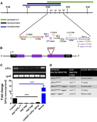
Three alleles ofangurwere obtained as second-site muta-tions through D2–3-mediated P-element mobilization of BL17200 and BL14472. All the heteroallelic mutant animals died at the third instar larval stage. The NMJ synapse in the mutants showed aberrant morphology resembling a bunch of grapes. Hence we named these mutants angur (a Hindi word meaning“bunch of grapes”). Whileangur7andangur2
were obtained throughP-element mobilization ofSyndEP877/
TM6 (BL17200) located 2416 bp upstream of the Syndapin ORF,angur5was obtained independently throughP-element
mobilization of KG06118b (BL14472) located 406,920 bp downstream of the Syndapin ORF. Complementation anal-ysis of these mutants showed that they belonged to the same complementation group. To ascertain the affected gene, we performed mapping with various deficiency lines spanning chromosome arm 3R. The deficiency lines BL9485, BL9487, and BL26529 (having chromosomal de-letion between 93B6and 93F14) failed to complement the
anguralleles (Figure 1A). We further checked for comple-mentation of angur alleles with smaller deficiencies and mutations in this region. Interestingly, one of these mutant lines, BL13478 (AP2sKG02457), which is aP-element
inser-tion in the third exon of thes2-adaptin ORF (Figure 1B),
did not complement the angur alleles. To find the new genomic position of the P-elements in angur alleles, we performed plasmid rescue followed by sequencing.angur2
andangur7were found to be present 147 and 153 bp
up-stream of a non-protein-coding gene, Heat shock RNA omega (Hsrv), respectively. Also, angur5 was found to
be present 136 bp upstream of Hsrv. Semiquantitative RT-PCR from these animals, however, showed the Hsrv
transcript to be comparable to controls (Figure S1).
Surprisingly, sequencing of thes2-adaptin genomic
re-gion in angur alleles revealed that the ORF was intact. However, RT-qPCR analysis showed a severe reduction in thes2-adaptin transcript level in all the heteroallelic
mu-tants or whens2-adaptin was ubiquitously knocked down
using an RNAi (Figure 1C). Furthermore, both the lethality and the NMJ morphological defects could be rescued by ubiquitous expression of thes2-adaptin ORF in heteroallelic
as well asAP2sKG02457homozygous mutant animals (Figure
1D). Pan-neuronal expression of thes2-adaptin ORF using
elavC155-Gal4 in angur heterozygous mutants (angur7/
AP2sKG02457orangur7/angur5) or inAP2sKG02457
homozy-gous mutants could rescue the synaptic phenotype but not the lethality. Thus, the upstream genomic region of Hsrv
appears to contain regulatory sequences that control the ex-pression ofs2-adaptin. To assess whether theP-element
in-sertions upstream ofHsrv affected the expression of other genes in this region, we performed RT-qPCR (data not shown) as well as semiquantitative RT-PCR and analyzed the expression of ETHR, SIFaR, Rab1, and CG5919, the genes
flankings2-adaptin. We did not detect any change in the
expression of these genes, suggesting that theP-element in-sertions specifically disrupteds2-adaptin expression (Figure S1). Conversely, we also assessed whether synaptic morphol-ogy was altered in two of the Hsrv alleles that have been reported previously to affect Hsrv transcript levels,Hsrv66
(1.6 kb deletion in theHsrvpromoter region and thefirst nine bases of the first exon) and EP93D (an EP insertion upstream ofHsrv) (Sengupta and Lakhotia 2006; Lakhotia
et al.2012). We found that the synaptic morphology in these larvae was comparable to that in controls, and thes2-adaptin
levels were unaltered (Figure S1), as assessed bya-adaptin staining. To avoid potential complications of the P-element insertions near theHsrvlocus, we also analyzed the allelic combinations ofAP2sKG02457with two of the deficiency lines,
Df(3R)ED10838 and Df(3R)ED10845. These larvae showed similar synaptic phenotypes and severely reduced synaptic
a-adaptin levels as AP2sKG02457 homozygous larvae or
AP2sKG02457/angur7 heteroallelic mutants (Figure S1). All
these data suggest thatanguris an allele ofs2-adaptin and
that the lethality and synaptic phenotype are due to the spe-cific loss ofs2-adaptin.
Mitotic clones of angur show a defect in eye morphology
Because angur mutants are homozygous lethal, we made mutant eye clones using the eyFLP system (Stowers and Schwarz 1999; Rikhy et al.2002). The homozygous angur
mutant eyes (EGUF/+; FRT82B,angur7/FRT82B, GMR-hid)
showed severe morphological defects with roughening and loss of bristles. The mutant eyes were also smaller than con-trol eyes (Figure 2, A and B). Scanning electron microscopy revealed severely disorganized ommatidia of uneven size. Bristles were sparsely present in the mutant clones compared to the evenly spaced bristles in control eyes (FRT82B,angur7)
could be partly restored by expressing the s2-adaptin
transgene in the mutant eyes (EGUF/+; FRT82B,angur7,
UAS-AP2s/FRT82B, GMR-hid) (Figure 2, C and F). To delve into the ultrastructural defects, we performed TEM on these genotypes. The photoreceptor cells R1–R7 could be distinctly visualized in control eyes (Figure 2G). Inter-estingly, the homozygous mutant eye clones lacked the photoreceptor cells (Figure 2H), and this could be restored in the rescued animals (Figure 2I). We counted the number of photoreceptors in all genotypes from four different car-tridges and found that it was always seven in control and rescue animals. In the mutant clones, however, we could not see any photoreceptor cells. Because photoreceptor cells were absent in the mutant clones, we hypothesized that synaptic transmission must be affected in them. To test this, we recorded ERGs and found thatangurmutant eye clones showed minimal depolarization, suggesting that they failed to perceive light (Figure 2J). The eye morphol-ogy and ERG defects were partially restored by expressing
thes2-adaptin transgene in the eyes. Rescue of the ERG defect
was significant compared to the mutant (P= 0.006) (Figure 2K). Taken together, these data suggest that s2
-adaptin-dependent endocytosis plays a crucial role in the biogenesis of rhabdomeres.
Subunits of AP2 are required for normal synapse morphology
The AP2 complex is believed to be essential for CME, and a block in the process results in defects in synaptic transmis-sion (Gonzalez-Gaitan and Jackle 1997; Gu et al. 2013). Similarly, defects in the CME pathway due to mutations in proteins implicated in the pathway also lead to morpho-logical defects at theDrosophilaNMJs (Rikhyet al.2002; Verstrekenet al.2002; Song and Zinsmaier 2003; Dickman
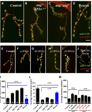
et al. 2006). Our observations revealed severe structural defects (Figure 3, A–C) ins2-adaptinmutants suggesting
that s2-adaptin-dependent endocytosis plays a crucial
role in synaptic development. We observed that boutons
Figure 1 angur is an allele of s2-adaptin, the
smallest subunit of adapter protein 2 (AP2) com-plex. (A) Deficiency mapping showed the angur
locus to be located in the region of 93D1–93D4
on the third chromosome ofDrosophila. The three deficiency lines that uncovered theangurlocus— Df(3R)BSC677, Df(3R)ED10838, and Df(3R) ED10845—are labeled. Solid bars mark the deleted regions in these deficiency lines. The 93D1–93D4
region of the chromosome contains at least 10 identified genes. Different colored arrows represent the relative positions and orientations of the genes in the 93D1–93D4region. Whileangur7andangur2
were obtained from mobilization of EP0877,angur5 was obtained from mobilization of KG06118b. The
P-element inanguralleles was located in the 59end of the hsrv gene but did not affect expression of thehsrvgene. (B) Genomic organization of the
s2-adaptin locus showing exons (represented by
solid boxes) and introns (represented by thin lines). The transcription start site is represented by an ar-row, and the untranslated regions are shown by black solid boxes. The insertion site of P-element
KG02457lies in the third exon of the gene. (C) Semi-quantitative (top panel) and Semi-quantitative RT-PCR (bot-tom panel) depicting transcript levels ofs2-adaptin
in controls, angur7/AP2sKG02457, angur7/angur2,
angur7/angur5, actin5C-Gal4-driven s
2-adaptin
RNAi, and rescued heteroallelic mutant (actin5C/+;
angur7/UAS-AP2s, AP2sKG02457), respectively.
s2-adaptin transcript level is dramatically reduced
in the heteroallelic mutants or actin5C -Gal4-driven s2-adaptin RNAi. rp49 transcript level
was used as an internal concentration control for messenger RNA (mRNA). The error bars in the bottom panel represent SEM. Statistical anal-ysis based on one-way ANOVA followed by post-hocTukey’s multiple comparison test. (D) Rescue
ofAP2sKG02457mutants and heteroallelic
ins2-adaptinmutants were clustered in contrast to controls,
where they are uniformly spaced. The s2-adaptin mutants
showed an over twofold increase in the number of boutons (Figure 3K) (control: 98 6 4.8;angur7/AP2sKG02457: 1856
5.9;angur7/angur5: 27468.6), while there was a significant
decrease in the bouton area (Figure 3L) (control: 8.760.5mm2;
angur7/AP2sKG02457: 2.360.13mm2; angur7/angur5: 2.56
0.1mm2). Both lethality and morphological defects were
res-cued by ubiquitously expressing as2-adaptin transgene in
ho-mozygous AP2sKG02457 or heteroallelic s
2-adaptin mutants
using theactin5C-Gal4 driver (Figure 3D). Synaptic morphology defects but not lethality also could be rescued by expressing a
s2-adaptin transgene using pan-neuronal driverselavC155-Gal4
ornSyb-Gal4, supporting the idea that morphological defects are due to a selective lack of s2-adaptin in neurons (Figure 1D).
Average number of boutons reverted to wild-type levels in mu-tant animals rescued with as2-adaptin transgene (actin5C/+;
angur7/AP2sKG02457, UAS-AP2s: 89.8864.06). Similarly, the
average size of an individual bouton also was restored to wild-type values in the rescued animals (actin5C/+; angur7/
AP2sKG02457, UAS-AP2s: 6.3860.42mm2).
To analyze the consequence of loss of other subunits of the AP2 complex, we next asked whether reducing other compo-nents of AP2 would result into similar morphological defects. Predictably, we observed that RNAi-mediated knockdown of any of the subunits of AP2 with various tissue-specific Gal4 drivers yielded a wide range of phenotypes (Table S1). Im-portantly, knocking down any of the subunits of the AP2 complex in neurons phenocopied the s2-adaptin mutation
(Figure 3, E–I and M, andFigure S2). Consistent with this observation, disrupting AP2 complex assembly in neurons by RNAi-mediated knockdown of phosphatidylinositol-4-kinase IIIa(PI4KIIIa), an enzyme that mediates the synthesis of PI (4)P, a precursor for biosynthesis of PI(4,5)P2, affected syn-apse morphology similar to other subunits of the AP2 com-plex (Figure 3J). The synaptica-adaptin staining and the
b2-adaptin staining (not shown) were significantly reduced
when the AP2 subunit or PI4KIIIawas neuronally knocked down (Figure S2). Taken together, these data suggest that all the subunits of AP2 along with phospholipid PI(4,5)P2 are obligate partners for regulating morphological plasticity atDrosophilaNMJs.
s2-adaptin facilitates basal synaptic transmission
The AP2 complex is believed to be the major adapter for clathrin assembly during CME at the synapses and hence is believed to be essential for SV retrieval and regeneration of SVs for sub-sequent rounds of neurotransmitter release (Gonzalez-Gaitan and Jackle 1997; Collins et al.2002; Heerssen et al. 2008; Cheung et al. 2010; Guet al.2013; Watanabeet al. 2014). Hence, in order to assess the need for the AP2 complex in endocytosis, we loadeds2-adaptinmutant and control
synap-tic terminals with FM1-43. Surprisingly, we found that dye uptake in angur7/AP2sKG02457was reduced only by 30%
compared to control synapses (Figure 4, A and B) (compared to controls, FM1-43 fluorescence intensity in angur7/
AP2sKG02457was 72.0362.2%;angur7/angur5was 49.736
1.6%; and actin5C/+; angur7/AP2sKG02457, UAS-AP2s
was 82.0862.6%). Next, to ascertain the functional impor-tance ofs2-adaptin in synaptic transmission, we performed
intracellular recordings at the NMJ of third instar larvae from
s2-adaptinmutants. We observed that both mEJP amplitude Figure 2 angurmutant eye clones show severe defects in rhabdomere
development. (A–C) Brightfield images of (A) control (FRT82B,angur7), (B) mutantangur7eye clone (EGUF/+; FRT82B,angur7/FRT82B, GMR-hid), and (C) transgene-rescued animal (EGUF/+; FRT82B,angur7, UAS-AP2s/ FRT82B, GMR-hid). (D–F) SEMs of (D) control (FRT82B,angur7), (E) mutant
and frequency were significantly higher ins2-adaptinmutants
compared to controls (Figure 4, C and E). However, EJP am-plitude ins2-adaptinmutants was reduced compared to
con-trol synapses (concon-trol: 43.2561.7 mV;angur7/AP2sKG02457:
31.6061.9 mV;angur7/angur5: 28.3461.1 mV) (Figure 4, D
and F). Moreover, the quantal content ins2-adaptinmutants
was significantly reduced (control: 67.63 6 3.37; angur7/
AP2sKG02457: 35.48 6 2.5; angur7/angur5: 34.54 6 2.3)
(Figure 4G). All the physiological characteristics were re-stored to normal levels when as2-adaptin transgene was
expressed in the mutant neurons (EJP amplitude inactin5C/+;
angur7/AP2sKG02457, UAS-AP2s: 42.0661.1 mV; quantal
content in actin5C/+; angur7/AP2sKG02457, UAS-AP2s:
64.54 6 5.2). Consistent with this observation, reducing
a-adaptin levels in neurons resulted in a similar reduction
in EJP amplitude and quantal content (Figure 4, D, F, and G) (EJP amplitude inelavC155/+;a-adaptin RNAi/+: 25.176
2.2 mV; quantal content in elavC155/+; a-adaptin RNAi/+:
27.26 62.53). Taken together, these data suggest that
s2-adaptin facilitates the basal synaptic transmission at
DrosophilaNMJ synapses.
The functional requirement of the AP2 complex in SV membrane retrieval at mammalian hippocampal synapses has been debated (Kononenko et al.2014; Watanabe et al.
2014). Hence, in order to test the functional requirement of
s2-adaptin in SV re-formation from the presynaptic
mem-brane, we directly measured the rate of re-formation of normal-pH synaptic vesicles using synaptopHluorin imag-ing (Poskanzer et al. 2006; Kumaret al. 2009). Interest-ingly, we found that the kinetics of membrane retrieval
Figure 3 Mutation ins2-adaptin causes aberrant
neuromuscular junction formation. (A–D) Confocal image of NMJ synapses at muscle 4 of (A) control animals, (B and C) heteroallelics2-adaptinmutants (angur7/AP2sKG02457andangur7/angur5), and (D) transgene-rescued animal (actin5C/+;angur7/
UAS-AP2s,AP2sKG02457) double immunolabeled with a
presynaptic marker CSP (green) and neuronal mem-brane marker HRP (red) to reveal the bouton outline. Compared to the control NMJ, the heteroallelic combination of s2-adaptinshows severely altered NMJ morphology. Scale bar, 15mm. (E–J) Confocal images of NMJ synapses at muscle 4 co-labeled with CSP (green) and HRP (red) of (E) control and (F–J) neuronally expressing RNAi against AP2 com-plex subunits or PI4KIIIa: (F)elavC155/+;a-adaptin RNAi/+, (G) elavC155/+; b
2-adaptin RNAi/
UAS-Dicer, (H)elavC155/+;m
2-adaptin RNAi/UAS-Dicer,
(I) elavC155/+;s
2-adaptin RNAi/UAS-Dicer, and (J)
elavC155/+; PIK4IIIaRNAi/+. Note that neuronal knockdown of any of the subunits of the AP2 com-plex or PIK4IIIaphenocopiess2-adaptinmutations. Scale bar, 15mm. (K) Histogram showing average number of boutons at muscles 6/7 of the A2 hemisegment in control animals (98.2 6 4.8), heteroallelic s2-adaptin mutants (angur7/
AP2sKG02457: 185.46 6;angur7/angur2: 233.46
7;angur7/angur5: 27468.5) and the rescued ani-mals (actin5C/+; angur7/UAS-AP2s, AP2sKG02457: 89.964.0). The numbers in columns represent the number of 6/7 NMJs of the A2 segment used for bouton quantification. ***P , 0.0001. Error bars represent standard error of the mean. Statistical anal-ysis based on one-way ANOVA followed bypost-hoc
Tukey’s multiple-comparison test. (L) Histogram showing average bouton area in control animals (8.76 0.5mm2), heteroallelic s
2-adaptin mutants (angur7/AP2sKG02457: 2.3 6 0.13 mm2; angur7/
angur2: 3.560.12mm2;angur7/angur5: 2.560.1
mm2) and rescued animals (actin5C/+; angur7/
UAS-AP2s,AP2sKG02457: 6.460.4mm2). The numbers in columns represent the number of boutons used for quantification.***P,0.0001. Error bars represent
standard error of the mean. Statistical analysis based on one-way ANOVA followed bypost-hocTukey’s multiple-comparison test. (M) Histogram showing quantification of average bouton numbers at muscles 6/7 of the A2 hemisegments of third instar larvae in control animals and neuronally expressing RNAi against AP2 subunits (control at 18°, 8563; control at 28°, 9862;a-adaptin, 167613;b2-adaptin, 14567.0;m2-adaptin, 13765;s2-adaptin, 11864)
and acidification was mildly but significantly different in
s2-adaptinmutants compared to control animals. The rates
of re-formation of synaptic vesicles weret= 6.960.17 sec andt= 7.660.17 sec for control anmals ands2-adaptin
mutants, respectively, when fit to a single exponential function (Figure 4H). Hence, taken together, our data suggest that the AP2 complex has a minimal role in SV retrieval from the presynaptic membrane at Drosophila
NMJ synapses.
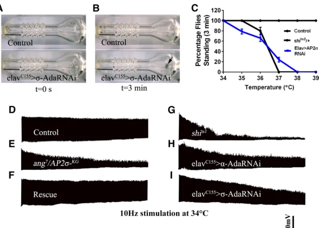
Compromised SV trafficking inDrosophilaleads to synap-tic fatigue at high-frequency nerve stimulation and also leads to temperature-dependent paralysis (Kosaka and Ikeda 1983; Zinsmaier et al.1994; Rikhyet al.2002; Verstreken
et al.2002; Kohet al.2004). Hence, we tested whether re-ducing s2-adaptin levels leads to paralysis and synaptic
fa-tigue at elevated temperature. Interestingly, we found that neuronal knockdown ofs2-adaptin (elavC155/+;s2-adaptin
RNAi/+) results in temperature-dependent paralysis above
Figure 4 s2-adaptin facilitates basal synaptic
transmission as well as synaptic vesicle endocytosis. (A) Representative images of FM1-43 dye uptake by control, heteroallelic s2-adaptin mutant (angur7/AP2sKG02457), and transgene-rescued (actin5C/+; angur7/UAS-AP2s, AP2sKG02457) syn-apses. (B) Relative synaptic FM1-43 fluorescence uptake in control, heteroallelics2-adaptinmutant (angur7/AP2sKG02457: 72.16 2.3% and angur7/
angur5: 49.73 6 1.7%) and transgene-rescued (actin5C/+;angur7/UAS-AP2s,AP2sKG02457: 82.1 62.6%) synapses.***P,0.0001;**P,0.001. Error bars represent standard error of the mean. Statistical analysis based on one-way ANOVA followed bypost-hocTukey’s multiple-comparison test. (C) Representative traces of mEJP in control animals, heteroallelics2-adaptinmutant combina-tions (angur7/AP2sKG02457 and angur7/angur5), transgenic-rescued animals (actin5C/+; angur7/
UAS-AP2s,AP2sKG02457), andelavC155/+;a-adaptin RNAi/+ animals. (D) Representative traces of EJPs in control animals, s2-adaptin heteroallelic mutant combinations (angur7/AP2sKG02457 and angur7/
angur5), transgene-rescued animals (actin5C/+;
angur7/UAS-AP2s, AP2sKG02457), and elavC155/+;
a-adaptin RNAi/+ animals. (E) Histogram showing average mEJP amplitude in control animals (0.656 0.03 mV), heteroallelics2-adaptinmutants (angur7/
AP2sKG02457: 0.9160.08 mV;angur7/angur5: 0.85 60.04 mV), transgene-rescued animals (actin5C/+;
angur7/UAS-AP2s,AP2sKG02457: 0.6960.04 mV), andelavC155/+;a-adaptin RNAi/+ (0.9460.06 mV) animals. At least 8 NMJ recordings of each genotype were used for quantification.***P,0.0001. Error bars represent standard error of the mean. Statistical analysis based on one-way ANOVA followed by
post-hocTukey’s multiple-comparison test. (F) Histo-gram showing average EJP amplitude in control an-imals (43.256 1.67 mV), heteroallelics2-adaptin mutants (angur7/AP2sKG02457: 31.60 6 1.9 mV;
angur7/angur5: 28.34 6 1.05 mV), transgene-rescued animals (actin5C/+; angur7/UAS-AP2s,
AP2sKG02457: 42.06 61.14 mV), and elavC155/+;
a-adaptin RNAi/+ (25.17 62.24 mV) animals. At least 8 NMJ recordings of each genotype were used for quantification.***P,0.0001. Error bars represent standard error of the mean. Statistical analysis based on one-way ANOVA followed bypost-hocTukey’s multiple-comparison test. (G) Quantification of quantal content in control animals (67.6363.37), heteroallelics2-adaptinmutants (angur7/AP2sKG02457: 35.4862.45;angur7/angur5: 34.5462.25), transgene-rescued animals (actin5C/+;angur7/UAS-AP2s,AP2sKG02457: 64.5465.18), andelavC155/+;a-adaptin RNAi/+ (27.2662.53) animals.***P,0.0001. Error bars represent standard error of the mean. Statistical analysis based on one-way ANOVA followed bypost-hocTukey’s multiple-comparison test. (H) Graph showing the SpHfluorescence response to a 50-Hz stimulus train for 10 sec in 2.0 mM Ca2+containing HL3 of control (blue,n= 37 boutons,
35°(Figure 5, A–C). We found that high-frequency nerve stim-ulation in angurmutants or in animals with neuronally re-duceds2- ora-adaptin at 34°leads to an activity-dependent
decline of EJP amplitude; however, unlike with shibirets
alleles, the neurotransmitter release was not completely abolished (Figure 5, D–I). These data suggest that unlike essential endocytic proteins such as Endophilin and Shibire, AP2-modulated vesicle trafficking and its activity are also required during elevated temperatures to sustain neuro-transmitter release.
s2-adaptin regulates synaptic vesicle regeneration
under high-frequency nervefiring
To further assess a requirement fors2-adaptin in SV recycling,
we stimulated the s2-adaptin mutant synapses in larval
body wall muscle for 5 min at 10 Hz and recorded EJPs. While the control and rescued synapses (actin5C/+; angur7/
AP2sKG02457, UAS-AP2s) did not show stimulation-dependent
decline in EJP amplitudes over 5 min, mutant synapses (angur7/AP2sKG02457andangur7/angur5) exhibited a signifi
-cant decline in EJP amplitude (Figure 6, A and B). The decline in EJP amplitude at the end of 5 min of 10 Hz of nerve stim-ulation was50% relative to thefirst stimulus. Similar results were obtained when a-adaptin was neuronally reduced (elavC155/+; a-adaptin RNAi/+) (Figure 6, A and B). Thus,
these data suggest that not only is AP2 required for maintaining basal synaptic transmission, but it is also crucial under high-frequency nerve firing to resupply SVs at Drosophila NMJ synapses.
Synaptic mutants implicated in CME, such asendophilin
anddap160, show complete recovery from synaptic depres-sion within a few minutes of rest after cessation of high-frequency stimulation, suggesting that synaptic membrane retrieval for vesicle regeneration in these mutants is stimu-lus dependent (Marieet al.2004; Dickmanet al.2005). In contrast, we found thats2-adaptinmutants or animals with
neuronally reduceda-adaptin do not recover from synaptic depression even after 4 min of rest following cessation of high-frequency stimulation. This observation suggests that in addition to its requirement in synaptic membrane re-trieval, AP2 complex function is also required during the much slower process of SV trafficking, possibly at one of the rate-limiting steps in SV regeneration (Figure 6B). Hence, we reasoned that the mutants do not recover from synaptic depression because of compromised SV regenera-tion. To address this directly, we labeled larval NMJ synap-ses of control and mutant animals (angur7/AP2sKG02457
and angur7/angur5) with FM1-43 before or after 4 min of
rest following cessation of high-frequency stimulation and estimated the ECP. Surprisingly, we found that even after 4 min of rest, the total recycling pool did not recover to its initial value in the mutants or at the synapses wherea-adaptin was reduced in the neurons (elavC155/+;a-adaptin RNAi/+)
(Figure 6C). Taken together, these data uncover a role for AP2 in the regeneration of synaptic vesicles under high-frequency stimulation inDrosophila.
To further investigate the underlying mechanism of syn-aptic fatigue in angur mutants, we estimated ECP and the total vesicle pool at the NMJ synapses. In order to measure ECP, synapses were continuously stimulated in the presence of 1mM Bafilomycin A1 at 3 Hz until synaptic depression was attained. The initial decline in quantal content during the short duration of neuronal activity or at 3 Hz stimulation mostly represents ECP; however, the later depletion reflects the mobilization of vesicles from the reserve pool (Kimet al.
2009). Consistent with our previous observations, the num-ber of vesicles in ECP was found to be significantly lower in
angurmutants (angur7/AP2sKG02457: 16,5906974;angur7/
angur5: 15,040 6870; controls: 25,050 61511). Similar
results were obtained whena-adaptin levels were neuronally reduced (elavC155-GAL4.a-adaptin RNAi: 15,73061033;
controls: 25,05061511) (Figure 6, D and E). The number of SVs in the ECP of rescued synapses (actin5C-GAL4/+;
angur7/UAS-AP2s, AP2sKG02457: 23,635 6 1358.75) was
comparable to that in control animals (Figure 6, D and E). The total vesicle pool was measured by continuously stimulat-ing the synapses at 10 Hz in the presence of 1mM Bafilomycin A1. We quantified the total vesicle pool by integrating the quantal content from the depression curve (Figure 6F) until remarkable depletion was attained. Our data suggest signif-icant reduction of the total vesicle pool in angur mutant (angur7/AP2sKG02457: 53,220 6 3156; angur7/angur5:
50,93062877) or neuronally reduceda-adaptin (elavC155
-GAL4.a-adaptin RNAi: 63,46063650) synapses compared to control (94,61065929) and rescued synapses ( actin5C-GAL4/+; angur7/UAS-AP2s, AP2sKG02457: 102,200 6
6315) (Figure 6G). Taken together, these data suggest that
s2-adaptin is required to maintain the vesicle pool at
Dro-sophila NMJ synapses. The synaptic depression observed under high-frequency stimulation is possibly due to less availability of SVs at thes2-adaptinmutant synapses. s2-adaptin is an obligate member for stabilizing the
AP2 complex and clathrin but not other synaptic proteins
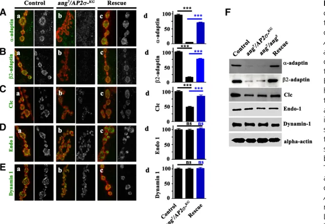
Because the AP2 complex acts as a hub for several molecular interactions during CME, we next asked whethers2-adaptin
affects localization or stability of other synaptic proteins (Schmid and McMahon 2007). To address this, we quantified the synaptic immunofluorescence levels of various synaptic proteins in s2-adaptin mutants. We observed a severe
re-duction in synaptic a-adaptin (compared to controls, the
a-adaptin fluorescence intensity in angur7/AP2sKG02457
was 5.660.2%; inangur7/angur5was 1.460.1%, and in
actin5C/+; angur7/AP2sKG02457, UAS-AP2swas 73.46 6
1.6%), andb2-adaptin immunoreactivity (compared to
con-trols, the b2-adaptin fluorescence intensity in angur7/
AP2sKG02457 was 16.53 6 1.4%; in angur7/angur5 was
31.97 6 1.6%; and in actin5C/+; angur7/AP2sKG02457,
UAS-AP2s was 76.07 6 2.0%) at the s2-adaptin mutant
levels of clathrin at the mutant synapses might be altered. Hence, to assess the levels of clathrin at the s2-adaptin
mutant synapses, we overexpressed an EYFP-tagged Clc
transgene in neurons of heteroallelic s2-adaptin mutants
(elavC155/+;UAS-Clc/+;angur7/AP2sKG02457) or in the
res-cue animals (elavC155/+;UAS-Clc/+;angur7/AP2sKG02457,
UAS-AP2s). Compared to controls (elavC155/+; UAS-Clc/
+), the EYFP fluorescence intensity at mutant (48.5 6 2.3%) synapses was dramatically reduced but was signifi -cantly restored in the rescue (84.764.1%) synapses (Figure 7C andFigure S3). Other synaptic proteins implicated in CME were not altered ins2-adaptinmutants (Figure 7, D and E, and Figure S3) Taken together, these data suggest that all the subunits of the AP2 complex are specifically essential for re-cruitment and/or stability of clathrin at the synapse.
The altered synaptic abundance ofa-adaptin,b2-adaptin, or
clathrin in s2-adaptinmutants could be the result of either
degradation of these proteins or a defect in synaptic targeting. To address this, we performed Western blots on lysates ppared from third instar larval brain. We observed a severe re-duction in the a-adaptin, b2-adaptin, and clathrin protein
levels ins2-adaptinmutants (Figure 7F). Consistent with the
immunofluorescence studies, the levels of other synaptic pro-teins, such as Endophilin1, Dynamin1, Dap160, Eps15, and Nwk, were not significantly altered in s2-adaptin mutants
(Figure 7, D and E, andFigure S3). Hence, these data suggest thats2-adaptin is critically important for stabilizing not only
the AP2 complex but also clathrin. Moreover, AP2 appears to be specific adapter for clathrin at the synapses ofDrosophilaNMJ.
Microtubule-based cytoskeleton is disorganized in
s2-adaptin mutants
It has been shown that synaptic growth is controlled via regulation of the synaptic microtubule cytoskeleton and that
any alteration of this cytoskeleton affects synaptic growth (Rooset al.2000). To examine whether the microtubule cytoskeleton was disrupted in the mutant NMJ synapses, we labeled the synapses with MAb 22C10. This antibody labels Futsch, which is associated with the dendritic, axo-nal, and nerve-terminal cytoskeleton (Rooset al.2000). In control boutons, the microtubules appeared continuous with periodic loop structures (Figure 8, A, B, and G) (24.7561.7% of type Ib boutons on muscles 6 and 7 of the A2 hemisegment). Contrary to the control boutons, the microtubules in the s2-adaptin mutant boutons
appeared fragmented, with almost total disappearance of the periodic loop structures (Figure 8, C, D, and G) (angur7/AP2sKG02457: 4.42 60.6% and angur7/angur5:
3.33 6 0.5% of type Ib boutons on muscles 6 and 7 of the A2 hemisegment). These periodic loop structures could be rescued by ubiquitouss2-adaptin transgene
ex-pression (Figure 8, E, F, and G) (actin5C/+; angur7/
AP2sKG02457, UAS-AP2s: 24.9862.9% of type Ib boutons
on muscles 6 and 7 of the A2 hemisegment). RNAi-mediated knockdown of any of the subunits of AP2 or PI4KIIIa in neurons also resulted in a significant decrease in the number of periodic loop structures (Figure S2). These data suggest thats2-adaptin regulates NMJ morphology by
regu-lating the cytoskeletal architecture.
Because BMP signaling is required for a normal microtu-bule cytoskeleton (Wanget al.2007), we assessed whether this signaling pathway was affected ins2-adaptinmutants.
We estimated the synaptic levels of pMAD and found that the mutant synapses had over threefold higher levels of pMAD compared to controls (Figure 8, H and I) (compared to con-trols, the pMADfluorescence intensity inendoD4was 332.56
16.7%; angur7/AP2sKG02457 was 345.8 6 12.6%; angur7/
angur5 was 362.8 6 14.6%, and actin5C/+; angur7/ Figure 5 Reducings2-adaptin levels in neurons
causes temperature-sensitive paralysis at elevated temperature. (A and B) Control (elavC155/+) 2- to 3-day-old adultflies do not suffer paralysis in 3 min at 38ο, whereas similarly aged adultflies expressing
s2-adaptin RNAi (elavC155/+;s2-adaptin RNAi/+) in
neurons showed complete paralysis in 3 min at 38ο.
These animals recovered within 2 min when shifted to 25ο(not shown). (C) Paralysis profile of
control animals and animals expressing RNAi against
s2-adaptin in neurons (elavC155/+; s2-adaptin
RNAi/+) andshits2/+. While the control animals do not suffer paralysis in 3 min, the RNAi-expressing animals are paralyzed above 35ο. Five trials were
performed for each genotype at each temperature in a sushi cooker (Ramaswamiet al.1993). Each trial contained 10–15 animals. Error bars repre-sent standard error of the mean. (D–I) Repre-sentative traces of EJPs under high-frequency stimulation of indicated genotypes stimulated at 10 Hz for 5 min at 34°in 1.5 mM Ca2+containing
HL3. Unlikeshibrets1(G), mutant (E) and neuronally reduceda- ors2- adaptin (H and I) animals do not
AP2sKG02457, UAS-AP2s was 102.2 6 5.1%). Because
pMAD has been shown to accumulate in the motor nuclei of synaptic mutants, we analyzed nuclear pMAD levels in
s2-adaptinmutants. Consistent with other endocytic
mu-tants (Vanlandinghamet al.2013), we found significantly elevated pMAD levels in the motor nuclei of s2-adaptin
mutants (angur7/AP2sKG02457: 162.5 6 5.6% and
angur7/angur5: 160.96 4.7%) (Figure 8, J and K). Our
data thus suggest that mutation in s2-adaptin causes a
BMP pathway-dependent aberrant NMJ morphology by altering the presynaptic cytoskeleton. We suggest that
s2-adaptin-dependent endocytosis is essential for
attenu-ating BMP-dependent synaptic growth signaling.
Discussion
AP2/CME has been proposed to play an essential role in SV endocytosis. Moreover, mutation in the proteins affecting CME also results in altered NMJ development inDrosophila, suggesting regulation of synaptic signaling by CME. In this study, we analyzed the role of AP2, one of the major adapters for clathrin at synapses, in the context of its physiological relevance and synaptic signaling. Our results demonstrate that AP2 facilitates basal synaptic transmission and SV endo-cytosis but also is essential during high-frequency nervefiring to regenerate SVs. Moreover, we provide evidence that AP2 regulates morphological plasticity at theDrosophilaNMJ by
Figure 6 s2-adaptin function is essential under
high-frequency stimulation to regenerate synaptic vesicles. (A) Normalized EJP amplitudes in control, heteroallelic s2-adaptin mutant (angur7/
AP2sKG02457and angur7/angur5), rescued larvae
(actin5C/+; angur7/UAS-AP2s, AP2sKG02457), and neuronally knocked down a-adaptin (elavC155/+;
a-adaptin RNAi/+) animals stimulated at 10 Hz for 5 min in 1.5 mM Ca2+. The mutant larvae showed
stimulus-dependent decline in the EJP amplitudes over time. (B) Representative traces of indicated ge-notypes under high-frequency stimulation at 10 Hz for 5 min in 1.5 mM Ca2+containing HL3. After
1.5 min of rest, test stimuli were given every 30 sec to assess the recovery of EJP from synaptic depres-sion. Note that the mutant synapses do not recover to thefirst stimulus levels even after 4 min of rest from high-frequency stimulation. (C) Estimation of endo-exo cycling pool at NMJ synapses of indicated genotypes before high-frequency stimulation and after 4 min of rest from high-frequency stimulation. While the controls and rescue synapses maintained the ECP pool to its initial value after 4 min of rest, thes2-adaptinmutant synapses and theelavC155/+;
a-adaptin RNAi/+ synapses show significant decline in the endo-exo cycling pool and do not recover to the initial value after 4 min of rest.n= 50 boutons from eight different NMJs of muscles 6/7 of the A2 hemisegment were counted. (D) Synaptic depres-sion curve showing the depletion in quantal con-tent over stimulus number in synapses stimulated at 3 Hz in the presence of 1mM Bafilomycin A1 to deplete ECP. (E) Histogram showing ECP estimates of control (25,05061511), heteroallelic mutants (angur7/AP2sKG02457: 16,590 6 974; angur7/
stabilizing the microtubule loops and attenuating the BMP signaling pathway.
Role ofs2-adaptin in regulating rhabdomere biogenesis
The biogenesis of rhabdomeres is regulated by endocytosis and intracellular trafficking (Schuck and Simons 2004). Components of the endocytic machinery, when perturbed, lead to defects in rhabdomere formation. One such example is the disruption of the rhabdomere base whenshitsflies are
grown at restrictive temperature for 4 hr (Pinal and Pichaud 2011). Rh1-Gal4-driven shits1 flies raised at 19°led to an
enlargement of photoreceptor cell bodies with the inter-rhabdomeric space being reduced (Gonzalez-Bellido et al.
2009). Similarly, whena-adaptin was knocked down using GMR-Gal4, rhabdomere biogenesis was disrupted, with smaller rhabdomeres (Raghu et al. 2009). In accordance with these observations, the TEM analysis ofangurmutant eye clones showed no detectable rhabdomeres. Consistent with the defect in rhabdomere formation, we also observed no membrane depolarization inangur mutant eye clones. The visual cascade inDrosophilabegins with rhodopsin be-ing converted to meta-rhodopsin by light, followed by a subsequent phosphorylation by GPRK1. The phosphorylated meta-rhodopsin binds to arrestins, where its activity is quenched. The rhodopsin-arrestin complex is endocytosed, and rhodopsin is degraded in the lysosomes (Wang and Montell 2007). Thus, endocytosis regulates rhodopsin turnover in the photoreceptor cells, and blocking of the endocytic pathway components by mutations in Arr1, Arr2, or AP2 leads to retinal degeneration and photorecep-tor cell death. This further establishes a strong link be-tween endocytosis and retinal degeneration. Thus, it is likely that defective endocytosis in angur mutant eye
clones affects meta-rhodopsin turnover, resulting into de-fective rhabdomeres and disorganized ommatidia. More-over, endocytosis of several signaling receptors also has been shown to regulate morphogenesis duringDrosophila
eye development. The Notch signaling pathway has been studied extensively, and it has been shown that the Notch extracellular domain is transendocytosed during this pro-cess into the Delta-expressing cells (Weinmaster 2000). The localization of Notch and Delta is disrupted when endocy-tosis is acutely blocked, and this prevents internalization of Notch in the Delta-expressing cells (Parkset al.2000). We thus speculate that the endocytic defect in angur mutant clones affects turnover and internalization of signaling mol-ecules that may lead to defective rhabdomere development.
s2-adaptin regulates regeneration of SVs during
high-frequency nervefiring
The functional analysis of thes2-adaptinmutants as well as of
other subunits of AP2 reveals its requirement in maintaining basal synaptic transmission and regeneration of synaptic ves-icles under high-frequency nerve firing at Drosophila NMJ synapses. Our data are consistent with previous reports showing the requirement for Drosophila a-adaptin in SV recycling at NMJ synapses (Gonzalez-Gaitan and Jackle 1997). However, in contrast to its role at mammalian central synapses, we found that loss of AP2 at Drosophila NMJ synapses affects basal synaptic transmission and SV endo-cytosis, albeit not very strongly. Moreover, the kinetics of SV re-formation in s2-adaptin mutants was only mildly
af-fected. Consistent with the recent observation that loss of the AP2 complex at mammalian central synapses or its subunits in C. elegans affects synaptic vesicle biogenesis, we observed compromised synaptic transmission and SV
Figure 7 s2-adaptin is required specifically for AP2
complex and clathrin stability. (A–E) Representa-tive images of third instar larval boutons from control, heteroallelic s2-adaptin mutant (angur7/
AP2sKG02457), and transgene-rescued (actin5C/+;
angur7/UAS-AP2s, AP2sKG02457) NMJ synapses co-labeled with HRP (red) and other synaptic pro-teins (green): (A) a-adaptin; (B) b2-adaptin; (C);
EYFP-Clc (D) Endophilin 1, and (E) Dynamin 1. The histograms on the right show quantification of these synaptic proteins in the boutons, expressed as percentage of control levels. The fluorescence intensity of at least 50 individual boutons was mea-sured and the background subtracted for the quan-tification. ***P , 0.0001. Error bars represent standard error of the mean. Statistical analysis based on one-way ANOVA followed by post-hoc
Tukey’s multiple-comparison test. (F) Western blot analysis of various synaptic proteins in larval brain of control, heteroallelic s2-adaptin mutant (angur7/
AP2sKG02457andangur7/angur5), and rescued
ani-mals (actin5C/+; angur7/UAS-AP2s, AP2sKG02457). Level of a-actin was used as loading control. The level of clathrin was assessed by probing the blots with anti-GFP antibody. For the EYFP-Clc experiment, the control genotype waselavC155/+;EYFP-Clc/+; the heteroallelic mutant genotype was
re-formation after high-frequency stimulation, suggesting that atDrosophilaNMJ synapses, AP2 function is required not only for SV trafficking but also for SV regeneration under high-frequency nerve stimulation (Guet al.2013; Kononenkoet al.2014). Our direct estimation of vesicle pool size ins2-adaptinmutants further supports its requirement in
the regulation of SV pool size and is consistent with a recent report (Gu et al.2013). Because loss of AP2 inDrosophila
does not completely abrogate SV re-formation, it remains possible that other known synaptic clathrin adapters such as AP180, Eps15, and Epsin might partially compensate for loss of the AP2 complex (Zhanget al.1998; Jakobssonet al.
2008; Cheung and Cousin 2012). This is corroborated by the observation that even a 96% reduction in synaptica-adaptin level caused only an50% reduction in clathrin, suggesting that other clathrin adapters might contribute to retention and/or stability of synaptic clathrin.
The striking physiological defect ins2-adaptinmutants or
neuronally reduceda-adaptin was their inability to recover
from synaptic depression even after 4 min of cessation of high-frequency stimulation. Consistent with these observa-tions, the endo-exo cycling pool did not recover to its initial value even after 4 min of rest following high-frequency nerve stimulation. This suggests that in addition to its requirement in synaptic vesicle endocytosis, AP2 complex functions at a relatively slower step downstream of membrane retrieval, possibly during membrane sorting from endosomes to regen-erate fusion-competent synaptic vesicles (Gu et al. 2013; Kononenkoet al.2014).
PI(4,5)P2 and subunits of the AP2 complex are obligate partners for synapse growth in Drosophila
Several studies in cell culture support a model that indicates that the AP2 complex is an obligate heterotetramer for subunit stability and function (Motley et al. 2003; Boucrot et al.
2010). However, studies of AP2 function inC. eleganssuggest thata/s2subunits and b2/m2subunits may constitute two
hemicomplexes that can carry out the minimal function of the
Figure 8 Mutations ins2-adaptin cause disruption
of the presynaptic cytoskeleton and upregulated BMP signaling. (A–F) Representative images of third instar larval NMJs from (A and B) control animals, heteroallelics2-adaptinmutants (C and D) (angur7/
AP2sKG02457), and transgene-rescued (E and F)
(actin5C/+; angur7/UAS-AP2s, AP2sKG02457) ani-mals co-labeled with HRP (red) and 22C10 (green). (G) Histogram showing quantification of the per-centage of boutons containing Futsch loops of the indicated genotypes.***P,0.0001. Error bars represent standard error of the mean. Statistical analysis based on one-way ANOVA followed by
post-hoc Tukey’s multiple-comparison test. n = 6 NMJ synapses of muscles 6/7 of the A2 hemisegment. (H) Representative images of third instar larval boutons from (a) control, (b) heteroallelics2-adaptinmutant (angur7/AP2sKG02457), and (c) transgene-rescued (actin5C/+; angur7/UAS-AP2s, AP2sKG02457) NMJ synapses co-labeled with HRP (red) and pMAD (green). (I) Histogram showing quantification of synaptic pMADfluorescence of the indicated geno-types. Thefluorescence intensity of at least 50 ind i v i d u a l b o u t o n s w a s m e a s u r e d a n d t h e background subtracted for the quantification.***P,
0.0001. Error bars represent standard error of the mean. Statistical analysis based on one-way ANOVA followed by post-hoc Tukey’s multiple-comparison test. (J) Representative images of motor nuclei of third instar larval VNC from (a) control, (b) heteroallelic
s2-adaptinmutant (angur7/AP2sKG02457), and (c) transgene-rescued (actin5C/+; angur7/UAS-AP2s,
AP2sKG02457) animals co-labeled with Elav (red) and
pMAD (green). (K) Histogram showing quantification of the nuclear pMADfluorescence of the indicated genotypes. Thefluorescence intensities of at least 50 motor nuclei were measured both for Elav and for pMAD, and the ratio of pMAD/elav represented in the quantification.***P,0.0001. Error bars repre-sent standard error of the mean. Statistical analysis based on one-way ANOVA followed by post-hoc