Limb development: an international model for
vertebrate pattern formation
CHERYLL TICKLE*
Department of Anatomy and Developmental Biology, University College London, London, United Kingdom
ABSTRACT Limb development is an excellent model for studying how patterns of differentiated cells and tissues are generated in vertebrate embryos. The cell interactions that mediate patterning have been discovered and, more recently, some of the molecules involved in these interactions have been identified. This has provided a direct link to genetics and thus to genes that cause human congenital limb defects.
KEY WORDS:
chick embryo, growth factors, retinoic acid, Hox genes0214-6282/2000/$20.00 © UBC Press
Printed in Spain www.ehu.es/ijdb
*Present and corresponding address for reprints: Department of Anatomy and Physiology, Wellcome Trust Biocentre at Dundee, University of Dundee, Dow Street, Dundee DD1 5EH, United Kingdom. FAX: 44 1382 345893. e-mail: [email protected]
Abbreviations used in this paper: BMP, Bone morphogenetic protein; Dpp, Decapentaplegic; FGF, Fibroblast growth factor; Shh, Sonic hedgehog.
Introduction
The study of vertebrate limb development has been truly inter-national. Important embryological work on limb development was carried out in the United States particularly in the late 1940’s and continuing into the 1950’s and 60’s. In Britain, much of the special contribution to the field is based on models of limb development which were formulated in the 1970’s and which provided a frame-work for further experimental frame-work. These models have assumed a new significance now that molecules which are important for limb development have been identified. The explosion of knowledge about the molecular biology of limb development has taken place in the space of less than ten years and has come not only from work in Europe but also from work in the United States and in Japan. As in other areas of active research, ideas about limb development have often caused controversy. This not withstanding, the field has been marked by cooperation between scientists in different coun-tries and with different expertise and this has often been the key to advances in understanding.
Embryology of the vertebrate limb
There is a wealth of information about embryology of vertebrate limbs (reviewed Saunders, 1977; Tickle and Eichele, 1994). The study of limb development in chick embryos has been particularly important because it is relatively easy to manipulate the developing limb through a window in the egg shell. Work by Saunders and colleagues in the USA was mainly responsible for uncovering the three main interactions that operate in the limb bud and are necessary for limb development; an interaction between the thick-ened ectodermal rim of the limb bud, the apical (ectodermal) ridge,
and underlying mesenchyme which mediates limb bud outgrowth: an ectodermal-mesenchymal interaction that controls dorso-ven-tral (extensor/flexor) patterning and a mesenchymal-mesenchy-mal interaction which controls antero-posterior patterning (e.g. in human hand, thumb to little finger).
Positional information and limb development
Another important model around this time was the polar co-ordinate model which emphasised short range interactions rather than long range signalling between adjacent cells (French et al., 1976). This model was also applied to limb development and stimulated much discussion (Iten and Murphy, 1980). The forma-tion of the periodic pattern of skeletal elements in the vertebrate limb has also been modelled in terms of reaction-diffusion systems. These self-organising systems could generate a prepattern with concentration peaks of activators prefiguring the position in which the skeletal elements develop (see for example, Wilby and Ede, 1975; Wolpert and Stein, 1984).
The progress zone model and proximo-distal patterning
Signalling by the apical ectodermal ridge is required for bud outgrowth. Limb bud outgrowth is accompanied by the successive laying down of structures along the proximo-distal axis of the limb, starting with proximal structures such as humerus and ending with digits. But how do cells know which structure along this axis to form? One possibility is that the apical ridge signal changes with time. However, when apical ridges from limb buds at different stages were recombined with mesenchyme, in all cases the limbs developed normally (Rubin and Saunders, 1972). Another pro-posal was that a timing mechanism, which operates in the mesen-chyme, controls proximo-distal pattern. According to this idea, the length of time that cells spend in the zone of undifferentiated mesenchyme, which is maintained by the apical ridge at the tip of the elongating bud, determines whether they will form proximal or distal structures. This region at the tip of the limb bud was called the progress zone (Summerbell et al., 1973).
The idea that a mesenchymal timing mechanism might operate at the tip of the limb bud came from experiments in which tips of undifferentiated mesenchyme were exchanged between old and young limb buds (Summerbell et al., 1973). The grafted tips
behaved autonomously. This ruled out the possibility that struc-tures already formed might dictate which strucstruc-tures formed next. In addition, if a graded signal from the apical ridge specified cell position along the proximo-distal axis, then one might have ex-pected a normal limb pattern to be re-established.
Recent work shows that Fibroblast Growth Factors (FGFs) mediate apical ridge signalling. When the apical ridge is removed, truncated limbs are produced (Saunders, 1948; Summerbell, 1974). FGFs are expressed in the apical ridge and application of FGFs (FGF8, FGF4, FGF2) can rescue development of limb buds from which the apical ridge has been removed (Niswander et al., 1993; Fallon et al., 1994; Crossley et al., 1996). The mechanisms of FGF signalling by the ridge and their relationship with progress zone function have not been characterised in any detail. It seems likely that FGF acts as a local signal and thus could not directly control the size of the progress zone. This suggests that secondary signals induced by FGFs secreted by the apical ridge are involved in maintaining the progress zone. It is also possible that mesenchy-mal cells measure time either directly or indirectly by the total amount of FGF to which they have been exposed.
Dorso-ventral patterning
Signalling by the ectoderm covering the sides of the limb controls patterning along the dorso-ventral axis (Patou and Kieny, 1973; MacCabe et al., 1974). This was shown by experiments in which limb buds were separated into mesenchyme and ectoderm components and these different tissues from right and left wing buds were recombined so that, for example, dorsal ectoderm was placed over ventral mesoderm. The distal part of the limb that developed after such an operation had reversed dorso-ventral pattern, in accordance with the change in ectoderm polarity. This dorso-ventral pattern was judged mainly by reference to muscle and tendon pattern which differs dorsally and ventrally (in flexor and extensor regions respectively) and by reference to epidermal differentiation which is controlled by the underlying dermis. When an apical ridge was grafted either to the dorsal surface or to the ventral surface of a wing bud, new limb outgrowths were induced with either a double-dorsal or a double-ventral pattern respectively. The simplest model to account for the effects of ectoderm is that both dorsal and ventral ectoderm produce graded signals that pattern each half of the limb (reviewed Tickle, 1995). Cartilage differentiation may be confined to the core of the limb bud by general inhibitory signals produced by the ectoderm (Solursh, 1984).
Signalling by dorsal ectoderm is mediated, at least in part, by Wnt7a (Parr and McMahon, 1995). The signalling molecule en-coded by this gene is a member of a family of vertebrate signalling molecules related to the product of the Drosophila gene, wingless. This family also comprises one of the int genes, genes at integra-tion sites of the mouse mammary tumour virus which lead to tumour formation. Wnt7a transcripts are confined to dorsal ectoderm in vertebrate limb buds and when Wnt7a was functionally inactivated in mice, the paws of these animals were found to have a double-ventral pattern. Ventral ectoderm expresses En-1, a gene encod-ing a transcription factor related to the product of the Drosophila gene, engrailed. When En-1 is functionally inactivated in mice, the paws of the mice now have a double-dorsal pattern (Loomis et al., 1996).
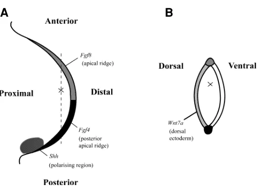
Fig. 1. Diagram to illustrate how the ideas of positional information have been applied in relation to a three-dimensional co-ordinate system to chick limb development and some of the signals that have been identified. (A) Dorsal view of limb bud showing position of cell (X) with respect to proximo-distal and antero-posterior axes. (B)Section of limb bud taken along dotted line in (A) showing position of cell with respect to dorso-ventral axis. Sources of several known signals are stippled.
It is not clear whether the product of Wnt7a itself can act as a positional signal to specify dorsal pattern or whether indeed that this type of long range signalling is needed. Cells that will form muscles and tendons originally lie close to the ectoderm and then take up more central positions later in development (Hurlé et al., 1990; Murray and Wilson, 1997). This displacement of muscles and tendons could be related to the formation of the dermis.
The phenotypes of the knockouts, just outlined above, showed that when genes expressed either dorsally or ventrally were functionally inactivated, dorso-ventral patterning was symmetrical. This resembles the pattern in outgrowths of chick limbs covered on both sides with either dorsal ectoderm or with ventral ectoderm. Indeed in the mice in which En-1 was functionally inactivated, Wnt7a was found to be expressed ventrally as well as dorsally. However, in contrast, En-1 is still expressed only ventrally in Wnt7a mutant mice. Thus, it seems that a signal independent of En-1 expression governs ventral patterning. Such a signal could be produced by all the ectoderm both dorsal and ventral in normal limb development and be over-riden by Wnt7a dorsally (Parr and McMahon, 1995). Another possibility is that a ventralising signal is produced at the dorso-ventral interface and generates a symmetri-cal gradient both dorsally and ventrally (Akita, 1996).
Positional signalling by the polarising region
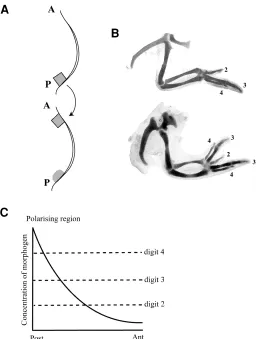
Patterning across the antero-posterior axis provides the best example of positional signalling in the limb bud. Grafting experi-ments by Saunders first revealed the signalling activity of mesen-chyme cells at the posterior margin of the chick limb (Saunders and Gasseling, 1968). When grafts of these cells were placed at the anterior margin of a second limb bud, mirror-image symmetrical patterns of digits resulted (Fig. 2A,B).
The rules that govern additional digit formation were explored in an extensive series of grafting experiments. These showed that the character of a digit depends on distance from the polarising region and a model in which the polarising region produces a diffusible morphogen was proposed (Tickle et al., 1975). Accord-ing to the model, a morphogen gradient would be established across the limb and cells at different distances from the polarising region would then be exposed to different concentrations of morphogen (Fig. 2C). Local morphogen concentration would then provide a measure of distance across the limb bud. The main features of this model are that the polarising region morphogen acts long range and in a dose dependent fashion. Low concentra-tions of morphogen would specify an anterior digit; high concen-trations a posterior digit. Such dose-dependent effects on digit specification were seen when different numbers of polarising cells were grafted (Tickle, 1981).
Identification of limb morphogens
The first molecule that was discovered which could provide a positional signal to the developing limb was retinoic acid, a vitamin A derivative. Retinoic acid was applied to the chick limb because of its reported inhibitory effects on cell-cell communication (Pitts et al., 1986). Completely unexpectedly it was found that when retinoic acid was applied to the anterior margin of a chick wing bud, this mimicked signalling of the polarising region (Tickle et al., 1982). Retinoic acid fulfils two main criteria for a positional signal; it acts
in a dose-dependent fashion and is readily diffusible in the limb (Tickle et al., 1985). In addition, retinoic acid can be extracted from chick limb buds and has been shown to be enriched posteriorly where the polarising region is located (Thaller and Eichele, 1987). However, there is no evidence that cells at a distance from a source of retinoic acid respond directly to the local retinoic acid concentra-tion. Moreover, when mesenchyme cells next to a retinoic acid source were implanted into a second wing bud, they were found to have acquired polarising activity and induced formation of addi-tional digits (Noji et al., 1991; Wanek et al., 1991).
More recently, peptide signalling molecules have been found to be expressed in the polarising region. Of these, Sonic Hedge-hog (Shh) a member of the vertebrate family of molecules related to the product of the Drosophila, hedgehog gene, is able to polarise the limb (Riddle et al., 1993). When sonic hedgehog was expressed anteriorly, mirror image duplicated patterns of digits Fig. 2. Diagram to illustrate how the signalling properties of the polarising region were identified and a model to explain polarising region signalling. (A). A cube of mesenchyme was cut out of the posterior margin of one chick wing bud and grafted to anterior margin of a second wing bud. A, anterior; P, posterior. (B) Normal wing skeleton (digit pattern 2 3 4) and, below, wing skeleton following operation in (A; digit pattern 4 3 2 3 4). Numbers refer to the digits. (C) Model for positional signalling by the polarising region. The proposal is that the polarising region secretes a diffusible morphogen that establishes a concentration gradient across the limb. Cells at different distances from the polarising region will be exposed to different morphogen concentrations. High morphogen concentrations will specify a digit 4 and lower concentrations a digit 2 .
A
B
resulted. Recent experiments suggest that Shh acts in a dose-dependent fashion and can exert long-range effects (Yang et al., 1997). Different numbers of sonic hedgehog expressing cells or beads soaked in different concentrations of the amino-terminal peptide of Shh produced dose-dependent changes in digit pat-tern. Furthermore, DiI labelling experiments showed that cells at some distance from a bead soaked in Shh contributed to the additional digits. Although widespread diffusion of the amino-terminal peptide from the bead can be detected, it is not clear that Shh produced by polarising region cells and which has undergone cholesterol modification is freely diffusible. Thus, Shh itself may not directly signal digit formation at a distance.
Cells in the polarising region express genes that encode members of the Bone Morphogenetic Protein (BMP) family, for example, the Bmp2 gene (Francis et al., 1994). The Bmp2 gene is a homologue of the Drosophila gene, dpp, and, in Drosophila, hedgehogsignalling is mediated by dpp. Furthermore, when Shh was applied to the anterior margin of wing buds, Bmp2 expression was activated in anterior mesenchyme (Laufer et al., 1994; Yang et al., 1997). Therefore, the idea that Bmps could mediate long range signalling by Shh in the vertebrate limb is attractive. However, when Bmps were applied to the anterior margin of a wing bud either by soaking beads in Bmp2 or by grafting cells
expressing Bmp2, mirror-image duplicated patterns of digits were not obtained. At best, an additional digit 2 or a branched digit 3 has been obtained (Duprez et al., 1996). In addition, when mesenchyme cells from next to a Shh bead are grafted to the anterior margin of a host limb bud, no additional digits were produced (Yang et al., 1997). These results are not those that would be expected if Bmp signalling specifies additional digits.
It seems likely that polarising region signalling involves a cas-cade of interacting signals. When Shh is functionally inactivated, the mice lack distal limb structures but some proximal development occurs (Chiang et al., 1996). Work with inhibitors of retinoid synthesis and with retinoid antagonists has shown that retinoic acid signalling is required for early limb bud initiation (Helms et al., 1996; Stratford et al., 1996). Thus it seems likely that retinoic acid patterns the proximal part of the limb and Shh, probably with Bmps, the distal parts. Another possibility is that there are parallel path-ways involving retinoic acid and Shh.
Molecular responses to signalling in the limb
Genes have been identified that are expressed in a position-dependent fashion in developing limbs and respond to patterning signals (Fig. 3; Izpisúa-Belmonte et al., 1991; Nohno et al., 1991). Genes in the 5' region of both the HoxA and HoxD gene clusters are expressed in overlapping domains in early limb buds of vertebrate embryos (Dollé et al., 1989; Yokouchi et al., 1991). Thus, cells at different positions express different combinations of Hox genes. Dorsal mesenchyme expresses Lmx-1, a gene en-coding a transcription factor that also contains a homeodomain (Riddle et al., 1995; Vogel et al., 1995). Experimental manipula-tions in chick limb buds have shown that the pattern of Hox gene expression can be regulated by cooperative signalling between the polarising region (retinoic acid, or Shh, or Bmp-2) and the apical ridge (FGF). Dorsal ectoderm signalling (Wnt7a) can regulate Lmx1 expression.
According to the ideas of positional information, these position-dependent patterns of gene expression might encode positional values and the results of ectopically expressing these transcription factors are to some extent consistent with this idea. When Lmx1 was ectopically expressed ventrally, this led to the local formation of dorsal structures (Riddle et al., 1995; Vogel et al., 1995) and similarly, when posterior Hoxd genes were ectopically expressed anteriorly (Morgan et al., 1992), this appeared to lead to the formation of posterior structures. However, it is clear from the limb phenotypes of mice in which individual Hox genes have been functionally inactivated, that there are complex interactions be-tween different Hox genes both within the same cluster and between clusters (reviewed Rijli and Chambon, 1997).
Signalling interactions in the limb are mutually regulated. Thus FGF signalling by the posterior apical ridge, not only plays a role in inducing Hoxd expression but also is necessary for maintenance of Shh expression in posterior mesenchyme (Vogel and Tickle, 1993; Laufer et al., 1994; Niswander et al., 1994). In turn, Shh signalling by posterior mesenchyme maintains Fgf4 expression in the overlying part of the apical ridge. Dorsal ectoderm via Wnt7a signalling also helps to maintain Shh expression (Yang and Niswander, 1995). Thus signalling along all three axes is coordinated and each “segment” of the limb pattern as it is laid down is thus correctly patterned along both antero-posterior and dorso-ventral axes.
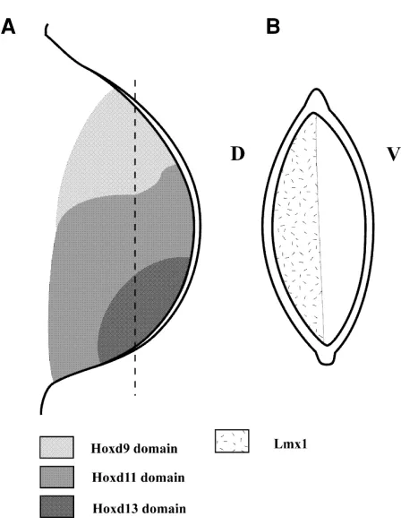
Fig. 3. Diagram to illustrate patterns of expression of genes encoding transcription factors; these patterns are established as a result of signalling in the chick limb bud. (A) Overlapping patterns of Hoxd gene expression in early chick limb bud. Only three domains shown for clarity. Cells in postero-distal region of the bud express Hoxd9, Hoxd10, Hoxd11, Hoxd12 and Hoxd13 while cells in antero-distal region of bud express Hoxd9 only. (B)Section through chick limb bud showing expression of Lmx1 in dorsal mesenchyme. D, dorsal; V, ventral.
Chick limb mutants
Chicken limb mutants have arisen and been studied in the UK, USA and Japan. The potential interest of these mutants was recognised by experimental embryologists, who examined cell interactions in mutant limb buds to try and identify the defective tissue(s) (reviewed Wolpert, 1976). More recently, gene expres-sion has also been analysed.
An important chicken mutant with respect to antero-posterior patterning is the talpid mutant and the talpid3 mutant has been studied in the UK (the US mutant is talpid2). In talpid mutants, the
limbs are polydactylous and up to 10 digits develop with uniform morphology (Hinchliffe and Ede, 1967). This uniformity of digit morphology in talpid3 has been shown to be associated with
uniformity of Hoxd gene expression at the tips of the limb buds (Izpisúa-Belmonte et al., 1992). Grafting experiments with both talpid2 and talpid3(the American and British talpids respectively)
showed that polarising activity is more widespread but it is now known that Shh gene expression is posteriorly restricted. However, Bmps are expressed uniformly throughout the mesenchyme and Fgf4 throughout the ridge (Francis-West et al., 1995). Recently, it has been shown that the defect in talpid3 is based on failure to express high levels of Ptc in response to Shh signalling (Lewis et al., 1999). Ptc is the gene that encodes the Shh receptor. It seems likely that this change in response of talpid 3cells to hedgehog
signalling could account for all the other developmental defects in the mutant embryos in addition to the limb defects.
Interpretation of positional information
The ways in which the expression of different Hox genes governs cell behaviour and controls tissue and cell arrangements are not known. There is only a limited repertoire of cell behaviour. Cells proliferate, die, change shape, move, adhere to other cells and/or extracellular matrix, and differentiate. These activities will have to be locally co-ordinated by, for example, gap junctional communication to produce specific local patterns of cells and tissues.
A major conceptual difficulty is how different positional values (such as those that lead to the development of each of the three different digits in a chick wing) lead to differentiation of the same cell type (cartilage). To some extent, this issue has been ad-dressed by the idea of non-equivalence (Lewis and Wolpert, 1976). This suggests, for example, that other cellular properties such as proliferation may depend on positional value. Non-equivalence can also explain how the same positional values may lead to different outcomes depending on the history of the cells. A good example is the development of the chick wing versus the leg. The signalling regions in both wing and leg buds were found to be interchangeable and the molecules produced in the apical ridge, polarising region and ectoderm appear to be the same. Moreover, dorsal mesenchyme in both wing and leg expresses Lmx1 (Vogel et al., 1995) and patterns of expression of most Hox genes are also similar in both wing buds and leg buds at early stages (Nelson et al., 1996). Nevertheless, cells, for example, at the anterior distal edge of the leg bud form a “big toe” while those in the equivalent position in the wing form a “thumb”. Thus, the origin of the cells from different axial levels –either wing or leg levels– affects how they interpret the same positional values in the limb bud. Molecules that
are responsible for these properties of “wingness” and “legness” have now been identified (see below).
Initiation of limb development
The development of four limbs is a hallmark of the tetrapod body plan. Thus issues about control of position, number and type of limbs in vertebrate embryos are fundamental (reviewed Cohn and Tickle, 1996). Fibroblast Growth Factors can induce ectopic limb development in chick embryos (Cohn et al., 1995) and specific family members have been identified that could act very early as endogenous limb initiation signals (Crossley et al., 1996; Ohuchi et al., 1997). Fgf10 , for example, is expressed very early in presumptive limb-forming regions of chick embryos (Ohuchi et al., 1997) and functional inactivation of Fgf10 in mouse embryos leads to failure of limb development (Min et al., 1998; Sekine et al., 1999).
The position in which factors operate to initiate forelimbs and hindlimbs must be part of the patterning process that governs the head to tail axis of the embryo. A number of different lines of evidence, both from transgenic mice and experimental manipula-tion in chicken embryos implicates Hox gene expression in the lateral plate mesoderm as encoding position, i.e., one combination of Hox gene expression specifies presumptive wing level, another combination, the interlimb level, and yet another, the leg level (Cohn et al., 1997). These differences will be set up very early long before the limb buds appear.
Genes that are expressed specifically in wing lateral plate mesoderm and in leg lateral plate mesoderm were first discovered in mice. These are Tbx genes which encode transcription factors and are related to the large T mouse gene (Chapman et al., 1996). It was shown in chick embryos that Tbx gene expression is stable when wing cells are transplanted to leg and vice versa (Gibson-Brown et al., 1998; Isaac et al., 1998; Logan et al., 1998; Ohuchi et al., 1998). Furthermore, work in the US and in Japan showed that, when the Tbx gene normally expressed in the leg is now expressed in the wing, this leads to development of ectopic leg structures (Logan and Tabin, 1999; Rodriguez-Esteban et al., 1999; Takeuchi et al., 1999).
An important feature of limb bud initiation is the positioning of the signalling regions. The apical ridge in both wing and leg forming regions in chick embryos arises at a dorsal-ventral compartment boundary in the ectoderm (Altabef et al., 1997) and work by two groups in the USA has shown that radical fringe signalling is involved (Laufer et al., 1997; Rodriguez-Esteban et al., 1997). This dorsal-ventral ectodermal compartment has also been detected in the interlimb region and thus can account for the positioning of ectopic limb buds in register with the normal limb buds along the sides of the body.
Chick limb development as a model for mammalian limb
development
The patterning mechanisms involved in limb development are conserved between different vertebrates. Thus, for example, the posterior margin of a mouse limb bud has polarising activity and can induce additional digits in a chick wing (Tickle et al., 1976) and the ectodermal jacket (including the apical ridge) from a rat limb bud can maintain development of chick wing mesenchyme (Jorquera and Pugin, 1971). Molecular analysis shows that Shh is expressed in the polarising region of both mouse and chick limb buds. Furthermore, the patterns of Hox gene expression at least in early buds are similar. This again shows the importance of interpretation of positional information, this time giving rise to the differences in morphology between vertebrates. Thus, grafts of mouse limb polarising region provide the same signal as grafts of the chick limb polarising region but when grafted to the chick wing lead to the formation of chick digits not mouse digits. A major challenge is to uncover the basis for the subtle differences in cell behaviour that lead to development of a chick “finger” rather than a mouse “finger”. There are over 100 different mutations known in mice that affect development of the limb. The ability to graft tissues from mouse limb buds into chicken wing buds and to monitor expression of genes that pattern the limb has given some interesting new insights into conditions, such as extra digits or loss of digits. Furthermore, the genes affected in such mouse mutants are rapidly being discovered. In many cases similarities to human conditions can be recognised (Winter, 1988). It has been shown, for example, that the mouse mutant, Hypodactyly, is due to a mutation in the Hoxa13 gene. A mutation in the same gene has been discovered in a patient with hand-genital syndrome (reviewed Scott, 1997). Other direct links have been made between experimental embryology and human clinical genetics.
With modern molecular techniques, the genetic bases of many human congenital malformations are rapidly being uncovered. Many inherited conditions are very rare and therefore the world-wide availability of families to study is limited. This is an instance where work in different centres and even in different countries can be particularly valuable. In Britain, a notable example has been the discovery in two different centres of the involvement of FGF receptors in a series of craniosynostosis conditions, some associ-ated with limb defects (Reardon et al., 1994; Wilkie et al., 1995). Another example is the identification of the involvement of a Tbx gene in Holt-Oram syndrome in a family in the UK and in a family in the USA (Basson et al., 1997; Li et al., 1997). These clinical discoveries provide a new dimension and stimulus to basic re-search on limb development.
Acknowledgements
The author’s own research has been supported by the MRC, BBSRC, Action Research, The Wellcome Trust, The Leverhulme Trust, Human Frontiers, The Anatomical Society and The Royal Society. I would like to thank my many collaborators over the years both in Britain and abroad for their most stimulating and invaluable contributions.
References
AKITA, K. (1996). The effects of the ectoderm on dorsoventral pattern of the epidermis, muscles and joints in the developing chick leg: a new model. Anat. Embryol. 193: 377-386.
ALTABEF, M., CLARKE, J. and TICKLE, C. (1997). Dorso-ventral compartments and origin of apical ectodermal ridge in developing chick limb. Development 124: 4547-4556.
BASSON, C.T., BACHINSKY, D.R., LIN, R.C., LEVI, T., ELKINS, J.A., SOULTS, J., GRAYSEL, D., KROUMPOUZOU, E., TRAILL, T.A., LEBLANC-STRACESKI, J., RENAULT, B., KUCHERLAPATI, R., SEIDMAN, J.G. and SEIDMAN, C.E. (1997). Mutations in human TBX5 cause limb and cardiac malformation in Holt-Oram syndrome. Nature Genet. 15: 30-35.
CHAPMAN, D.L., GARVEY, N., HANCOCK, S., ALEXIOU, M., AGULNIK, S.I., GIBSON-BROWN, J.J., CEBRA-THOMAS, J., BOLLAG, R.J., SILVER, L.J. and PAPAIOANNOU, V.E. (1996). Expression of the T-box family genes, Tbx1-Tbx5, during early mouse development. Dev. Dyn. 206: 379-390.
CHARITÉ, J., DE GRAAF, W., SHEN, S. and DESCHAMPS, J. (1994). Ectopic expression of Hoxb8 causes duplication of the ZPA in the forelimb and homeotic transformation of axial structures. Cell 78: 589-601.
CHIANG, C., LITINGTUNG, Y., LEE, E., YOUNG, K.E., CORDENT, J.L., WESTPHAL, H. and BEACHEY, P.A. (1996). Cyclopia and defective axial patterning in mice lacking Sonic hedgehog gene function. Nature 383: 407-413.
COHN, M. and TICKLE, C. (1996). Limbs: a model for pattern formation within the vertebrate body plan. Trends Genet. 12: 253-257.
COHN, M., IZPISÚA-BELMONTE, J.C., ABUD, H., HEATH, J.K. and TICKLE, C. (1995). Fibroblast growth factors induce additional limb development from the flank of chick embryos. Cell 80: 739-746.
COHN, M., PATEL, K., CLARKE, J., WILKINSON, D., KRUMLAUF, R. and TICKLE, C. (1997). Hox-9 genes and specification of vertebrate limbs. Nature 387: 97-101.
CROSSLEY, P.H., MINOWADA, G., MACARTHUR, C.A. and MARTIN, G.R. (1996). Roles for FGF-8 in the induction, initiation and maintenance of chick limb development. Cell 84: 127-136.
DOLLÉ, P., IZPISÚA-BELMONTE, J.C., FALKENSTEIN, H., RENUCCI, A. and DUBOULE, D. (1989). Co-ordinate expression of the murine Hox-5 complex homoeobox-containing genes during limb pattern formation. Nature 342: 767-772.
DUPREZ, D., KOSTAKOPOULOU, K., FRANCIS-WEST, P.H., TICKLE, C. and BRICKELL, P.M. (1996). Activation of Fgf4 and HoxD gene expression by BMP-2 expressing cells in the developing chick wing. Development 122: 1821-1828.
FALLON, J., LOPEZ, A., ROS, M., SAVAGE, M., OLWIN, B. and SIMANDL, B. (1994). FGF-2, apical ectodermal ridge growth signal for chick limb development. Science 264: 104-107.
FRANCIS, P.H., RICHARDSON, M.K., BRICKELL, P.M. and TICKLE, C. (1994). Bone morphogenetic proteins and a signalling pathway that controls pattern in the developing chick limb. Development 120: 209-218.
FRANCIS-WEST, P.H., ROBERTSON, K.E., EDE, D.A., RODRIGUEZ, C., IZPISÚA-BELMONTE, J.C., HOUSTON, B., BURT, D.W., GRIBBIN, C., BRICKELL, P.M. and TICKLE, C. (1995). Expression of genes encoding bone morphogenetic proteins and sonic hedgehog in talpid (ta3) limb buds: their relationship to the
signalling cascade involved in limb patterning. Dev. Dyn. 203: 187-197.
FRENCH, V., BRYANT, P.J. and BRYANT, S.V. (1976). Pattern regulation in epimorphic fields. Science 193: 969-981.
GIBSON-BROWN, J.J., AGULNIK, S., SILVER, L.M., NISWANDER, L. and PAPAIOANNOU, V. (1998). Involvement of T-box genes Tbx-2-Tbx-5 in verte-brate limb specification and development. Development 125: 2499-2509.
HELMS, J.A., KIM, C.H., EICHELE, G. and THALLER, C. (1996). Retinoic acid signalling is required during early chick limb development. Development 122: 1385-1394.
HINCHLIFFE, J.R. and EDE, D.A. (1967). Limb development in the polydactylous talpid3muatnt of the fowl. J. Embryol. Exp. Morphol. 17: 385-40.
HURLÉ, J.M., ROS, M.A., GANAN, Y., MACIAS, D., CRICTHLOW, M. and HINCHLIFFE, J.R. (1990). Experimental analysis of the role of ECM in the patterning of the distal tendons of the developing limb bud. Cell Differ. Dev. 30: 97-108.
ISAAC, A., RODRIGUEZ-ESTEBAN, C., RYAN, A., ALTABEF, M., TSUKUI, T., PATEL, K., TICKLE, C. and IZPISÚA-BELMONTE, J.C. (1998). Tbx genes and limb identity in chick embryo development. Development 125: 1867-1875.
ITEN, L.E. and MURPHY, D.J. (1980). Pattern regulation in the embryonic chick limb: supernumerary limb formation with anterior (non-ZPA) limb bud tissue. Dev. Biol. 75: 373-385.
mis-expression of posterior Hox4 genes in talpid (ta3) mutant wings correlates with
the absence of anteroposterior polarity. Development 114: 959-963.
IZPISÚA-BELMONTE, J.C., TICKLE, C., DOLLÉ‚ P., WOLPERT, L. and DUBOULE, D. (1991). Expression of the homeobox Hox-4 genes and the specification of position in chick wing development. Nature 350: 585-589.
JORQUERA, B. and PUGIN, E. (1971). Sur le comportement du mesoderme et de l’ectoderme du bourgeon de membre dans les échanges entre le poulet et le rat. C.R. Acad. Sci. Paris 272: 1522-1525.
LAUFER, E., DAHN, R., OROZCO, O.E., YEO, C.Y., PISENTI, J., HENRIQUE, D., ABBOTT, U.K., FALLON, J.F. and TABIN, C. (1997). Expression of RadicalFringe in limb-bud ectoderm regulates apical ectodermal ridge formation. Nature 386: 366-373.
LAUFER, E., NELSON, C.E., JOHNSON, R.L., MORGAN, B.A. and TABIN, C. (1994). Sonic hedgehog and Fgf4 act through a signalling cascade and feedback loop to integrate growth and patterning of the developing limb bud. Cell 79: 993-1003.
LEWIS, J. and WOLPERT, L. (1976). The principle of non-equivalence in develop-ment. J. Theor. Biol. 62: 479-490.
LEWIS, K.E., DROSSOPOULOU, G., PATON, I.R., MORRICE, D.R., ROBERTSON, K.E., BURT, D.W., INGHAM, P.W. and TICKLE, C. (1999). Expression of ptc and gli genes in talpid3suggests bifurcation in Shh pathway. Development 126:
2397-2407.
LI, Q.Y., NEWBURY-ECOB, R.A., TERRET, J.A., WILSO, D.I., CURTIS, A.R., YI, C.H., GEBURH, T., BULLEN, P.J., ROBSON, S.C., STRCHAN, T., BONNET, D., LYONNET, S., YOUNG, I.D., RAEBURN, J.A., BUCKLER, A.J., LAW, D.J. and BROOK, J.D. (1997). Holt-Oram syndrome is caused by mutations in TBX5, a member of the Brachyury (T) gene family. Nature Genet. 15: 21-29.
LOGAN, M. and TABIN, C. (1999). Role of Pitx1 upstream of Tbx-4 in specification of hindlimb identity. Science 283: 1736-1739.
LOGAN, M., SIMON, H.G. and TABIN, C. (1998). Differential regulation of T-box and homeobox transcription factors suggests roles in controlling chick limb-type identity. Development 125: 2825-2835.
LOOMIS, C.A., HARRIS, E., MICHAUD, J., WURST, W., HANKS, M. and JOYNER, A.L. (1996). The mouse Engrailed-1 gene and ventral limb patterning. Nature 382: 360-363.
LU, H.C., REVELLI, J.P., GOERING, L., THALLER, C. and EICHELE, G. (1997). Retinoid signalling is required for the establishment of a ZPA and for the expression of Hoxb-8, a mediator of ZPA formation. Development 124: 1643-1651.
MACCABE, J.A., ERRICK, J. and SAUNDERS, J.W. (1974). Ectodermal control of the dorso-ventral axis in the leg bud of the chick embryo. Dev. Biol. 39: 69-82.
MIN, H., DANILENKO, D.M., SCULLY, S.A., BOLON, B., RING, B.D., TARPLEY, J.E., DEROSE, M. and SIMONET, W.S. (1998). Fgf-10 is required for both limb and lung development and exhibits striking functional similarity to Drosophila branch-less. Genes Dev. 12: 3156-3161.
MORGAN, B.A., IZPISÚA-BELMONTE, J-C., DUBOULE, D. and TABIN, C.J. (1992). Targeted misexpression of Hox-4.6 in the avian limb causes apparent homeotic transformations. Nature 358: 236-239.
MURRAY, B. and WILSON, D.J. (1997). Muscle patterning, differentiation and vascularisation in the chick wing bud. J. Anat. 190: 261-273.
NELSON, C.E., MORGAN, B.A., BURKE, A.C., LAUFER, E., DI NAMBRO, E., MURTAUGH, L.C., GONZALES, E., TESSAROLLO, L., PARADA, L.F. and TABIN, C. (1996). Analysis of Hox gene expression in the chick limb bud. Development 12: 1449-1466.
NISWANDER, L., JEFFERY, S., MARTIN, G.R. and TICKLE, C. (1994). Signalling in vertebrate limb development: a positive feedback loop between sonic hedgehog and FGF4. Nature 371: 609-612.
NISWANDER, L., TICKLE, C., VOGEL, A., BOOTH, I. and MARTIN, G.R. (1993). FGF-4 replaces the apical ectodermal ridge and directs outgrowth and patterning of the limb. Cell 75: 579-587.
NOHNO, T., NOJI, S., KOYAMA, E., OHYAMA, K., MYOKAI, F., KURIOWA, A., SAITO, T. and TANIGUCHI, S. (1991). Involvement of the Chox-4 chicken homeobox genes in determination of antero-posterior axial polarity during limb development. Cell 64: 1197-1205.
NOJI, S., NOHNO, T., KOYAMA, E., MUTO, K., OHYAMA, K., AOKI, Y., TAMURA, K., OHSUGI, K., IDE, H., TANIGUCHI, S. and SAITO, T. (1991). Retinoic acid induces polarizing activity but is unlikely to be a morphogen in the chick limb bud. Nature 350: 83-86.
OHUCHI, H., NAKAGAWA, T., YAMMOTO, A., ARAGA, A., OHATA, T., ISHIMARU, Y., YOSHIOKA, H., KUWANA, T., NOHNO, T., YAMASAKI, M., ITOH, N. and NOJI, S. (1997). The mesenchymal factor, FGF10, initiates and maintains the outgrowth of the chick limb bud through interaction with FGF8, an apical ectoder-mal factor. Development 124: 2235-2244.
OHUCHI, H., TAKEUCHI, J., YOSHIYASU, H., ISHIMARU, Y., OGURA, K., TAKAHASHI, N., OGURA, T. and NOJI, S. (1998). Correlation of wing-leg identity in ectopic Fgf-induced chimeric limbs with the differential expression of chick Tbx5 and Tbx4. Development 125: 51-60.
PARR, B.A. and MCMAHON, A.P. (1995). Dorsalising signal Wnt-7a required for normal polarity of D-V and A-P axes of mouse limb. Nature 374: 350-353.
PATOU, M.P. and KIENY, M. (1973). Interaction ecto-mesodermique dans l’éstablissement de la polarité dorso-ventrale du pied de l’embryon de poulet. C.R. Acad. Sci. Paris D 277: 1225-1228.
PITTS, J.D., HAMILTON, A.E., KAM, E., BURK, R.R. and MURPHY, J.P. (1986). Retinoic acid inhibits gap junctional communication between cells. Carcinogenesis 7: 1003-1010.
REARDON, W., WINTER, R.M., RUTLAND, P., PULLEYN, L.J., JONES, B.M. and MALCOLM, S. (1994). Mutations in the fibroblast growth factor receptor 2 gene cause Crouzon syndrome. Nature Genet. 8: 98-103.
RIDDLE, R.D., ENSINI, M., NELSON, C., TUSCHIDA, T., JESSELL, T.M. and TABIN, C. (1995). Induction of the LIM homeobox gene Lmx-1 by Wnt7a establishes dorsoventral pattern in the vertebrate limb. Cell 83: 631-640.
RIDDLE, R.D., JOHNSON, R.L. and TABIN, C. (1993). Sonic hedgehog mediates the polarizing activity of the ZPA. Cell 75: 1401-1416.
RIJLI, F.M. and CHAMBON, P. (1997). Genetic interactions of Hox genes in limb development: learning from compound mutants. Curr. Opin. Genet. Dev. 7: 481-487.
RODRIGUEZ-ESTEBAN, C., SCHWABE, J.W.R., DE LA PENA, J., FOYS, B., ESHELMAN, F. and IZPISÚA-BELMONTE, J.C. (1997). Radical fringe positions the apical ectodermal ridge at the dorso-ventral boundary of the vertebrate limb. Nature 386: 360-365.
RODRIGUEZ-ESTEBAN, C., TSUKUI, T., YONEI, S., MAGALLON, J., TAMURA, K. and IZPISÚA-BELMONTE, J.C. (1999). The T-box genes Tbx4 and Tbx5 regulate limb outgrowth and identity. Nature 398: 814-818.
RUBIN, L. and SAUNDERS, J.W. (1972). Ectodermal-mesodermal interactions in the growth of limb buds in the chick embryo: constancy and temporal limits of the ectodermal induction. Dev. Biol. 28: 94-112.
SAUNDERS, J.W. (1948). The proximo-distal sequence of origin of limb parts of the chick wing and the role of the ectoderm. J. Exp. Zool. 108: 363-404.
SAUNDERS, J.W. (1977). The experimental analysis of chick limb bud development. In Vertebrate limb and somite morphogenesis (Eds. D.A. Ede, J.R. Hinchliffe and M. Balls). Cambridge University Press, Cambridge. pp. 1-24.
SAUNDERS, J.W. and GASSELING, M.T. (1968). Ectodermal-mesenchymal inter-actions in the origin of limb symmetry. In Epithelial-mesenchymal interactions (Ed. R. Fleishmajer and R.E. Billingham) Baltimore: Williams & Wilkins. pp. 78-97.
SAUNDERS, J.W., GASSELING, M. and CAIRNS, J.M. (1959). The differentiation of prospective thigh mesoderm grafted beneath the apical ectodermal ridge of the wing bud in the chick embryo. Dev. Biol. 1: 281-301.
SCOTT, M.P. (1997). Hox genes, arms and the man. Nature Genet. 15: 117-118.
SEKINE, K., OHUCHI, H., FUJIWARA, M., YAMASAKI, M., YOSHIZAWA, T., SATO, T., YAGASHITA, N., MATSUI, D., KOGA, Y., ITOH, N. and KATO, S. (1999). Fgf10 is essential for limb and lung formation. Nature Genet.21: 138-141.
SOLURSH, M. (1984). Ectoderm as a determinant of early tissue pattern in the limb bud. Cell Differ. 15: 17-18.
STRATFORD, T., HORTON, C. and MADEN, M. (1996). Retinoic acid regulates sonic hedgehog expression and initiates outgrowth in the chick limb bud. Current Biology 6: 1124-1133.
STRATFORD, T., KOSTAKOPOULOU, K. and MADEN, M. (1997). Hoxb-8 has a role in establishing early antero-posterior polarity in chick forelimb but not hindlimb. Development 124: 4225-4234.
SUMMERBELL, D. (1974). A quantitative analysis of the effect of excision of the AER from the chick limb bud. J. Embryol. Exp. Morphol. 32: 651-660.
TAKEUCHI, J.K., KOSHIBA-TAKEUCHI, K., MATSUMOTO, K., VOGEL-HOPKER, A., NAITOH-MATSUO, M., OGURA, K., TAKAHASHI, N., YASUDA, K. and OGURA, T. (1999). Tbx5 and Tbx4 genes determine the wing/leg identity of limb buds. Nature 398: 810-814.
THALLER, C. and EICHELE, G. (1987). Identification and spatial distribution of retinoids in the developing chick limb bud. Nature 327: 625-628.
TICKLE, C. (1981). The number of polarizing region cells required to specify additional digits in the developing chick wing. Nature 289: 295-298.
TICKLE, C. (1995). Vertebrate limb development. Curr. Opin. Genet. Dev. 5: 478-484.
TICKLE, C. and EICHELE, G. (1994). Vertebrate limb development. Annu. Rev. Cell Biol. 10: 121-152.
TICKLE, C., ALBERTS, B., WOLPERT, L. and LEE, J. (1982). Local application of retinoic acid to the limb bud mimics the action of the polarizing region. Nature 296: 564-566.
TICKLE, C., LEE, J. and EICHELE, G. (1985). A quantitative analysis of the effect of all-trans-retinoic acid on the pattern of chick wing development. Dev. Biol. 109: 82-95.
TICKLE, C., SHELLSWELL, G., CRAWLEY, A. and WOLPERT, L. (1976). Positional signalling by mouse limb polarizing region in the chick limb bud. Nature 259: 396-397.
TICKLE, C., SUMMERBELL, D. and WOLPERT, L. (1975). Positional signalling and specification of digits in chick limb morphogenesis. Nature 254: 199-202. VOGEL, A. and TICKLE, C. (1993). FGF-4 maintains polarizing activity of posterior
limb bud cells in vivo and in vitro. Development 119: 199-206.
VOGEL, A., RODRIGUEZ, C., WARNKEN, W. and IZPISÚA-BELMONTE, J.C. (1995). Dorsal cell fate specified by chick Lmx1 during vertebrate limb develop-ment. Nature 378: 716-720.
WANEK, N., GARDINER, D.M., MUNEOKA, K. and BRYANT, S.V. (1991). Conver-sion by retinoic acid of anterior cells into ZPA cells in the chick wing bud. Nature 350: 81-83.
WILBY, O.K. and EDE, D.A. (1975). A model generating the pattern of cartilage skeletal elements in the embryonic chick limb. J. Theor. Biol. 52: 199-217.
WILKIE, A.O.M., SLANEY, S.F., OLDRIDGE, M., POOLE, M.D., ASHWORTH, G.J., HOCKLEY, A.D., HAYWARD, R.D., DAVID, D.J., PULLEYN, L. and RUTLAND, P. (1995). Apert syndrome results from localised mutations of FGFR2 and is allelic with Crouzon syndrome. Nature Genet. 9: 165-172.
WINTER, R.M. (1988). Malformation syndromes: a review of mouse / human morphol-ogy. J. Med. Genet. 25: 480-487.
WOLPERT, L. (1969). Positional information and the spatial pattern of cellular differentiation. J. Theor. Biol. 25: 1-47.
WOLPERT, L. (1976). Mechanisms of limb development and malformation. Br. Med. Bull. 32: 65-70.
WOLPERT, L. (1996). One hundred years of positional information. Trends Genet. 9: 359-364.
WOLPERT, L. and STEIN, W. (1984). Positional information and pattern formation. In Pattern Formation (Eds. G. Malacinski and S.V. Bryant). Macmillan Publishing Co. Inc. pp.3-21.
YANG, Y. and NISWANDER, L. (1995). Interaction between the signalling molecules Wnt-7a and Shh during vertebrate limb development- dorsal signals regulate antero-posterior patterning. Cell 80: 939-947.
YANG, Y., DROSSOPOULOU, G., CHUANG, P.T., DUPREZ, D., MARTI, E., BUMCROT, D., VARGESSON, N., CLARKE, J., NISWANDER, L., MCMAHON, A. and TICKLE, C. (1997). Relationship between dose, distance and time in sonic hedgehog mediated regulation of antero-posterior patterning in chick limb. Development 124: 4393-4404.