BIOCHEMICAL CHARACTERISATION OF THE EUKARYOTIC CELL CYCLE REGULATORY PROTEINS. E2F AND pRB
Nadeem Ali-Khan
A thesis submitted in partial fulfilment of the requirements of University College London for the degree of Doctor of Philosophy.
June 2002
Division of Protein Structure,
ProQuest Number: U643133
All rights reserved
INFORMATION TO ALL USERS
The quality of this reproduction is dependent upon the quality of the copy submitted.
In the unlikely event that the author did not send a complete manuscript and there are missing pages, these will be noted. Also, if material had to be removed,
a note will indicate the deletion.
uest.
ProQuest U643133
Published by ProQuest LLC(2016). Copyright of the Dissertation is held by the Author.
All rights reserved.
This work is protected against unauthorized copying under Title 17, United States Code. Microform Edition © ProQuest LLC.
ProQuest LLC
789 East Eisenhower Parkway P.O. Box 1346
ABSTRACT
Control of the cell cycle is partly mediated by a transcriptional regulatory mechanism whose components include the pRb family of tumour suppressors (pRb, p i 30, p i 07) and the E2F/DP heterodimeric transcription factors. Each of these heterodimers consists of one member of the E2F family of proteins (E2Fs 1- 6) and one of the DP family (DPs 1 and 2). E2F/DP activation of cell cycle genes is negatively regulated by cyclin A-CDK2-mediated phosphorylation of DP. The formation of a complex between E2F/DP and a pRb family protein leads to anti proliferative transcriptional repression. Binding of the Human Papillomavirus (HPV) E7 oncoprotein to pRb blocks the interaction of the tumour suppressor with E2F/DP as part of a viral cell transformation mechanism.
Fragments of pRb, E2F-1 and DP-1 were over-expressed and purified by chromatographic means prior to their biochemical characterisation. Using the SELEX (Systematic Evolution of Ligands by Exponential Enrichment) procedure, attempts were made to determine high affinity DNA-binding sites for E2F homodimers. Although such sites were not identified, important considerations relating to the SELEX protocol are highlighted by these experiments.
ACKNOWLEDGEMENTS
I gratefully acknowledge my supervisor Dr. Steve Gamblin for his help, advice and support throughout the course of this study. I would also like to thank other members of the Division of Protein Structure, particularly Dr. B. Xiao, Dr. D. Emery and Dr. I. Tews for their assistance and guidance with experimental aspects of the work, as well as Dr. P. Walker for help with the computing. I am also grateful to members of the Photographic Department at the NIMR, particularly Frank, for their help with the figures presented in this thesis.
CONTENTS
ABSTRACT 3
ACKNOWLEDGEMENTS 5
CONTENTS 6
LIST OF FIGURES 11
LIST OF TABLES 13
ABBREVIATIONS 14
CHAPTER 1 INTRODUCTION 16
1.1 The Eukaryotic Cell Cycle 18
1.1.1 Checkpoints and Feedback Controls 20
1.2 Transcriptional Regulation of the Cell Cycle 22 1.2.1 Cancer and the Transcriptional Regulatory
Mechanism 27
1.3 The Retinoblastoma Tumour Suppressor Gene
Product (pRb) 29
1.3.1 Structural Aspects of pRb 29
1.3.2 Regulation of pRb 33
1.3.3 Mechanisms of pRb-Induced Transcriptional
Repression 35
1.3.3.1 Repression o f R N A polymerase Il-directed Transcription 35 1.3.3.2 pRb-induced Repression o f R N A polymerases I and III 43
1.4 The E2F Family of Transcription Factors 45
1.4.1 Structural Aspects 45
1.4.2 The Regulation of E2F 48
1.4.2.1 Synthesis o f E2F Transcription Factors 48
1.4.2.2 Pocket Proteins and C /E B P a 49
1.4.2.3 Phosphorylation o f E2F 50
1.4.2.4 Degradation and Stabilisation o f E2F 51
1.4.2.5 Subcellular Localisation o f E2F 52
1.4.2.6 Acétylation o f E2F 52
1.4.3.1 E 2F ’s Target Genes 5 3 1.4.3.2 E2F as an Activator and Repressor o f Transcription 55 1.4.3.3 Specificity o f E2F Family Members 5 8 1.4.3.4 Oncogenic, Tumour Suppressive and Apoptotic Functions
o fE 2 F 58
1.4.4 Mechanism of E2F Activity 59
1.4.4.1 Transcriptional Activation 59
1.4.4.2 Transcriptional Repression 61
1.4.4.3 Promoter Specificity 62
1.4.4.4 Apoptosis 63
1.5 Inactivation of pRb by Viral Oncoproteins 65
1.5.1 Structure 66
1.5.2 pRb-inactivating Function 68
1.5.3 Mechanism of pRb-inactivation 69
1.6 Presentation of Experimental Work 71
CHAPTER 2 METHODS AND MATERIALS 72
2.1 General 73
2.1.1 Media for Bacterial Cell Growth 73
2.1.2 Cloning 74
2.1.2.1 Restriction Enzyme Digestions 7 4
2.1.2.2 Ligation 75
2.1.2.3 Transformation 76
2.1.2.4 Selection and Purification of Recombinant Plasmids 7 6
2.1.3 Protein Analysis 77
2.1.3.1 Polyacrylamide Gel Electrophoresis (PAGE) 7 7 2.1.3.2 Determination of Protein Concentration 7 7
2.1.3.3 Electrospray Mass Spectrometry 7 7
2.1.4 DNA Analysis and Purification 79
2.1.4.1 Agarose Gel Electrophoresis 79
2.1.4.2 Nondenaturing Polyacrylam ide Gel Electrophoresis 79
2.1.4.5 Determination o f D N A Concentration 81 2.2 Identification of Aptamers for the E2F-1 Homodimer
(Methods and Materials) 82
2.2.1 Systematic Evolution of Ligands by Exponential
Enrichment (SELEX) 82
2.2.1.1 Preparation o f Double-Stranded Degenerate
O ligonucleotide (degOLIGO) 83
2.2.2 SELEX Method 1 83
2.2.2.1 Selection and A m plification 83
2 2 . 2 . 2 Cloning and Sequencing 85
2.2.2.5 EM SA (Electrophoretic M obility Shift A ssay) 85
2.2.3 SELEX Method 2 86
2.2.3.1 Preparation o f Control Oligonucleotide (conOLIGO) 8 6
2.2.3.2 Selection and Amplification 8 7
2.3 Regulation of E2F/DP DNA-Binding By Cyclin-CDK
Dependent Phosphorylation (Methods and Materials) 89
2.3.1 Purification of E2F-1 eye 89
2.3.1.1 Strategy 8 9
2.3.1.2 Protocol 89
2.3.2 Purification of DP-lggg and DP-lAs9s 91
2.3.2.1 Strategy 91
2.3.2.2 Protocol 91
2.3.3 Electrophoretic Mobility Shift Assays (EMSAs) 93
2.3.3.1 Probe 93
2.3.3.2 Assay 9 4
2.4 Investigation of the Interactions Between pRb, E2F
and HPV E7 (Methods and Materials) 97
2.4.1 Purification of pRbAB 97
2.4.1.1 Strategy 97
2.4.1.2 Protocol 9 8
2.4.2 Isothermal Titration Calorimetry (TTC) 100
2.4.2.1 General Points 100
CHAPTER 3 IDENTIFICATION OF APTAMERS
FOR THE E2F-1 HOMODIMER 104
3.1 Outline of the Systematic Evolution of Ligands
by Exponential Enrichment (SELEX) 105
3.1.1 General Procedure 105
3.1.2 Isolation of Protein-Aptamer Complexes 107
3.2 Introduction to Experiments 110
3.3 Results 113
3.3.1 Method 1 113
3.3.2 Method 2 118
3.4 Discussion 122
3.4.1 SELEX Method 1 122
3.4.2 SELEX Method 2 124
3.4.3 General Points Concerning SELEX 126
3.4.4 Crystal Structure of the E2F/DP-DNA Complex 127
3.4.5 SELEX and E2F - The Next Step 136
CHAPTER 4 REGULATION OF E2F/DP
DNA-BINDING BY
CYCLIN-CDK DEPENDENT
PHOSPHORYLATION 138
4.1 Outline of the Electrophoretic Mobility Shift Assay
(EMSA) 139
4.1.1 Principle of the EMSA 139
4.2 Introduction to Experiments 142
4.3 Results 150
4.3.1 Purification of E2F-1 eye 150
4.3.2 Purification of DP-ls98 and DP-1As98 153 4.3.3 Phosphorylation of the E2F Transcription Factor
by Cyclin A-CDK2 158
CHAPTER 5 INVESTIGATION OF THE
INTERACTIONS BETWEEN pRb,
E 2FA N D H PV E 7 169
5.1 Outline of Isothermal Titration Calorimetry (ITC) 170 5.1.1 The ITC Apparatus and Mode of Operation 170 5.1.2 Determination of Thermodynamic Binding
Parameters 173
5.1.3 The Thermodynamic Parameters and How Interactions
Influence Them 174
5.2 Introduction to Experiments 181
5.3 Results 187
5.3.1 Purification of pRbAB 187
5.3.2 Isothermal Titration Calorimetry Experiments 191
5.4 Discussion 208
5.4.1 Determination of the Minimal Fragments of
pRb and E2F-1 Required for Binding 208
5.4.2 Investigation of the Specificity of the E2F/pRb
Interaction 211
5.4.2.1 Crystal Structure o f p R b 1 (409.426) C om plex 2 1 2 5.4.2.2 Structural Role o f Non-Conserved Residues Betw een
E 2 F - 1 (409-426) &nd E 2 F -5 (3 2 3 -3 4 0 ) 2 1 7
5.4.3 Investigation of HPV E7-mediated Inhibition of
pRb-E2F Complex Formation 219
5.4.4 Conclusions in Terms of AH° and AS° 222
CHAPTER 6 CONCLUSION 227
LIST OF FIGURES
Figure 1 The Eukaryotic Cell Cycle 19
Figure 2 Functional Regions of the Retinoblastoma Tumour Suppressor
Gene Product (pRb) 30
Figure 3 Elements of Eukaryotic Transcriptional Initiation for
Genes Transcribed by RNA Polymerase II 37
Figure 4 Proposed Mechanisms for Repression of RNA Polymerase
Il-directed Transcription by pRb 38
Figure 5 Functional Regions of the Mammalian E2F and DP Proteins 47 Figure 6 Model for E2F-Mediated Transcriptional Regulation 57 Figure 7 Structural Organisation of the HPV E7 Oncoprotein 67
Figure 8 General Selex Procedure 106
Figure 9 Separation of Protein-Aptamer Complexes Using the
Electrophoretic Mobility Shift Assay (EMSA) 109 Figure 10 E2F-1 Protein Fragment Used in SELEX Experiments
(Methods 1 and 2) 111
Figure 11 Oligonucleotide Derived from Adenoviral Promoter 112 Figure 12 Degenerate Oligonucleotides (degOLIGO) Used in
SELEX Procedure (Methods 1 and 2) 114
Figure 13 Assessment of SELEX (Method 1) Procedure by EMSA 116 Figure 14 Alignment of DNA Sequences Obtained from SELEX
(Method 1) Procedure 117
Figure 15 Control Oligonucleotide (conOLIGO) Used in SELEX
(Method 2) 119
Figure 16 SELEX (Method 2): Isolation of E2F-DNA Complexes 120 Figure 17A Non-denaturing PAGE Anlaysis: Amplification of Recovered
DNA (Selex Method 2) 121
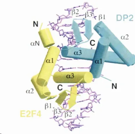
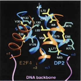
Figure 17B Crystal Structure of the E2F-4/DP-2 Heterodimer Bound to DNA 128 Figure 17C Schematic Showing Interactions Between E2F-4/DP-2 and DNA 128 Figure 17D Ribbons Representation of the Interface Between E2F-4 and DP-2 132 Figure 18 Principle of the Electrophoretic Mobility Shift Assay 140 Figure 19 Surface Representation of Recruitment Peptide-Binding
Site of Cyclin A 144
Figure 22 Molecular Mass Determination of Purified E2F-lcyc by ESMS 152 Figure 23 Purification of DP-lsgg: SDS-PAGE Analysis 154 Figure 24 Molecular Mass Determination of Purified DP-lggg by ESMS 156 Figure 25 Molecular Mass Determination of Purified DP-1 Aggg by
ESMS 157
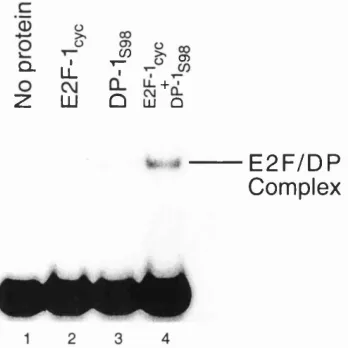
Figure 26 Binding of Recombinant E2F-1 and DP-1 to DNA 159
Figure 27 E2F-1/DP-1 Complex Formation 159
Figure 28 Interaction of Cyclin A-CDK2 with E2F/DP 161 Figure 29 Abolition of E2F/DP DNA-binding Activity by
Cyclin A-CDK2 161
Figure 30 Effect of Serine 98 Mutation on Abolition of E2F/DP
DNA-binding Activity 163
Figure 31 Subcloning Strategy for DP-lggg 165
Figure 32 Basic Components of the VP-ITC Microcalorimeter 171 Figure 33 E2F Protein Fragments Used in ITC Experiments 184 Figure 34 Fragments of pRb Used in ITC Experiments 185 Figure 35 HPV 16 E7 Protein Fragment Used in ITC Experiments 186 Figure 36 Purification of pRbAs: SDS-PAGE Analysis 189 Figure 37 Molecular Mass Determination of Purified pRbAB by
ESMS 190
Figure 38 ITC Titration Showing Interaction of pRbAB with E2F-1(409-426) 2 0 0
Figure 39 ITC Titration Showing Interaction of pRbAB with E2F-l(3gQ.437) 201
Figure 40 ITC Titration Showing Interaction of pRFab with E2F-1(243.437) 202
Figure 41 ITC Titration Showing Interaction of pRbAB with E2F-5(323-34Q) 203 Figure 42 ITC Titration Showing Interaction of pRbAB with E7(i7_9g) 204
Figure 43 ITC Titration Showing Interaction of pRbAB/E7 Complex with
E2F-1(409-426) 205
Figure 44 ITC Titration Showing Interaction of pRbAB/E7 Complex with
E2F-1 (3 8 0 -437) 206
Figure 45 ITC Titration Showing Interaction of pRbAB/E7 Complex with
E2F-1(243-437) 207
Figure 46 E2F Residues Critical for pRb Binding 213
Figure 47 Crystal Structure of pRb Pocket Domain Bound to E2F-1
and E7 Peptides 214
Figure 48 Schematic Showing Interactions Between E2F-1 Peptide
LIST OF TABLES
Table 1 Major Human Cycling and Cyclin-Dependent Kinases 23
Table 2 E2F-regulated Genes 54
Table 3 Compositions of the Media Used for Bacterial Cell Growth 73 Table 4 Dissociation Constants and Stoichiometries Determined by ITC
for Interaction of pRb with E2F-1 193
Table 5 Dissociation Constants and Stoichiometries Determined by TTC
for Interaction of pRb with E2F-1 and E2F-5 194 Table 6 Dissociation Constants and Stoichiometries Determined by ITC
for Interaction of pRb with E7 195
Table 7 Dissociation Constants and Stoichiometries Determined by TTC
for Interaction of pRb/E7 Complex with E2F-1 196 Table 8 Dissociation Constants for Interaction of pRb and pRb/E7
Complex with E2F-1 197
Table 9 Thermodynamic Parameters for the Interaction of pRb, pRb/E7 and E7 198 with E2F-1
ABBREVIATIONS
pME p-mercaptoethanol
Ad Adenovirus
ATP Adenosine 5’-triphosphate AU Absorbance units
CDK Cyclin-dependent kinase
CDKI Cyclin-dependent kinase inhibitor conOLIGO Control oligonculeotide
CR( 1-3) Conserved region (1-3)
Da Daltons
degOLIGO Degenerate oligonucleotide DHFR Dihydrofolate Reductase
dNTP 2’-deoxynucleotide 5’-triphosphate DP Differentiation Protein
dsDNA Double-stranded DNA DTT Dithiothreitol
El A Early 1 A Protein E2A Early 2 A gene
E2F Early 2 Factor (Also a generic term for E2F/DP Heterodimeric Transcription Factors
E4 Early 4 protein E7 Early 7 protein
EDTA Ethylenediaminetetra acetic acid EMSA Electrophoretic mobility shift assay ESMS Electrospray mass spectrometry
G Gap phase
GST Glutathione-S-transferase HPV Human Papillomavirus
IPTG Isopropyl-P-D-thiogalactopyranoside ITC Isothermal Titration Calorimetry Kd Dissociation constant
kDa Kilo daltons
M Mitotic Phase
N Stoichiometry
PIC Pre-initiation complex
pol RNA polymerase
PP Pocket protein
pRb Retinoblastoma tumour suppressor gene product recOLIGO Recovered oligonucleotide
S Synthesis Phase
SDS Sodium dodecyl sulphate
SELEX Systematic Evolution of Ligands by Exponential Enrichment selOLIGO Selected oligonucleotide
SV40 Simian virus 40 TAE Tris acetate EDTA TAg SV40 Large T Antigen TBP TATA-binding protein TFII Transcription Factor II
Tris Tris(hydroxymethyl)methy lamine
UV Ultra-violet
CHAPTER 1
CHAPTER 1 INTRODUCTION
The cell cycle is a process comprising distinct and ordered cellular events, culminating in the duplication of a cell. This process is necessary for growth, development and homeostasis. The cell cycle is regulated by a plethora of complex, partially overlapping and degenerate mechanisms that seek to maintain strict control of cellular division. A devastating consequence of uncontrolled cell proliferation is cancer, responsible for one in six deaths in the developed world.
1.1.
TH E EUKARYOTIC CELL CYCLE
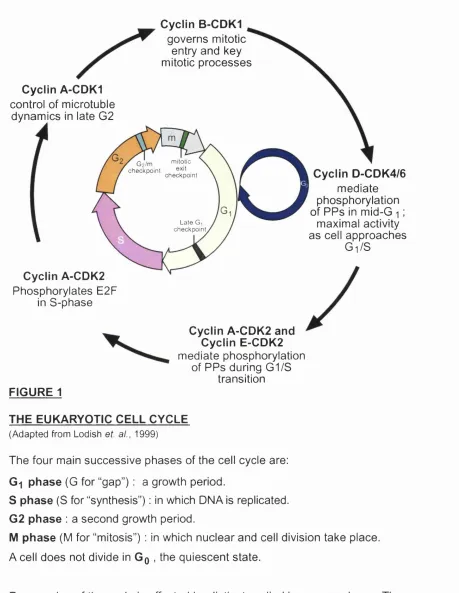
Figure 1 depicts the four main successive phases into which the cell cycle is generally divided. The Go phase (G for “gap”) is a quiescent state in which a cell does not divide. Certain terminally differentiated cells assume this state permanently and thus never replicate, e.g. neurons. Gq is sustained when there are low levels of nutrients or through contact inhibition, when the cell is in touch physically with other ones.
The cell’s entry into Gi can be induced by extracellular signals (such as growth factors and hormones). The Gi phase is the longest in the cycle and is a period of growth. The cycle’s duration in a multicellular organism ranges between eight hours and over one hundred days depending on the cell type, with most of this variation being attributable to the length of Gi.
The S phase (S for “synthesis”) is the period in which DNA is replicated such that the number of chromosomes in the cell (compared to that in Gq/Gi) is doubled. The S phase is followed by another period of growth, the G% phase (Alberts et al,
1994; Voet and Voet, 1995).
Cyclin B-CDK1
governs mitotic entry and key mitotic processes
Cyclin A-CDK1
control of microtuble dynamics in late G2
Cyclin A-CDK2
Phosphorylates E2F in S-phase
Gz/m niitotic
checkpoint ^ , checkpoint Late G checkpoint
Cyclin D-CDK4/6
mediate phosphorylation of PPs in mid-G i ;
maximal activity as cell approaches
Gi/S
Cyclin A-CDK2 and Cyclin E-CDK2
mediate phosphorylation of PPs during G1/S
transition
FIGURE 1
THE EUKARYOTIC CELL CYCLE
(Adapted from Lodish et. al., 1999)
The four main successive phases of the cell cycle are:
Gi phase (G for “gap”) : a growth period.
S phase (S for “synthesis”) : in which DNA is replicated.
G2 phase : a second growth period.
M phase (M for “mitosis”) : in which nuclear and cell division take place. A cell does not divide in Gq , the quiescent state.
Progression of the cycle is effected by distinct cyclin-kinase complexes. The cycle can be arrested at checkpoints if the cell is not in a satisfactory
physiological state (see text).
PPs : pocket proteins
chromosomes align on the mitotic spindle, a structure formed by the reorganisation of micro tubules. In anaphase, the chromosomes move to opposite poles of this spindle such that the original and duplicate DNA are segregated. Soon after the onset of mitosis in most higher eukaryotes, the nuclear envelope disintegrates into several vesicles and then reassembles around these separated chromosomes while they undergo decondensation in telophase. Two daughter cells are produced at the end of mitosis by the division of the cytoplasm, a process known as cytokinesis. However in yeast, the nucleus divides at the time of cytokinesis without disintegration of the nuclear envelope (Alberts et al, 1994; Lodish et al, 1999).
1.1.1
Checkpoints and Feedback Controls
G2/M checkpoint. Exit from mitosis represents another juncture that is subject to
L2
TRANSCRIPTIONAL REGULATION OF THE CELL CYCLE
The repression or activation of genes whose products are involved in cell cycle progression often depends upon the phosphorylation status of the proteins that regulate the transcription of these genes, that is, whether they are hypo- or hyperphosphorylated. Such transcriptional control proteins include the retinoblastoma (Rb) family of repressors and activators as well as transcription factors such as E2F. Phosphorylation and dephosphorylation can enhance or inhibit interactions between these proteins and can affect the localisation of some of them. The phosphorylation status of many proteins involved in the cell cycle is regulated by cyclin-CDK (cyclin-dependent kinase) complexes, CDKIs (cyclin- dependent kinase inhibitors) and phosphatases.
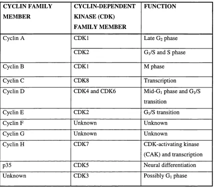
CYCLIN FAMILY M EM BER
CYCLIN-DEPENDENT KINASE (CDK)
FAMILY MEMBER
FUNCTION
Cyclin A CDKI Late G2 phase
CDK2 Gi/S and S phase
Cyclin B CDKI M phase
Cyclin C CDK8 Transcription
Cyclin D CDK4 and CDK6 Mid-Gi phase and Gi/S
transition
Cyclin E CDK2 Gi/S transition
Cyclin F Unknown Unknown
Cyclin G Unknown Unknown
Cyclin H CDK7 CDK-activating kinase
(CAK) and transcription
p35 CDK5 Neural differentiation
Unknown CDK3 Possibly Gi phase
TABLE 1
M A JO R HUMAN CYCLINS AND CYCLIN-DEPENDENT KINASES (Adapted from (Morgan, 1997))
The synthesis of D-type cyclins, as well as their association with CDK4 or 6, is reliant on growth factor stimulation that causes quiescent cells to enter Gi. These cyclin-kinase pairs are active in mid-Gi, with maximal activity occurring as the cell approaches the Gi/S transition. Cyclin D-kinase activity is positively and negatively regulated by cytokines and thus these particular cyclin-kinase pairs couple extracellular signals to the transcriptional control mechanism (Reynisdottir
et a l , 1995; Sherr, 1996).
The pRb-E2F association results in the inhibition of RNA polymerase II (pol Un directed mRNA synthesis, as discussed above. pRb is also able to repress pol I and III that mediate rRNA and tRNA production respectively. The mechanisms that have been proposed regarding pRb-induced transcriptional repression are described later (section 1.3.3). It is sufficient to point out at this stage that negative regulation of the cell cycle by pRb involves its suppression of the three RNA polymerases and consequent restraint of protein biosynthesis (Sanchez and Dynlacht, 1996; Fussenegger and Bailey, 1998). Furthermore, pRb plays a crucial role at the late Gi checkpoint, when the cell’s entry into S phase can be aborted in response to DNA damage that is detected by the p53 tumour suppressor. p53 induces the p21 CDK inhibitor whose abolition of cyclin-kinase activity prevents the phosphorylation of pRb family proteins, resulting in continued gene repression and arrest of the cell cycle in the Gi phase (Dictor et al, 1999).
CDKIs belong to either the INK4 or CIP families. Expression of specific family members is induced by contact inhibition, senescence, transforming growth factor (TGFp, an anti-mitogenic cytokine) as well as p53. CIP family members bind to and inhibit cyclin-kinase pairs and restrain a wider range of CDKs than INK4 proteins. Members of the INK4 family abolish the activity of CDKs active in G%. They are able to prevent cyclin-kinase complex formation by binding to solitary CDK and have also been shown to bind and inhibit intact cyclin-kinase pairs (Pavletich, 1999).
into S phase and CDK2 binds to cyclin A. The cyclin A-CDK2 complex is responsible for S-phase progression and its phosphorylation of pRb renders the tumour suppressor inactive from the S-phase onward. The same cyclin-kinase pair also phosphorylates E2F during the S-phase, causing its dissociation from promoters and consequently the abolition of transcription from genes under its control. Since the cyclin A gene is among these, cyclin A-CDK2 complex formation is promoted by E2F, culminating in the deactivation of the transcription factor by a negative feedback loop mechanism (Dynlacht et al, 1994; Krek et al,
1994; Xu et al, 1994; Fussenegger and Bailey, 1998).
Cyclin A binds to CDKl during the late G2 phase. This complex may contribute to
the control of microtubule dynamics as part of the cell’s preparation for mitosis. Another checkpoint occurs at the G2/M transition with entry into the M phase
being governed by the activity of cyclin B-CDKl. This cyclin-kinase pair regulates key mitotic processes including mitotic spindle formation, chromosome condensation and disintegration of the nuclear envelope. For example, cyclin B- CDKl-mediated phosphorylation of the lamin proteins, that comprise the nuclear lamina, causes them to depolymerise (Fussenegger and Bailey, 1998).
Premature mitotic entry is avoided by the inhibition of cyclin B-CDKl. This occurs through phosphorylation of a threonine and a tyrosine residue, both of which are conserved among the CDK family members (Thr 14 and Tyr 15 in CDKl) (Morgan, 1995; Fussenegger and Bailey, 1998). During S and G2,
shown to phosphorylate both Thr 14 and Tyrl5 (Fussenegger and Bailey, 1998). Dephosphorylation of these residues is brought about by the CDC25 phosphatase, resulting in activation of cyclin B-CDKl (Morgan, 1995). It is thought that this cyclin-kinase mediates inhibitory and stimulatory phosphorylation of W eel and CDC25 respectively, forming a positive feedback loop (Coleman and Dunphy, 1994; Morgan, 1995). The Chkl protein kinase and p53 tumour suppressor participate in the inhibition of CDC25 activity in response to DNA damage, resulting in cell cycle arrest at the G2/M checkpoint (Adams and Kaelin, 1998).
Cyclins A and B are degraded shortly before the onset of anaphase, with cyclin A degradation preceding that of cyclin B. It is thought that incomplete spindle assembly inhibits the ubiquitin-dependent destruction of cyclin B, thus preventing mitotic exit (Murray, 1992; Fussenegger and Bailey, 1998).
1.2.1
Cancer and the Transcriptional Regulatory Mechanism
Given their role in regulating the cell cycle, it is not surprising that the transcriptional regulators described in this section are subject to alterations that are associated with carcinogenesis. The gene encoding p53 is the most commonly mutated one in human cancer. The next most frequent genetic alterations relate to the p l6 gene, encoding a CDKl that targets CDK4. p l6 abnormalities are found in approximately one third of human cancers. One cell cycle regulator among pRb, cyclin D l, p l6 or CDK4 is aberrant in the majority of malignant tumours (Dictor
cycle control mechanism by virtue of oncoproteins that inactivate pRb (discussed later) and p53.
Inactivation of pRB following the overexpression of pRb-inactivating viral oncoproteins or disruption of the RB-1 (pRb gene) locus, is known to induce apoptosis. Cell death has also been shown to occur as a result of E2F-1 overexpression. Studies reveal the involvement of the p53 tumour suppressor in apoptotic induction and some details regarding the relevant mechanism have emerged. The fact that p53 plays a role in apoptosis may be the basis of the following observations. Both p53 and pRb, rather than either protein alone, are inactivated by tumour viruses. Inactivation of both proteins, by direct or indirect means, is also a frequent characteristic of other human tumours. It is interesting to note, however, that while E2F-1 overexpression can lead to p53-dependent apoptosis, E2F-1 can also induce cell death in the absence of p53 (Kaelin, 1999). The latter is partly mediated by the p53 homologue, p73 (Irwin et al, 2000; Lissy
LÀ
THE RETINOBLASTOMA TUMOUR SUPPRESSOR GENE PRODUCT (pRb)
The retinoblastoma tumour suppressor gene product (pRb) was so-named because alterations in its encoding gene were correlated with a cancer of the developing retina (retinoblastoma) (Benedict et al, 1983; Cavenee et al, 1983; Godbout et al, 1983; Sparkes et al, 1983). However, this name is perhaps misleading, since it does not convey the extent to which the protein is associated with other human cancers and cellular mechanisms in general. pRb is a component of the systems governing cell cycle control and differentiation. We have seen in section 1.2 how current knowledge pertaining to cell cycle regulation partly attributes pRb’s growth repressive function to its association with E2F. This section will describe pRb in more detail, including structural aspects of the protein related to its activities and proposed mechanisms for the transcriptional repression it induces.
1.3.1
Structural Aspects of pRb
The term “pocket proteins” is applied to members of the pRb family that comprises p i 07, p i 30 and pRb, since they possess a common structural feature known as the pocket domain (Hu et al, 1990; Huang et al, 1990; Kaelin et al,
1990; Ewen et al, 1992; Hannon et al, 1993; Huang et al, 1993; Mayol et al,
1993; Chow and Dean, 1996; Lee et al, 1998). This is divided into A and B
E2F binding Pocket Domain
c-abi binding MDM2 binding
LO
O CD CM CO LO CM CM CM
(0(01
-CD CO LO N CO CO
I f
L I
FIGURE 2
N 0 0 CM CO O T - LO CÛ CO
(0 (0 ( 0
V L
O 0 0 LO T - T - C O 0 0 0 0 CJ) O T-CM CM
D- N -00 00 00 00
( 0 ( 0 ( 0 ( 0 ( 0 ( 0 CO
I
X X
_ i
g
FUNCTIO NAL REGIONS OF THE R ETIN O B LA STO M A TU M O U R
SUPPRESSO R GENE PRODUCT pRb
(Adapted from Kaelin, 1999)
A and B: subdom ains of the pocket dom ain
S: spacer region
KXLKXL: cyclin-CDK docking site
(single letter amino acid code where x is any residue)
J : potential serine/threonine phosphoacceptor site
are connected by intramolecular associations along a common interface and, covalently, by a spacer region (Lee et al, 1998) (figure 2). The pocket region is the main focus of tumour-associated genetic changes in pRb (Hu et al, 1990; Huang et al, 1990; Kaelin et al, 1990). This region is sufficient and necessary for the induction of transcriptional repression when fused to a heterologous DNA- binding domain (Sellers et al, 1995; Weintraub et al, 1995). Restraint of cell growth depends on the pocket, although an additional region of pRb is also required in this context (Hiebert et al, 1992; Qin et al, 1992). The principal interactions, between pRb and most of the viral and cellular proteins to which it binds, involve the pocket region (Hu et al, 1990; Huang et al, 1990; Kaelin et al,
1990; Helin et al, 1992; Kaelin et al, 1992).
Among the viral proteins able to interact with pRb (discussed in section 1.5) is the human papillomavirus (HPV) E7 oncoprotein (Dyson et al, 1989). E7 is able to disrupt pRb-E2F complexes (Chellappan et al, 1992) and cooperate with an activated ras oncogene to transform primary rodent cells (Matlashewski et al,
members. It also appears from this structure that the A-B interface of the pocket is necessary for stable folding of the B subdomain (Lee et al, 1998). This is probably the reason why the A subdomain is required for the binding of proteins containing LXCXE sequences to pRb (Hu et al, 1990; Huang et al, 1990; Kaelin
et al, 1990). Some cellular proteins that interact with pRb also possess an
LXCXB-like motif, including HDACl (Brehm et al, 1998; Luo et al, 1998; Magnaghi-Jaulin e t a l , 1998).
All tumour-associated pRb mutants investigated so far are unable to bind to B2F (Sellers et al, 1998). B2B does not possess an LXCXB motif in its pRb-binding region (Helin et al, 1992) (figure 46). Furthermore, it has been demonstrated that certain mutant viral oncoproteins that still possess the LXCXB motif, are able to bind pRb without inhibiting the formation of a pRb-B2F complex (Fattaey et al,
indeed bind to the A-B interface. This structure will be discussed in more detail later.
The N-terminus of pRb as well as the pocket domain contain BRCT-related motifs (Yamane et al, 2000). BRCT regions bind double-strand breaks of DNA and are found in several proteins involved in DNA repair and cell cycle checkpoints (Yamane et al, 2000).
The region C-terminal to the pocket of pRb interacts with the c-Abl tyrosine kinase. This is an S phase-activated kinase that can enhance transcription and is inhibited by the tumour suppressor (Welch and Wang, 1993; Welch and Wang,
1995). The C-terminal region of pRb also associates with MDM2, a cellular oncoprotein that can negatively regulate p53 and pRb (Xiao et al, 1995).
1.3.2
Regulation of pRb
pRb is phosphorylated on amino acid residues within consensus target sites for cyclin-CDKs (Lees et al, 1991) (Lin et al, 1991) (figure 2). Upon phosphorylation by the latter, pRb is inactivated, resulting in cell cycle progression (Chen et al, 1989; Hinds et al, 1992; Lundberg and Weinberg,
Ewen et al, 1993; Dynlacht et al, 1994; Lundberg and Weinberg, 1998; Harbour
et al, 1999; Ezhevsky et al, 2001). When the cell enters S phase, cyclin A-CDK2
continues to phosphorylate pRb to disrupt the association of the tumour suppressor with E2F (Pines and Hunter, 1990; Ewen et al, 1993; Dynlacht et al,
1994).
Each of the cyclin-kinases phosphorylates distinct sites on the tumour suppressor (Kitagawa et al, 1996; Zarkowska and Mittnacht, 1997; Zarkowska et al, 1997). The phosphorylation of specific sites appears to regulate distinct activities of pRb. For example, the phosphorylation of threonine 821 and 826 in pRb inhibits its interaction with proteins containing the LXCXE motif (Knudsen and Wang, 1996). The binding of E2F to pRb can be inhibited by phosphorylation of serine 608 and 612 in the spacer region of the tumour suppressor. The phosphorylation of several sites in pRb’s C-terminus can also achieve the same effect (Knudsen and Wang, 1997).
Overexpression of gankyrin, an oncogenic protein, results in increased phosphorylation of pRb and activation of E2F-1 (Higashitsuji et al, 2000). Gankyrin binds to pRb through an LXCXE motif (section 1.3.1) and accelerates the degradation of the tumour suppressor in vitro and in vivo (Higashitsuji et al,
Internal cleavage of pRb abolishes its ability to bind E2F-1 (Fattman et al, 1997). It has been demonstrated that such cleavage can be brought about during apoptosis by caspase-3 and -7 (Fattman et al, 2001; Katsuda et al, 2002). Taken together, these results suggest the following model for the regulation of pRb during apoptosis. pRb is functionally inactivated during apoptosis by caspases that cleave the tumour suppressor internally, leading to the release of E2F. The latter is then able to participate in the apoptotic process (section 1.4.3.4)
1.3.3
Mechanisms of pRb-induced Transcriptional Repression
pRb is able to negatively regulate transcription by repressing all three RNA polymerases as outlined below.
1.3.3.1
Repression of RNA polymerase Il-directed transcription
The binding of pocket proteins (PPs) to E2F results in the repression of E2F- responsive genes (Hiebert et al, 1992; Weintraub et al, 1992; Hagemeier et al,
1993; Helin et al, 1993a; Chow and Dean, 1996; Chow et al, 1996; Ferreira et al, 1998).
masking its transactivation domain, thereby interfering with E2F’s interaction with components of the basal transcriptional machinery (Pearson and Greenblatt, 1997; Ross et al, 1999) (figures 3 and 4). Such repression of E2F is termed direct inhibition. In vitro transcription assays show that E2F-induced transcription is repressed by pRb if the tumour suppressor is present before PIC assembly (Ross et al, 1999; Ross et al, 2001). pRb’s ability to repress such transcription however, is greatly diminished if the tumour suppressor is present after formation of a partial (TFIIA-TFIID) or complete PIC (Ross et al, 1999; Ross et al, 2001).
The tethering of pRb to a promoter (either through association of the tumour suppressor with E2F or through fusion of pRb to a heterologous DNA binding domain) results in the repression of transcriptional activators that have proximal DNA binding sites (Weintraub et al, 1992; Adnane et al, 1995; B remuer et al,
1995; Sellers et al, 1995; Weintraub et al, 1995; Zhang et al, 1999) (figure 4). The term active repression refers to this particular ability of the tumour suppressor. However, the mechanism by which this is achieved is distinct from that of pRb-mediated direct inhibition of E2F. Ross and colleagues have shown that active repression by pRb occurs whether the tumour suppressor is present before or after PIC assembly. This suggests that in contrast to direct E2F inhibition, active repression by pRb may involve perturbation of a process that takes place after PIC assembly, such as recruitment of cofactors (Ross et al,
The Pre-initiation Complex (Adapted from Voet and Voet, 1995)
HD IIP
IIB
p o l II IIH iTATA
In order to find the correct transcriptional start site and initiate transcription, RNA polymerase II (pol II) requires General Transcription Factors (G IF s), of which many are multiprotein complexes. These are known as TFIIA, TFIIB etc. (TF; Transcription Factor). Pol II and the GTFs sequentially associate to form a pre-initiation complex (PIC), also referred to as the basal transcriptional machinery, that allows transcription to take place at a “basal” rate
(Lodish e t a\., 1999) (Voet and Voet, 1995). In the illustration above, the gene’s promoter has a TATA-box, a conserved DNA sequence commonly found in the core promoters of genes transcribed by pol II (Lemon and Tjian, 2000) (Lodish etal., 1999). The TATA-box binding protein (TBP) binds to this sequence and interacts with TBP-associated factors (TAFs) to form TFIID (Voet and Voet, 1995). TFIIH incorporates enzymatic activities that are involved in steps of transcription occurring soon after the initiation of mRNA synthesis. The kinase activity of this GTF may serve to regulate elongation of the mRNA chain (Blau e t al., 1996).
B Activators (Adapted from Lodish e t a l . , 1999) Proximal enhancer/ Distal promoter-proximal
enhancer region Gene
= / A— I— 1
I-I
Several Kbs of DNA
i
I
TATA-box
Distal ^ enhancer
Proximal enhancer/ — Sequence-specific
promoter-proximal transcription factor (activator)
region ^
Activation domain
Transcription is stimulated by transcription factors known as activators (Lodish e t a\., 1999). A simplified model for the role of these proteins is illustrated above. Activators bind
specific nucleic acid sequences in enhancer regions of DNA (Lemon and Tjian, 2000). It is thought that such transcription factors stimulate the assembly of the PIC and control the frequency of transcriptional re-initiation by new pol II molecules. Their activation domains (ADs) associate with components of the basal machinery. These interactions are partly responsible for looping of the DNA and formation of a stable functional PIC (Lodish e t al., 1999). Several co-regulators (e.g. CBP, HDACl - see text) are required to mediate the effects of sequence-specific transcription factors on the basal machinery (Lemon and Tjian, 2000). Co-regulators and other elements of pol 11-directed transcription are described
in detail elsewhere (Lodish e t a i , 1999) (Lemon and Tjian, 2000) (Voet and Voet, 1995).
FIGURE 3: ELEMENTS OF EUKARYOTIC TRANSCRIPTIONAL INITIATION FOR GENES TRANSCRIBED BY RNA POLYMERASE II
PIC
components
pRb masks the transactivation domain of E2F. This interferes with E2F’s interaction with components of the PIC (pre-initiation complex.)
pRb represses proximally-binding activator proteins (APs), once tethered to a promoter. pRb recruits co-repressors (CRs) such as HDACs. It can also block the activator-PIC interaction.
TAF250 pRb binds and inhibits TAF(1I)250
kinase, a component of the PIC.
pRb partially reverses E2F-induced changes in DNA topography,
disrupting interaction between PIC and transcriptional activators.
FIGURE 4
PROPOSED MECHANISMS FOR REPRESSION OF RNA POLYMERASE ll-DIRECTED TRANSCRIPTION BY pRb
(Adapted from Kaelin, 1999)
In the context of the mechanism of pRb-mediated active repression, chromatin structure is thought to be of particular importance. pRb is able to recruit different types of chromatin remodeling factors, that is, factors that alter chromatin structure to facilitate or prevent access of transcription factors to nucleosomal DNA (Workman and Kingston, 1998). Among the chromatin remodeling factors with which pRb interacts are histone deacetylase 1 (HDACl) (Brehm et al, 1998;
Luo et al, 1998; Magnaghi-Jaulin et al, 1998) and HDACs 2 and 3 (Dahiya et al,
However, pRb-mediated repression of a proximal SV40 enhancer occurs independently of HD AC (Luo et al, 1998). Furthermore, the study conducted by Ross and colleagues referred to earlier, showed that although pRb-mediated active repression was chromatin-dependent, it was not HD AC-dependent in their in vitro assay (Ross et al, 2001). Thus in such settings, HD AC-independent mechanisms of pRb-mediated active repression must be invoked. In this context, pRb’s ability to recruit other co-repressors may be the means by which the tumour suppressor achieves its effect. For example, pRb is able to recruit the CtBP co-repressor via CtIP (Meloni et al, 1999). CtBP possesses both HD AC-dependent (Criqui-Filipe
et al, 1999; Postigo and Dean, 1999) and -independent (Meloni et al, 1999;
Koipally and Georgopoulos, 2000; Phippen et al, 2000) repressive activities. The co-repressor interacts with the hPc2 polycomb group protein (Sewalt et al, 1999) and recruits the latter to pRb (Dahiya et al, 2001). It has been postulated that polycomb proteins repress gene expression by co-ordinating the formation of densely-packaged, inactive chromatin that is inaccessible to transcription factors (heterochromatin) (Pirrotta, 1997). Thus formation of the pRb-CtBP-hPc2 complex may, at least in part, be responsible for pRb-mediated active repression by HD AC-independent means. However, much further work is required to elucidate the mechanisms of CtBP-mediated repression (Chinnadurai, 2002). HD AC-dependent and -independent repressive activities are also exhibited by another pRb-binding co-repressor, RBPl (Lai et al, 1999; Lai et al, 2001).
that interacts with pRb’s pocket domain in vivo and cooperates with the tumour suppressor to repress the cyclin E promoter (Nielsen et ai, 2001). pRb also associates with BRGl and BRM, two components of human SWI/SNF (hSWI/SNF) that belongs to a family of ATP-dependent nucleosome remodeling complexes (Dunaief et ai, 1994; Strober et ai, 1996; Tronche et ai, 1997). Expression of BRGl and BRM in cells that are deficient for both of these proteins results in pRb-dependent growth arrest (Dunaief et ai, 1994; Strober et al, 1996). pRb-mediated repression of the cyclin A promoter is compromised in cell lines lacking BRGl, while ectopic expression of BRGl in these cells restores the tumour suppressor’s ability to effect such repression (Strobeck et ai, 2000). BRM has been shown to cooperate with pRb, both to repress cyclin A (Reisman et ai,
2002), and to repress E2F-1 activity in transient transfection assays (Trouche et ai, 1997). A study conducted by Zhang and colleagues (Zhang et ai, 2000) suggests that pRb-HDAC-hSWI/SNF and pRb-hSWI/SNF complexes contribute to the temporal regulation (section 1.2) of cyclins A and E. pRb also binds DNM Tl, a DNA methyltransferase that cooperates with the tumour suppressor to repress transcription in an HD AC-dependent and -independent manner (Robertson et ai, 2000).
requires the recruitment of pRb to the promoter by E2F, or by means of a DNA binding domain fused to the tumour suppressor (Weintraub et al, 1995; Luo et al,
1998). It appears that the affinity of the proximal activators for pRb is not high enough to permit them to recruit the tumour suppressor (Weintraub et al, 1995;
Luo et al, 1998). Thus it seems that pRb has to be concentrated at the promoter
by a relatively high affinity interaction with E2F, before the comparatively low affinity interaction between pRb and the proximal activator can be established (Weintraub eta l, 1995; Luo eta l, 1998).
activators, for which pRb has some affinity, are repressed by pRb-binding after E2F-mediated recruitment of the tumour suppressor.
Other mechanisms by which pRb may repress transcription include a partial reversal in E2F-induced DNA bending (Huber et ah, 1994), possibly perturbing interactions between transcriptional activators and the PIC. Furthermore, the tumour suppressor has been shown to bind and inhibit TAF(II)250 kinase (Siegert and Robbins, 1999), a component of the basal transcriptional machinery (figure 4). Defective TAF(II)250 appears to cause arrest of the cell cycle in GI (Hayashida gr aZ., 1994).
1.3.3.2
pRb-induced repression of RNA polymerases I and III
pRb represses pol I-directed transcription by interacting with the architectural upstream binding factor (UBF) (Cavanaugh et al, 1995; Voit et al, 1997). The latter enhances pol I-directed transcription (Smith et al, 1990; Hannan et al,
1996; Hannan et al, 1999). UBF is acetylated by the CBP acetyltransferase, promoting pol I-directed transcription (Pelletier et al, 2000). pRb is able to repress pol I by binding to UBF and preventing its association with CBP (Pelletier
et al, 2000). The tumour suppressor also recruits HDACl (as discussed
1995; Moss and Stefanovsky, 1995; Hempel et al, 1996). This interaction is also blocked by the formation of a pRb-UBF complex (Hannan et al, 2000). Endogenous pRb and p i 30 appear to exhibit redundancy in their negative regulation of pol I, whereas endogenous p i 07 does not suppress rRNA synthesis (Ciarmatori et al, 2001).
It has been demonstrated that pRb represses pol III activity in vivo (White et al,
lA
THE E2F FAMILY OF TRANSCRIPTION FACTORS
The discovery that E2F associates with pRb (Bandara and La Thangue, 1991; Chellappan et ai, 1991; Chittenden et al, 1991) initiated extensive study of this family of transcription factors that are key regulators of cell proliferation. This has given rise to a wealth of information owing to which an intriguing picture of E2F as both an activator and repressor of transcription has begun to emerge. The apparently dual nature of E2F is discussed here along with the structure, mode of action and regulation of the family members.
1.4.1
Structural Aspects
The E2F family of transcription factors comprises heterodimeric proteins. Each heterodimer consists of one member of the E2F family of proteins and one from the DP family (Huber et al, 1993; Wu et al, 1995; Cartwright et al, 1998). Thus the term “E2F” is used generically to signify such a heterodimer. Members of the E2F family of proteins are able to homodimerise and bind DNA, as are DP proteins, but the DNA-homodimer interaction is much weaker than that between a heterodimer and DNA (Bandara et al, 1993; Girling et al, 1993; Huber et al,
1993; Krek et al, 1993). E2F-1 and DP I bave been shown to cooperatively transactivate an E2F-responsive promoter (Helin et al, 1993b).
al, 1993; Lees et al, 1993), E2F-3 (Lees et al, 1993), E2F-4 (Beijersbergen et al, 1994; Ginsberg et al, 1994; Sardet et al, 1995), E2F-5 (Hijmans et al, 1995; Sardet et al, 1995) and E2F-6 (Morkel et al, 1997; Cartwright et al, 1998; Trimarchi et al, 1998). Two mammalian DP family members have been identified, namely DP-1 (Girling et al, 1993) and DP-2 (Ormondroyd et al, 1995;
Wu et al, 1995; Zhang and Chellappan, 1995; Rogers et al, 1996). E2F proteins
have a number of functional regions that are highly conserved (figure 5). They all possess N-terminal DNA-binding (Kaelin et al, 1992; Cress et al, 1993; Ivey- Hoyle et al, 1993; O ’Connor and Hearing, 1994) and dimérisation regions (Helin
et al, 1993) that precede a “marked box” (Lees et al, 1993). This latter region is
bound by the adenovirus E4 (ORF6/7) protein as part of the viral replication process (O'Connor and Hearing, 1994). It may also be involved in dimérisation and DNA bending (Cress and Nevins, 1996; Vidal et al, 1996). All E2F proteins, with the exception of E2F-6 have a C-terminal transactivation region in which a section for binding pocket proteins is embedded (Helin et al, 1992; Kaelin et al,
E2F -1
E2F -2
E2F -3
E2F -4
E2F -5
E2F -6
DP-1
DP-2
FIGURE 5
Nuclear Localisation Signal and Cyclin A binding
DNA binding
Dimerization
Pocket protein binding Marked Box Transactivation
DNA
binding Dimerization
Alternate Splicing
FUNCTIONAL REGIONS OF THE M AM M ALIAN E2F AND DP
PROTEINS.
(Adapted from Black et al., 1999)
The DP family members (figure 5) are distantly related to the E2F proteins, with the greatest homology occurring in the DNA binding and dimérisation regions (Girling et al, 1993). Furthermore, DP-2 variants are produced through alternative splicing of the encoding gene, some of which have a nuclear localisation signal (Ormondroyd et al, 1995; Zhang and Chellappan, 1995; de la Luna et al, 1996; Rogers et al, 1996). The different characteristics of the DP and E2F proteins, together with the ability of any E2F to dimerise with any DP (Wu et al, 1995; Cartwright et al, 1998), gives rise to heterodimers that are distinct in terms of how they are regulated and the functions they perform.
1.4.2
The Regulation of E2F
A number of inter-related mechanisms have been identified to date that contribute to the control of E2F activity. Their elucidation has helped to construct a model of E2F function that will be discussed later.
1.4.2.1
Synthesis of E2F Transcription Factors
Transcriptional and protein levels of E2Fs 4 and 5 are, on the whole, constant throughout the cell cycle while transcription and translation of E2Fs 1 -3 occur in mid-late Gi (Kaelin et al, 1992; Slansky et al, 1993; Ginsberg e ta l , 1994; Sardet
et al, 1995; Moberg et al, 1996; Sears et al, 1997; Leone et al, 1998). Activated
induce an increase in the levels of E2F-1 protein and mRNA (Berkovich and Ginsberg, 2001). In contrast to E2Fs 1-3, E2F-3B (section 1.4.1) is detectable throughout the cell cycle and its levels peak in the Go phase, during which it is bound to pRb (He et al, 2000; Leone et al, 2000; He and Cress, 2002). It is therefore thought that the primary function of E2F-3B may be to mediate transcriptional repression (in conjunction with pRb). Expression of DPs 1 and 2 does not fluctuate to a great extent during the replicative cycle (Bandara et al,
1994).
1.4.2.2
Pocket Proteins and C/EBPa
The binding of pocket proteins (PPs) to E2F results in the repression of E2F- responsive genes (Hiebert et al, 1992; Weintraub et al, 1992; Helin et al, 1993a; Chow and Dean, 1996; Chow et al, 1996; Ferreira et al, 1998). The ability of pRb to both inhibit E2F-induced transcription, as well as to actively repress proximally-binding activators, is discussed fully in section 1.3.3.1 E2Fs 1-3 bind preferentially to pRb, E2F-4 interacts with greatest affinity with p i 07 and p i 30 (as well as pRb under certain conditions) and E2F-5 binds only to p i 30 (Lees et al, 1993; Beijersbergen et al, 1994; Ginsberg et al, 1994; Hijmans et al, 1995; Vairo et al, 1995; Wu gr al, 1995; Moberg et al, 1996). Different complexes predominate in different phases of the cell cycle. Quiescent and differentiated cells principally contain pl30-E2F complexes, pRb-E2F is most evident as cells progress from G1 to S and pl07-E2F is most prevalent in S phase (Mudryj et al,
1993; Cobrinik et al, 1993; Ikeda et al, 1996; Moberg et al, 1996). Cyclin-CDK- mediated phosphorylation of the PPs, that begins in mid-Gl, results in disruption of the PP-E2F interaction (Beijersbergen et al, 1995; Smith et al, 1996; Xiao et al, 1996; Alevizopoulos et al, 1997) (also see section 1.3.2).
The C/EBPa transcription factor is required for the differentiation o f white adipose tissue (Wang et al, 1995) and neutrophil granulocytes (Zhang et al,
1997). It has been found in complexes (from nuclear extracts) that contain E2F (Slomiany et al, 2000; Porse et al, 2001). C/EBPa inhibits the induction of an E2F/DP-responsive promoter in transient transfection assays (Slomiany et al,
2000) and negatively regulates c-Myc in a manner that depends on an E2F- binding site in the c-myc promoter (Johansen et al, 2001). Repression of E2F by C/EBPa can take place independently of pRb (Slomiany et al, 2000; Johansen et al, 2001; Porse et al, 2001). The ability of C/EBPa to repress E2F is crucial for C/EBPa-induced terminal differentiation (Porse et al, 2001). The most commonly observed C/EBPa mutation in victims of acute myeloid leukaemia, results in the overexpression of a form of C/EBPa which does not repress E2F (Pabst et al,
2001; Porse et al, 2001).
1.4.2.3
Phosphorylation of E2F
1994; Krek et al, 1994; Xu et al, 1994; Kitagawa et al, 1995). E2F-1 DNA binding is downregulated in S phase by cyclin A-kinase-mediated phosphorylation (Krek et al, 1994) (as discussed previously in section 1.2). The physiological importance of this was demonstrated by the expression of mutant E2F-1, defective in cyclin A binding, that caused cells to arrest in S phase and induced apoptosis (Krek et al, 1995). Consistent with the absence of a cyclin A binding region in E2Fs 4 and 5 (section 1.4.1), cyclin A-CDK2 does not promote a loss of DNA binding by heterodimers comprising these E2Fs (Dynlacht et al, 1997).
1.4.2.4
Degradation and Stabilisation of E2F
Overexpressed E2F-1 and 4 are subject to degradation by the ubiquitin- proteasome pathway for which they are targeted by sequences in their C-terminus (Hateboer et al, 1996; Hofmann et al, 1996). It is thought that this may provide an additional way to downregulate E2F activity. The binding of pocket proteins to E2Fs 1 and 4 stabilises E2F since it increases its half-life from approximately two hours to over eight hours, as shown by pulse-chase experiments (Hateboer et al,
1.4.2.5
Subcellular Localisation of E2F
In Go and early Gi, E2Fs-4 and 5 are present in the nucleus and cytoplasm but during other periods of the cell cycle, they are predominantly located in the cytoplasm (Allen et al, 1997; Lindeman et al, 1997; Muller et al, 1997; Verona
et al, 1997). Consistent with the finding that E2Fs-l to 3 possess a nuclear
localisation signal (section 1.4.1), these proteins are predominantly nuclear (Magae et al, 1996; Muller et al, 1997; Verona et al, 1997). The mechanisms governing the localisation of E2Fs-4 and 5 still require elucidation and may involve DP-2 splice variants possessing a nuclear localisation signal (de la Luna et al, 1996), as well as the formation of E2F-PP complexes (Allen et al, 1997; Lindeman et al, 1997).
1.4.2.6
Acétylation of E2F
be recruited by p300 and CBP (Yang et al, 1996), which also display intrinsic histone acetylase activity themselves (Bannister and Kouzarides, 1996; Ogryzko
et al, 1996). However, in addition to acetylating E2F, p300 CBP and PCAF also
acetylate other transcription factors, demonstrating that their substrate specificity is not limited to histones (Gu and Boeder, 1997; Imhof et al, 1997; Boyes et al,
1998; Munshi et al, 1998; Zhang and Bieker, 1998). HDACl, (the histone deacetyl ase mentioned in section 1.3.3.1), can reverse the acétylation of E2F-1 (Martinez-Balbas et al, 2000; Marzio et al, 2000). This suggests that HDACl- mediated deacetylation of E2F contributes to the downregulation of the transcription factor, given the effect acétylation has upon E2F’s activity (this section) and its stability (section 1.4.2.4).
1.4.3
Functions of E2F
1.4.3.1
E2F’s Target Genes
Genes whose promoters contain E2F-binding sites can be broadly categorised as those involved in cell cycle control as well as DNA synthesis (table 2). Current data portray E2F as a regulator that activates or, when associated with a pocket protein, actively represses transcription of a target gene. This is discussed in the next section (1.4.3.2).
Table 2 : E2F-REGULATED GENES Adapted from Helin, 1998.
GENE/PRODUCT NUMBER
OF E2F SITES
EFFECT
OF E2F
SITES
REFERENCES
Growth Regulatory Genes Cyclin E pl07 B-Myb E2F-1 CDKl Cyclin A c-myc
One to six Repressing
Two One palindrome Two palindromes One One One Genes Essential for DNA Replication
ORCl One
palindrome Thymidylate Synthase One
Dihydrofolate Reductase Thymidine kinase DNA polymerase a
One palindrome One palindrome One Activating Repressing Repressing Repressing Repressing Not reported Repressing Not reported Activating Activating Not reported
(Ohtani et al, 1995; Botz et a l , 1996; Geng et a l ,
1996)
(Zhu et al, 1995)
(Lam and Watson, 1993; Zwicker et a l, 1996)
(Hsiao et a l , 1994; Johnson
et a l, 1994; Neuman et a l ,
1994)
(Tommasi and Pfeifer, 1995)
(Schulze et a l, 1995; Huet
et a l, 1996)
(Thalmeier et a l , 1989; Plet et a l , 1992)
(Ohtani et a l , 1996)
(Jenh et a l , 1985; Ayusawa
et a l, 1986; Jolliff et a l ,
1991)
(Wells et al, 1996; Fry et a l , 1997)
The list of genes in the above table is not exhaustive and is only intended to demonstrate the nature and range of promoters containing E2F sites and the effect of such sites. Further information in this context can be found elsewhere (Helin, 1998; Lavia and Jansen-Durr, 1999). Evidence is also beginning to emerge that E2F plays a role in the regulation of other genes whose products are involved in mitosis, apoptosis, differentiation and development (Ishida et al, 2001; Muller et ai, 2001), as well as DNA repair and checkpoints (Ren et al, 2002).
1.4.3.2
E2F as an Activator and Repressor of Transcription
Several lines of evidence demonstrate that E2F acts as a transcriptional activator. For example, E2F has been shown to activate transcription of reporter constructs harbouring its cognate binding sites (Helin et al, 1992; Shan et al, 1992). A close correlation between E2F’s ability to effect transcription and to promote cell cycle progression has been demonstrated (Johnson et al, 1993; Shan and Lee, 1994;
Qin et al, 1995). The transactivation regions of the E2F proteins are conserved
(section 1.4.1) and functional when fused to other DNA-binding regions (Kaelin
et al, 1992). Furthermore, the combined loss of E2Fs 1-3 in mouse embryonic
fibroblasts, severely impairs the induction of several E2F target genes (Wu et al,
2001).
et al, 1994). This suggests that the transcription factor participates in transcriptional repression when bound to the DNA. These results are consistent with those obtained from in vivo footprinting analysis. For example such analysis of the B-myb promoter showed that the E2F site is specifically occupied in early G l when the B-myb promoter is inactive (Zwicker et al, 1996). The two E2F proteins expressed in Drosophila, dE2F-l and dE2F-2, exhibit opposing functions. dE2F-l serves as an activator of transcription while dE2F-2 acts as a repressor (Frolov et al, 2001).
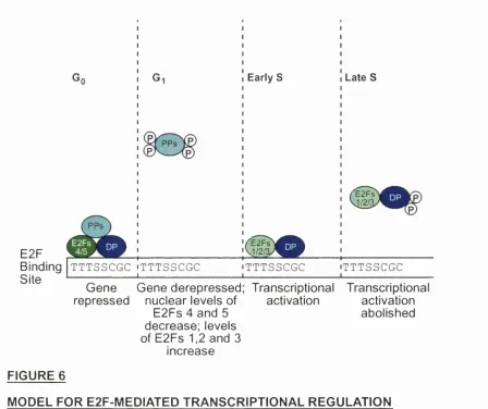
E2F
Binding | t t t s s c g c Site
TTTSSCGC
Early S
^E2Fs
J/2/%
TTTSSCGC
Late S
'^E2Fs
J/2/:
T TTSSCGC Gene Gene derepressed; Transcriptional Transcriptional repressed nuclear levels of
E2Fs 4 and 5 decrease; levels of E 2 F s 1 ,2 a n d 3
increase
activation activation
abolished
FIGURE 6
MODEL FOR E2F-MEDIATED TRANSCRIPTIONAL REGULATION
(Adapted from Helin, 1998)
TTTSSCGC : the E2F DNA-binding consensus site where 8 denotes C or G bases
PPs : pocket proteins
(P) : phosphorylation by cyclin-dependent kinases
1.4.3.3
Specificity of E2F Family Members
The existence of several E2F family members and the differences in their regulation (as discussed previously) implies that distinct heterodimers target specific genes. Accordingly, variations were observed with regard to gene induction when the individual E2F proteins were overexpressed in cells. For example, upon overexpression in REF52 cells, E2F-2 emerged as the most potent activator of the DHFR and thymidine kinase genes (as determined by mRNA levels) compared to the other E2F proteins (DeGregori et al, 1997). Furthermore, experiments involving formaldehyde cross-linking followed by immunoprécipitation have revealed that different E2F proteins interact preferentially with specific promoters (Takahashi et al, 2000; Wells et al, 2000;
Kel et al, 2001).
1.4.3.4
Oncogenic, Tumour Suppressive and Apoptotic Functions of E2F
Several studies suggest that E2F is oncogenic. For example, E2Fs-l to 3 have been shown to induce S-phase in serum-starved REF52 cells and in serum-starved or pl6-arrested Rati fibroblasts (Lukas et al, 1996; DeGregori et al, 1997). The combined loss of E2Fs 1-3 in mouse embryonic fibroblasts renders these cells unable to enter S phase, progress through mitosis and proliferate (Wu et al,