2-(2-Naphthyl)-1,3-dioxane
Damien Thevenet,aReinhard Neiera* and Helen Stoeckli-Evansb
a
Institute of Chemistry, University of Neuchaˆtel, rue Emile-Argand 11, 2009 Neuchaˆtel, Switzerland, andbInstitute of Physics, University of Neuchaˆtel, rue Emile-Argand 11, 2009 Neuchaˆtel, Switzerland
Correspondence e-mail: [email protected]
Received 30 December 2009; accepted 6 January 2010
Key indicators: single-crystal X-ray study;T= 173 K; mean(C–C) = 0.002 A˚;
Rfactor = 0.024;wRfactor = 0.061; data-to-parameter ratio = 7.6.
The title compound, C14H14O2, crystallizes in the chiral monoclinic space group P21. This acetal is composed of a planar naphthalene ring with a 1,3-dioxane ring substituent, which has a chair conformation. In the crystal structure, symmetry-related molecules are connected via a weak C— H O interaction to form a helical chain propagating in [010]. While there are no –stacking interactions present, there are weak C—H interactions involving the naphthalene aromatic rings, which link the helical chains to form a two-dimensional network in the (011) plane.
Related literature
For information on commonly used protecting groups for carbonyl compounds, see: Kocienski (1994); Showler & Darley (1967). For methods for their deprotection, see: Cordes & Bull (1974); Fujiokaet al.(2004); Ateset al.(2003). For kinetic and thermodynamic studies of acetals and ketals in the naphtha-lene series and other physical data, see: Newman & Dickson (1970); Carmichael & Hug (1986). For the synthesis of 2-naphthaldehyde acetal, see Gopinath et al. (2002). For details of the new photochemical reaction to hydrolyse the acetal into an aldehyde, see Thevenet & Neier (2010). For information on 1,3-dioxane ring related compounds, see: Buys & Eliel (1970). For the synthesis and crystal structure of a related compound, see: Borbas et al. (2002). For normal geometric parameters for molecular compounds, see: Allen (2002).
Experimental
Crystal data
C14H14O2
Mr= 214.25 Monoclinic, P21
a= 7.5351 (6) A˚
b= 7.8575 (8) A˚
c= 9.4057 (9) A˚
= 92.839 (11)
V= 556.20 (9) A˚3
Z= 2
MoKradiation
= 0.08 mm1
T= 173 K
0.380.300.08 mm
Data collection
Stoe IPDS diffractometer 4461 measured reflections 1098 independent reflections
951 reflections withI> 2(I)
Rint= 0.024
Refinement
R[F2> 2(F2)] = 0.024
wR(F2) = 0.061
S= 1.05 1098 reflections 145 parameters
1 restraint
H-atom parameters constrained max= 0.13 e A˚
3
min=0.11 e A˚
[image:1.610.313.565.356.414.2]3
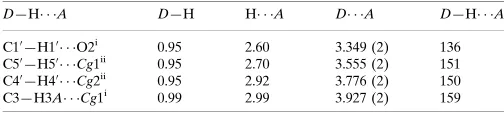
Table 1
Hydrogen-bond geometry (A˚ ,).
Cg1 andCg2 are the centroids of the C10 –C40
/C90 /C100
and C50 –C100
rings, respectively.
D—H A D—H H A D A D—H A
C10 —H10
O2i
0.95 2.60 3.349 (2) 136
C50—H50 Cg1ii
0.95 2.70 3.555 (2) 151
C40—H40 Cg2ii
0.95 2.92 3.776 (2) 150
C3—H3A Cg1i
0.99 2.99 3.927 (2) 159
Symmetry codes: (i)xþ2;y1
2;zþ1; (ii)xþ2;yþ 1 2;zþ2.
Data collection: EXPOSE in IPDS-I (Stoe & Cie, 2000); cell refinement: CELL in IPDS-I; data reduction: INTEGRATE in IPDS-I; program(s) used to solve structure:SHELXS97(Sheldrick, 2008); program(s) used to refine structure:SHELXL97(Sheldrick, 2008); molecular graphics: PLATON (Spek, 2009) and Mercury (Macraeet al., 2006); software used to prepare material for publica-tion:SHELXL97andPLATON.
HSE is grateful to the XRD Application Laboratory, Microsystems Technology Division, Swiss Center for Elec-tronics and Microtechnology, Neuchaˆtel, for access to the X-ray diffraction equipment.
Supplementary data and figures for this paper are available from the IUCr electronic archives (Reference: CV2685).
References
Allen, F. H. (2002).Acta Cryst.B58, 380–388.
Ates, A., Gautier, A., Leroy, B., Plancher, J. M., Quesnel, Y., Vanherck, J. C. & Marko, I. E. (2003).Tetrahedron,59, 8989–8999.
Borbas, A., Szoba, Z. B., Szilagyi, L., Benyei, A. & Liptak, A. (2002).
Tetrahedron,58, 5723–5732.
Buys, H. R. & Eliel, E. L. (1970).Tetrahedron Lett.32, 2779–2782. Carmichael, I. & Hug, G. L. (1986).J. Phys. Chem. Ref. Data,15, 1–250. Cordes, E. H. & Bull, H. G. (1974).Chem. Rev.74, 581–603.
Fujioka, H., Sawama, Y., Murata, N., Okitsu, T., Kubo, O., Matsuda, S. & Kita, Y. (2004).J. Am. Chem. Soc.126, 11800–11801.
Gopinath, R., Haque, S. J. & Patel, B. K. (2002).J. Org. Chem.67, 5842–5845.
organic compounds
Acta Cryst.(2010). E66, o473–o474 doi:10.1107/S1600536810000644 Thevenetet al.
o473
Acta Crystallographica Section EStructure Reports Online
New York: Thieme Medical Publishers.
Macrae, C. F., Edgington, P. R., McCabe, P., Pidcock, E., Shields, G. P., Taylor, R., Towler, M. & van de Streek, J. (2006).J. Appl. Cryst.39, 453–457. Newman, M. S. & Dickson, R. E. (1970).J. Am. Chem. Soc.92, 6880–6884.
Showler, A. J. & Darley, P. A. (1967).Chem. Rev.67, 427–440. Spek, A. L. (2009).Acta Cryst.D65, 148–155.
supporting information
sup-1
Acta Cryst. (2010). E66, o473–o474
supporting information
Acta Cryst. (2010). E66, o473–o474 [https://doi.org/10.1107/S1600536810000644]
2-(2-Naphthyl)-1,3-dioxane
Damien Thevenet, Reinhard Neier and Helen Stoeckli-Evans
S1. Comment
Acetals are the most commonly used protecting groups for carbonyl compounds in organic synthesis (Kocienski, 1994;
Showler & Darley, 1967), and many methods have been developed for their deprotection (Cordes & Bull, 1974; Fujioka
et al., 2004; Ates et al., 2003). The title 2-naphthaldehyde acetal (Newman & Dickson, 1970; Carmichael & Hug, 1986)
was synthesized to investigate the scope of a new photochemical reaction capable of hydrolysing the acetal into an
aldehyde (Thevenet & Neier, 2010). The NMR spectra of the unsubstituted 1,3-dioxane ring displays a complicated
AA′BB′MN system (Buys & Eliel, 1970), and the X-ray crystal structure was helpful for the interpretation of the NMR
spectra (Thevenet & Neier, 2010).
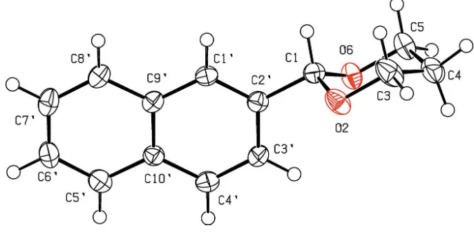
The structure of the title compound is illustrated in Fig. 1, and the geometrical parameters are given in the
Supplementary information and the archived CIF. The bond lengths and angles are close to those in three similar
compounds located in the Cambridge Crystal Structure Database (CSD, V 5.30, last update Sept. 2009; Allen, 2002). For
example, methyl 2,3-di-O-acteyl-4,6-O-(2-naphthyl)methylene-α-D-galactopyranoside (Borbas et al., 2002), which also
crystallized in the monoclinic space group P21, and where the naphthalene ring is planar and the two six-membered rings
in the galactopyranoside unit have chair conformations.
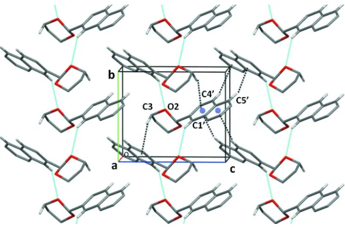
In the crystal of the title compound symmetry related molecules are connected via a C—H···O interaction (Table 1)
giving rise to the formation of helical chains propagating in [010]. These chains are further linked via weak C—H···π
interactions to form a two-dimensional network in (011) - see Fig. 2 and Table 1 for details.
S2. Experimental
The title compound was synthesized using a modified strategy described by (Gopinath et al., 2002). To a solution of
2-naphthaldehyde (0.64 mmol), trimethylorthoformate (1.41 mmol) and 1,3-propanediol (5.12 mmol) in dry nitromethane
(2 ml) was added tetrabutylammonium tribromide (0.025 mmol). The homogeneous reaction mixture was stirred at r.t.
and the progress of the reaction monitored by TLC and GC. After completion of the reaction the mixture was poured into
a solution of NaHCO3 (10 ml) and the products were extracted with diethyl ether (3 × 10 ml). The organic layer was
separated, dried over anhydrous Na2SO4 and concentrated. The white solid obtained was purified by recrystallization in
MeOH, giving colourless thin plate-like crystals of the title compound.
1H NMR 400 MHz (CDCl
3) δ 7.97 (br s, 1H, H1′), 7.85 (m, 3H, H4′,5′,8′), 7.60 (dd, 1H, 3J3′-4′ = 8.5 Hz, 3J3′-1′ = 1.7 Hz, H3′),
7.48 (m, 2H, H6′,7′), 5.68 (s, 1H, H1), 4.33 (dddd, 2H, 2J3e-3a;5e-5a = -11.7 Hz, 3J3e-4a;5e-4a = 5.0 Hz, 3J3e-4e;5e-4e = 1.5 Hz, 4J3e-5e =
3.0 Hz, H3e,5e), 4.06 (ddd, 2H, 2J3a-3e;5a-5e = -11.7 Hz, 3J3a-4a;5a-4a = 12.4 Hz, 3J3a-4e;5a-4e = 2.7 Hz, H3a,5a), 2.29( dtt, 1H, 2J4a-4e =
-13.5 Hz, 3J
4a-3a;4a-5a = 12.4 Hz, 3J4a-3e;4a-5e = 5.0 Hz, H4a), 1.50 (dtt, 1H, 2J4e-4a = -13.5 Hz, 3J4e-3a;4e-5a = 2.7 Hz, 3J4e-3e;4e-5e = 1.5
Hz, H4 e); 13C NMR 100 MHz (CDCl3) δ 136.1 (C2′), (133.6, 133.1) (C9′,10′), (128.4, 128.1, 127.7) (C4′,5′,8′), (126.2, 126.0)
(C6′,7′), 125.3 (C1′), 123.8 (C3′), 101.8 (C1), 67.5 (C3,5), 25.9 (C4); HRMS (ESI, +): [M + Na]+ = 237.09. Note: The same
S3. Refinement
In the final cycles of refinement, in the absence of significant anomalous scattering effects, 944 (93%) Friedel pairs were
merged and Δf" set to zero. The H-atoms could all be located in difference electron-density maps. In the final cycles of
refinement they were included in calculated positions and treated as riding atoms: C—H = 0.95–1.0 Å, with Uiso(H) =
1.2Ueq(parent C-atom). Using the one-circle Stoe Image Plate Diffraction System it is not always possible to measure
100% of the Ewald sphere, and here only 93.7% of the data were accessible out to 50° in 2θ. This has little effect on the
bond distances and angles when comparing their values with those of the related structure mentioned above (Borbas et
[image:4.610.132.475.223.396.2]al., 2002).
Figure 1
supporting information
sup-3
[image:5.610.137.475.68.292.2]Acta Cryst. (2010). E66, o473–o474
Figure 2
A view along the a axis of the crystal packing of the title compound. The C—H···O and C—H···π interactions are shown
as dotted cyan and black lines, respectively. [The blue balls represent the centroids of the two aromatic rings; H-atoms
not involved in the C—H···O and C—H···π interactions have been omitted for clarity; the C—H···π interactions are
shown for one molecule only; see Table 1 for details].
2-(2-Naphthyl)-1,3-dioxane
Crystal data
C14H14O2 Mr = 214.25 Monoclinic, P21 Hall symbol: P 2yb a = 7.5351 (6) Å b = 7.8575 (8) Å c = 9.4057 (9) Å β = 92.839 (11)° V = 556.20 (9) Å3 Z = 2
F(000) = 228 Dx = 1.279 Mg m−3
Mo Kα radiation, λ = 0.71073 Å Cell parameters from 4553 reflections θ = 2.1–26.0°
µ = 0.08 mm−1 T = 173 K Plate, colourless 0.38 × 0.30 × 0.08 mm
Data collection
Stoe IPDS diffractometer
Radiation source: fine-focus sealed tube Graphite monochromator
φ rotation scans
4461 measured reflections 1098 independent reflections
951 reflections with I > 2σ(I) Rint = 0.024
θmax = 26.0°, θmin = 2.2° h = −8→8
Refinement on F2 Least-squares matrix: full R[F2 > 2σ(F2)] = 0.024 wR(F2) = 0.061 S = 1.05 1098 reflections 145 parameters 1 restraint
Primary atom site location: structure-invariant direct methods
Secondary atom site location: difference Fourier map
Hydrogen site location: inferred from neighbouring sites
H-atom parameters constrained w = 1/[σ2(F
o2) + (0.0412P)2] where P = (Fo2 + 2Fc2)/3 (Δ/σ)max < 0.001
Δρmax = 0.13 e Å−3 Δρmin = −0.11 e Å−3
Special details
Geometry. Bond distances, angles etc. have been calculated using the rounded fractional coordinates. All su's are estimated from the variances of the (full) variance-covariance matrix. The cell e.s.d.'s are taken into account in the estimation of distances, angles and torsion angles
Refinement. In the final cycles of refinement, in the absence of significant anomalous scattering effects, 944 (93%) Friedel pairs were merged and Δf " set to zero. The H-atoms could all be located in difference electron-density maps. In the final cycles of refinement they were included in calculated positions and treated as riding atoms: C—H = 0.95 - 1.0 Å, with Uiso(H) = 1.2Ueq(parent C-atoms). Using the one-circle Stoe Image Plate Diffraction System it is not always possible to measure 100% of the Ewald sphere, and here only 93.7% of the data were accessible out to 50° in 2θ.
Fractional atomic coordinates and isotropic or equivalent isotropic displacement parameters (Å2)
x y z Uiso*/Ueq
O2 1.16506 (19) 0.57058 (16) 0.43163 (12) 0.0337 (4)
O6 1.33418 (18) 0.38156 (15) 0.57014 (12) 0.0295 (4)
C1 1.1616 (2) 0.4392 (2) 0.53276 (17) 0.0251 (5)
C1′ 0.9100 (2) 0.46504 (19) 0.69218 (16) 0.0239 (5)
C2′ 1.0796 (2) 0.5075 (2) 0.66364 (16) 0.0240 (5)
C3 1.2305 (3) 0.5057 (3) 0.30168 (18) 0.0417 (7)
C3′ 1.1796 (3) 0.6163 (2) 0.75664 (17) 0.0277 (6)
C4 1.4131 (3) 0.4321 (3) 0.32815 (19) 0.0400 (7)
C4′ 1.1073 (3) 0.6764 (2) 0.87667 (18) 0.0297 (6)
C5 1.4128 (3) 0.3067 (3) 0.44944 (18) 0.0348 (6)
C5′ 0.8555 (3) 0.6908 (2) 1.03543 (18) 0.0309 (6)
C6′ 0.6865 (3) 0.6467 (2) 1.06380 (18) 0.0316 (6)
C7′ 0.5852 (3) 0.5438 (2) 0.96939 (18) 0.0328 (6)
C8′ 0.6551 (2) 0.4853 (2) 0.84779 (18) 0.0284 (5)
C9′ 0.8310 (2) 0.52609 (19) 0.81562 (16) 0.0236 (5)
C10′ 0.9334 (2) 0.63231 (19) 0.91039 (17) 0.0239 (5)
H1 1.08840 0.34250 0.49320 0.0300*
H1′ 0.84350 0.39320 0.62810 0.0290*
H3′ 1.29720 0.64750 0.73540 0.0330*
H3A 1.14900 0.41670 0.26250 0.0500*
H3E 1.23480 0.59870 0.23090 0.0500*
H4′ 1.17550 0.74940 0.93860 0.0360*
H4A 1.45060 0.37410 0.24110 0.0480*
H4E 1.49890 0.52450 0.35160 0.0480*
supporting information
sup-5
Acta Cryst. (2010). E66, o473–o474
H5A 1.53630 0.27170 0.47590 0.0420*
H5E 1.34500 0.20400 0.41910 0.0420*
H6′ 0.63660 0.68620 1.14860 0.0380*
H7′ 0.46700 0.51460 0.99020 0.0390*
H8′ 0.58470 0.41640 0.78400 0.0340*
Atomic displacement parameters (Å2)
U11 U22 U33 U12 U13 U23
O2 0.0500 (9) 0.0289 (6) 0.0225 (6) 0.0088 (6) 0.0049 (5) 0.0012 (5)
O6 0.0283 (8) 0.0376 (7) 0.0228 (5) 0.0058 (6) 0.0027 (5) −0.0007 (5)
C1 0.0269 (11) 0.0239 (8) 0.0244 (8) −0.0013 (6) 0.0008 (7) 0.0000 (6)
C1′ 0.0245 (11) 0.0228 (8) 0.0240 (8) −0.0016 (6) −0.0023 (7) 0.0007 (6)
C2′ 0.0265 (11) 0.0229 (8) 0.0225 (8) −0.0003 (7) 0.0012 (7) 0.0021 (7)
C3 0.0679 (17) 0.0358 (9) 0.0220 (8) 0.0095 (10) 0.0084 (9) 0.0013 (8)
C3′ 0.0224 (11) 0.0303 (9) 0.0307 (9) −0.0050 (7) 0.0031 (7) −0.0032 (7)
C4 0.0533 (16) 0.0374 (10) 0.0306 (9) −0.0015 (9) 0.0158 (9) −0.0061 (8)
C4′ 0.0273 (12) 0.0299 (9) 0.0317 (9) −0.0050 (7) −0.0007 (7) −0.0058 (7)
C5 0.0371 (13) 0.0395 (10) 0.0283 (9) 0.0069 (8) 0.0077 (8) −0.0053 (8)
C5′ 0.0345 (14) 0.0286 (9) 0.0298 (9) 0.0016 (7) 0.0026 (8) −0.0023 (7)
C6′ 0.0322 (12) 0.0327 (9) 0.0309 (8) 0.0066 (8) 0.0105 (7) 0.0024 (7)
C7′ 0.0230 (12) 0.0382 (11) 0.0377 (9) 0.0031 (7) 0.0063 (8) 0.0075 (8)
C8′ 0.0222 (11) 0.0316 (9) 0.0313 (8) −0.0035 (8) 0.0004 (7) 0.0029 (7)
C9′ 0.0222 (11) 0.0226 (8) 0.0257 (8) 0.0007 (6) −0.0005 (7) 0.0051 (6)
C10′ 0.0242 (11) 0.0211 (7) 0.0264 (8) 0.0010 (7) 0.0013 (7) 0.0007 (6)
Geometric parameters (Å, º)
O2—C1 1.405 (2) C8′—C9′ 1.411 (2)
O2—C3 1.434 (2) C9′—C10′ 1.421 (2)
O6—C1 1.405 (2) C1—H1 1.0000
O6—C5 1.433 (2) C1′—H1′ 0.9500
C1—C2′ 1.504 (2) C3—H3A 0.9900
C1′—C2′ 1.360 (2) C3—H3E 0.9900
C1′—C9′ 1.415 (2) C3′—H3′ 0.9500
C2′—C3′ 1.414 (2) C4—H4A 0.9900
C3—C4 1.502 (3) C4—H4E 0.9900
C3′—C4′ 1.362 (3) C4′—H4′ 0.9500
C4—C5 1.508 (3) C5—H5A 0.9900
C4′—C10′ 1.407 (3) C5—H5E 0.9900
C5′—C6′ 1.359 (3) C5′—H5′ 0.9500
C5′—C10′ 1.417 (2) C6′—H6′ 0.9500
C6′—C7′ 1.399 (3) C7′—H7′ 0.9500
C7′—C8′ 1.363 (2) C8′—H8′ 0.9500
C1—O2—C3 109.55 (14) C9′—C1′—H1′ 119.00
C1—O6—C5 110.36 (13) O2—C3—H3A 110.00
O6—C1—C2′ 108.84 (13) C4—C3—H3E 110.00
C2′—C1′—C9′ 121.09 (14) H3A—C3—H3E 108.00
C1—C2′—C1′ 120.15 (14) C2′—C3′—H3′ 120.00
C1—C2′—C3′ 119.63 (14) C4′—C3′—H3′ 120.00
C1′—C2′—C3′ 120.22 (15) C3—C4—H4A 110.00
O2—C3—C4 110.27 (15) C3—C4—H4E 110.00
C2′—C3′—C4′ 119.98 (19) C5—C4—H4A 110.00
C3—C4—C5 109.96 (18) C5—C4—H4E 110.00
C3′—C4′—C10′ 121.12 (17) H4A—C4—H4E 108.00
O6—C5—C4 110.33 (18) C3′—C4′—H4′ 119.00
C6′—C5′—C10′ 120.74 (16) C10′—C4′—H4′ 119.00
C5′—C6′—C7′ 120.65 (17) O6—C5—H5A 110.00
C6′—C7′—C8′ 120.42 (19) O6—C5—H5E 110.00
C7′—C8′—C9′ 120.64 (16) C4—C5—H5A 110.00
C1′—C9′—C8′ 122.48 (14) C4—C5—H5E 110.00
C1′—C9′—C10′ 118.47 (14) H5A—C5—H5E 108.00
C8′—C9′—C10′ 119.06 (14) C6′—C5′—H5′ 120.00
C4′—C10′—C5′ 122.42 (15) C10′—C5′—H5′ 120.00
C4′—C10′—C9′ 119.09 (14) C5′—C6′—H6′ 120.00
C5′—C10′—C9′ 118.49 (15) C7′—C6′—H6′ 120.00
O2—C1—H1 110.00 C6′—C7′—H7′ 120.00
O6—C1—H1 110.00 C8′—C7′—H7′ 120.00
C2′—C1—H1 110.00 C7′—C8′—H8′ 120.00
C2′—C1′—H1′ 119.00 C9′—C8′—H8′ 120.00
C3—O2—C1—O6 64.83 (17) O2—C3—C4—C5 51.9 (2)
C3—O2—C1—C2′ −175.74 (14) C2′—C3′—C4′—C10′ −0.1 (3)
C1—O2—C3—C4 −58.4 (2) C3—C4—C5—O6 −50.8 (2)
C5—O6—C1—O2 −64.14 (17) C3′—C4′—C10′—C5′ 179.07 (16)
C5—O6—C1—C2′ 176.74 (14) C3′—C4′—C10′—C9′ −1.3 (2)
C1—O6—C5—C4 56.5 (2) C10′—C5′—C6′—C7′ −0.6 (3)
O2—C1—C2′—C1′ 104.47 (17) C6′—C5′—C10′—C4′ 179.42 (16)
O2—C1—C2′—C3′ −75.40 (18) C6′—C5′—C10′—C9′ −0.2 (2)
O6—C1—C2′—C1′ −134.75 (15) C5′—C6′—C7′—C8′ 0.4 (3)
O6—C1—C2′—C3′ 45.38 (19) C6′—C7′—C8′—C9′ 0.6 (2)
C9′—C1′—C2′—C1 179.19 (14) C7′—C8′—C9′—C1′ 178.60 (15)
C9′—C1′—C2′—C3′ −0.9 (2) C7′—C8′—C9′—C10′ −1.4 (2)
C2′—C1′—C9′—C8′ 179.59 (15) C1′—C9′—C10′—C4′ 1.6 (2)
C2′—C1′—C9′—C10′ −0.5 (2) C1′—C9′—C10′—C5′ −178.80 (14)
C1—C2′—C3′—C4′ −178.90 (15) C8′—C9′—C10′—C4′ −178.48 (15)
C1′—C2′—C3′—C4′ 1.2 (2) C8′—C9′—C10′—C5′ 1.2 (2)
Hydrogen-bond geometry (Å, º)
D—H···A D—H H···A D···A D—H···A
C1′—H1′···O2i 0.95 2.60 3.349 (2) 136
supporting information
sup-7
Acta Cryst. (2010). E66, o473–o474
C4′—H4′···Cg2ii 0.95 2.92 3.776 (2) 150
C3—H3A···Cg1i 0.99 2.99 3.927 (2) 159