warwick.ac.uk/lib-publications
Manuscript version: Author’s Accepted Manuscript
The version presented in WRAP is the author’s accepted manuscript and may differ from the
published version or Version of Record.
Persistent WRAP URL:
http://wrap.warwick.ac.uk/107893
How to cite:
Please refer to published version for the most recent bibliographic citation information.
If a published version is known of, the repository item page linked to above, will contain
details on accessing it.
Copyright and reuse:
The Warwick Research Archive Portal (WRAP) makes this work by researchers of the
University of Warwick available open access under the following conditions.
Copyright © and all moral rights to the version of the paper presented here belong to the
individual author(s) and/or other copyright owners. To the extent reasonable and
practicable the material made available in WRAP has been checked for eligibility before
being made available.
Copies of full items can be used for personal research or study, educational, or not-for-profit
purposes without prior permission or charge. Provided that the authors, title and full
bibliographic details are credited, a hyperlink and/or URL is given for the original metadata
page and the content is not changed in any way.
Publisher’s statement:
Please refer to the repository item page, publisher’s statement section, for further
information.
Local surface structure and composition control the hydrogen
evolution reaction on iron nickel sulfides
Cameron L. Bentley,
‡[a]Corina Andronescu,
‡[b,c]Mathias Smialkowski,
‡[d]Minkyung Kang,
[a]Tsvetan
Tarnev,
[b]Bernd Marler,
[e]Patrick R. Unwin,*
[a]Ulf-Peter Apfel,*
[d]Wolfgang Schuhmann*
[b]Abstract: In order to design more powerful electrocatalysts, develo-ping understanding of the role of surface atomic-structural composi-tion of widely abundant bulk materials is crucial. This is particularly true in the search for alternative hydrogen evolution reaction (HER) catalysts that can replace platinum. We report scanning electrochemi-cal cell microscopy (SECCM) measurements of the (111)-crystal
planes of Fe4.5Ni4.5S8, a highly active HER catalyst. In combination
with structural characterization methods, we show that this technique can reveal differences in activity arising from even the slightest com-positional changes. By probing electrochemical properties at the na-noscale level, in conjunction with complementary atomic/structural information, novel design principles emerge to be applied for rational material synthesis.
In recent years, hydrogen (H2) has attracted increasing attention
for energy storage and utilization.[1,2] While it is currently
genera-ted predominantly from fossil fuels,[3–5] water splitting by
electro-catalysis is the desired method for sustainably generating H2, as
this would enable energy storage from various renewable
sour-ces.[6] Among the most active electrocatalysts for the hydrogen
evolution reaction (HER) is platinum and its alloys,[7] which are
expensive, rare and require the use of non-corrosive
(non-poi-soning) electrolyte solutions.[8] For these reasons, there has been
a drive to develop alternative (electro)catalysts, such as metal
chalcogenides,[9–13] which have been shown to efficiently catalyze
the HER, sustaining high current densities at relatively low
over-potentials.[1,5,14,15]
We recently reported on highly conductive Fe4.5Ni4.5S8 as an
efficient HER catalyst that is able to operate under otherwise
“poi-soning” conditions.[16,17] Notably, in its bulk form (i.e., without any
need for elaborate nanoparticle preparation), this iron nickel
sul-fide achieved a current density (J) of 10 mA cm-2 at an
over-potential (η) of as low as 190 mV for surface activated samples
(280 mV for the as synthesized material) and further sustained a
catalytic rate of 2.14 mmol H2 h-1 cm-2 for more than 170 h at high
J (> 600 mA cm-2). Operando phonon studies on this material also
showed that upon long-term electrolysis, surface sulfur is re-placed by protons, leading to the formation of a highly reactive
FeNi-hydride surface.[18] Furthermore, it was shown
electrochemi-cally that while non-uniform Fe/Ni distributions on the electrode surface produced no notable alteration of the bulk electrocatalytic performance in the freshly synthesized material, long-term
elec-trolysis (i.e., days timescale) led to a decreased HER activity at
disparate Fe/Ni ratios. These observations suggest that the ato-mic surface composition is more important for high catalytic acti-vity than nanostructuring in this class of material.
Electrocatalytic investigations are usually performed with macroscopic voltammetric techniques to reveal the “average”
pro-perties of a bulk material (as in Fe4.5Ni4.5S8) or nanoparticle
en-semble. By contrast, scanning electrochemical cell microscopy (SECCM), a high resolution electrochemical imaging technique, is able to directly probe catalytic activity at the nanoscale by targe-ting characteristic surface sites, and, when combined with infor-mation from other imaging and spectroscopic techniques, allows structure to be unequivocally related to function, an omnipresent
goal in materials science.[19–23] Indeed, the power of this technique
was recently highlighted for the HER catalyst molybdenum
disul-fide (MoS2)[8,24], where moderate (albeit, much higher than
previ-ously reported[15,25,26]) and high catalytic activity was measured
directly at the basal and edge planes, respectively. Such an ap-proach aims to identify the structural features which constitute an
active surface (e.g., edge plane of MoS2) at the micro-/nanoscale,
which can guide catalyst design and synthesis at the macroscale. In order to obtain an in-depth correlation between structural
para-meters (i.e., exposed crystallographic facet and/or atomic
compo-sition) and the electrochemical properties (e.g., catalytic activity)
of the complex bimetallic HER catalyst, Fe4.5Ni4.5S8, we report
spatially-resolved SECCM studies performed on the bulk crystal-line material, where the location of the measurement can be determined.
Bulk Fe4.5Ni4.5S8 was synthesized following our previously
reported procedure starting from the constituent elements in
sealed quartz ampules.[16,17] Subsequent high temperature
treatment at 860˚C of finely grounded Fe4.5Ni4.5S8 containing 26 %
iodine afforded two sets of different crystals over a period of 16 days (Figure 1A and B). Both the platelets (Figure 1A) and hexagonal crystals (Figure 1B), reveal the same powder X-ray diffraction patterns showing the same overall crystallographic structure (Supporting Information, Figure S1). In addition, electron backscatter (EBSD) diffraction patterns suggest the preferential formation of (111)-surfaces (Supporting Information, Figure S2). The connectivity of the elements was further verified by single crystal X-ray analysis on the hexagonal crystals (Figure 1C) and
[a] Dr. C. L. Bentley, Dr. M. Kang, Prof. Dr. P. R. Unwin Department of Chemistry; University of Warwick Coventry CV4 7AL (U.K.)
E-mail: [email protected]
[b] Dr. C. Andronescu, T. Tarnev, Prof. Dr. W. Schuhmann Analytical Chemistry - Center for Electrochemical Sciences (CES) Ruhr-Universität Bochum; Universitätsstrasse 150,
D-44780 Bochum (Germany) E-mail: [email protected] [c] Dr. C. Andronescu
University Politehnica of Bucharest, Department of Bioresources and Polymer Science, 1-7 Gh. Polizu Street, Bucharest (Romania) [d] M. Smialkowski, Dr. U.-P. Apfel
Inorganic Chemistry I: Ruhr-Universität Bochum Universitätsstrasse 150, D-44780 Bochum (Germany) E-mail: [email protected]
[e] Dr. B. Marler; Department of Geology, Mineralogy and Geophysics Ruhr-Universität Bochum; Universitätsstrasse 150,
D-44780 Bochum (Germany)
is in line with previous reports on its mineral crystal structure.[27]
While the platelets are comprised of layers of Fe4.5Ni4.5S8 (Figure
[image:3.595.51.284.168.305.2]1D), such layers are not visible in the hexagonal crystals and no notable surface structuring is observed (Figure 1E). The different morphology can be explained by slightly altered crystal formation processes and an inhomogeneous temperature distribution over the course of the 16 days synthesis.
Figure 1. Images of A) platelet and B) hexagonal shaped Fe4.5Ni4.5S8 crystals.
Fe4.5Ni4.5S8 crystal structure with a top view of the (111)-plane (C) along with the
electron micrographs of the platelet (D, scale bar 1 µm) and hexagonal (E, scale bar 2 µm) shaped crystals. The nickel and iron sites (orange) are equally distri-buted within the crystal and are bridged by sulphur (yellow).
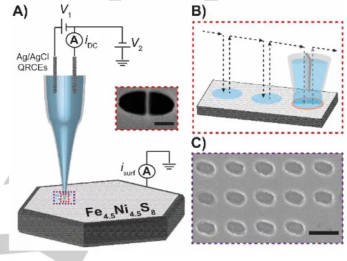
After crystal growth, the intrinsic catalytic activity of the
crystalline Fe4.5Ni4.5S8 material was evaluated using SECCM (see.
Figure 2A for the schematic experimental set-up). By means of SECCM, amperometric or voltammetric measurements were performed in the confined area defined by the meniscus (droplet) cell created between the pulled double-barrel nanopipet probe (SECCM tip; electron micrograph shown in Figure 2A, inset) and substrate (working electrode) surface. Local voltammetric measurements were performed using a previously described
“hopping-mode” protocol,[24,28–30] as represented schematically in
Figure 2B, in which the SECCM tip was approached to the sample sequentially at a series of predefined locations. Upon each landing of the microdroplet a linear-sweep (LSV) or cyclic voltammogram (CV) was recorded. The geometric area of the
electrochemical cell (i.e., the evaluated active catalyst area),
defined by the “footprint” of the meniscus (droplet) cell, was readily visualized, post-experiment, using scanning electron microscopy (SEM), as shown in Figure 2C.
A representative LSV measured on a hexagonal-shaped
Fe4.5Ni4.5S8 (111) single-crystal basal surface [electron
back-scat-ter diffraction (EBSD) data shown in the Supporting Information, Figure S2] using SECCM is shown in Figure 3A. This LSV, which is the average of 98 individual measurements at different
loca-tions, is shifted by ca. ‒60 mV compared to that measured
macro-scopically of the bulk material (Supporting Information, Figure S3). This is unsurprising, because, as previously alluded to, the macro-scopic measurement reveals the “average” properties of all ex-posed surface sites and features of the bulk material, whereas the
SECCM measurement targets a specific surface site, i.e., the
(111) crystallographic plane in this case. This indicates that the
“enhanced” bulk activity found for the Fe4.5Ni4.5S8 (111)
single-crystal surface is likely attributable to the presence of defect sites.
The SECCM meniscus probe was landed on a macroscopic
feature (‘crack’) on the Fe4.5Ni4.5S8 (111) surface (SEM image
shown in Figure S4A) in order to simulate a surface “defect” site; a representative LSV is shown in Figure 3A. Clearly, the HER is
facilitated at the macroscopic defect site, evident from the ca.
4-fold increase in J at a given E or ca. +100 mV shift in the LSV (i.e.,
compare the pink and blue curves in Figure 3A). We propose that the different reactivity at the defect sites stems from a faster sulfur release rate at such edges, resulting in a faster substitutional pro-tonation and therefore a higher activity, which is in agreement with
results recently obtained by operando phonon spectroscopy.[18]
Figure 2.A) Schematic showing the spatially-resolved electrocatalytic measurements obtained using SECCM. A bias voltage (+0.05 V) of V1 was
applied between the two Ag/AgCl wire quasi-reference counter electrodes (QRCEs) and the resulting ion conductance current (iDC) was measured and
employed for tip positioning and monitoring the status of the meniscus cell. A voltage of V2 was applied to one of the QRCEs to control the working electrode
(e.g., Fe4.5Ni4.5S8) potential (Es), where Es= ‒(V1/2 + V2) and the working
electrode current (isurf) was measured. A SEM image of the end of a
representative nanopipet probe is shown in the inset (scale bar 200 nm). B)
Voltammetric hopping-mode SECCM protocol, where the arrows show the movement of the nanopipet probe along the substrate surface. C) SEM image of the droplet “footprints” left on the surface of the sample after a cathodic polarization experiment (scale bar 1 µm).
Verifying this behavior on the macroscale would be challen-ging, necessitating the preparation of samples with engineered surface defects. It should be noted that it is assumed that the
presence of the surface defect (“crack”) does not significantly
influence the probed surface area (i.e., effective electrode area).
This is because the dimensions of the feature are relatively minor on the scale of the probed area (Figure S4A) and the fact that the
DC and AC ion conductance currents (iDC and iAC, induced by V1
in Figure 2A), which indicate on the morphology of the meniscus cell, do not vary significantly from that measured on the [111] ba-sal surface (Supporting Information, Figure S4B to E). In addition,
as mass transport (i.e., proton diffusion) would be expected to be
greatly hindered within the confined dimensions of the ca. 40 nm
wide “crack”, the effective electrode area (caused by electrolyte “wicking” into the crack) would need to increase by more than a factor of 4 to explain the enhanced currents in Figure 3A. Given
that droplet spreading on the basal surface is minimal (i.e., the
dimensions of the droplet footprints are comparable to the tip
Fotos of different Pentlandite Single Crystals
Sample 3 Sample 4 Sample 9
• These pictures are taken from the first samples we provided to Pat Unwin et. al.
• All have the ratio of (Fe,Ni)9S8, but remember:
3& 4seemed to have higher iron content (Fe/Ni>1), while9was okay (Fe/Ni=1)
A) Fotos of different Pentlandite Single Crystals
Sample 3 Sample 4 Sample 9
• These pictures are taken from the first samples we provided to Pat Unwin et. al.
• All have the ratio of (Fe,Ni)9S8, but remember:
3& 4seemed to have higher iron content (Fe/Ni>1), while9was okay (Fe/Ni=1) B)
XRD of SM015 – Pentlandite Single Crystals
(111)-plane (spacefill model) Crystal structure
C) SEM of SM015 – Pentlandite Single Crystal (first time)
• All pictures showthe surface of a SM015-crystal at different magnitudes.
• Impurities on the surface glass (see EDX)
[image:3.595.298.549.213.402.2]diameter, see Figure 1), such an increase in the effective surface
area is very unlikely and thus the enhanced J in Figure 3A is
attri-buted to a legitimate increase in activity (HER kinetics), as discus-sed above.
Figure 3.A) Averaged LSVs (scan rate, ν = 250 mV s‒1, [HClO4] = 0.1 M)
obtained from a hexagonal Fe4.5Ni4.5S8 (111) crystal basal surface (blue trace)
and macroscopic defect site (‘crack’, pink trace) and; Fe-rich areas of platelet-shaped crystals (red and green traces). B) Histograms showing the distribution in E at a J of 500 mA cm‒2. The number of measurements (N) in A) and B) were
98, 4, 97 and 101 for the blue, pink, red and green traces, respectively. C)
Averaged CVs (ν = 250 mV s‒1, [HClO4] = 0.1 M) obtained from hexagonal
Fe4.5Ni4.5S8(111) (N = 240, blue trace) and an Fe-rich area of a platelet-shaped
crystal (N = 297, green trace). The first and second cycles are indicated by solid and dashed lines, respectively. The arrows indicate the direction of the first sweep. Note that the probed surface area (effective electrode area) was assu-med to be unperturbed by the presence of the macroscopic defect (crack).
The enhanced activity at the macroscopic defect site on
Fe4.5Ni4.5S8 (111) is also clearly evident in Figure 3B, which shows
the distribution in potential at a current density J of 500 mA cm‒2.
Mean values of ‒0.59 V (N = 98) and ‒0.48 V (N = 4) were derived
for the Fe4.5Ni4.5S8 (111) basal surface and macroscopic defect
site, respectively. These microscopic data indicate that defect-rich
Fe4.5Ni4.5S8 (111) would be more active than the pristine bulk
material. It is worth noting that the values measured at the
Fe4.5Ni4.5S8 (111) basal surface follow a normal distribution, with a
standard deviation of 0.01 V. This clearly demonstrates the repro-ducibility of the SECCM technique for performing nanoscopic electrocatalytic measurements at single-crystal surfaces, which allows tens to hundreds of spatially-resolved LSVs to be recorded
in different areas of a surface in a matter of minutes.[8,24]
Additional platelet-shaped crystalline Fe4.5Ni4.5S8 samples
were also evaluated using the SECCM technique; representative LSVs are shown in Figure 3A. Evidently, the two samples possess
enhanced HER activity, with the LSVs (i.e., red and green traces)
each being shifted by ca. +100 mV compared to the hexagonal
Fe4.5Ni4.5S8 (111) crystal basal surface discussed above (i.e., blue
trace). In addition, there is a clear difference at the foot of the LSV, which was further investigated by cyclic voltammetry with SECCM,
as shown in Figure 3C. While the Fe4.5Ni4.5S8 (111) hexagonal
crystal gives rise to a relatively featureless CV, with a positive hysteresis on the reverse sweep, there is a prominent shoulder in the CV of the “high activity” platelet-shaped sample. These diffe-rences suggest that there are either different crystallographic orientations exposed or there is a difference in the atomic compo-sition of the sample surfaces. The former was ruled out by EBSD analysis and the latter was confirmed by energy-dispersive X-ray spectroscopy (EDX; Supporting Information, Figure S5). While most spots investigated on the sample surfaces possess the
ex-pected Fe : Ni : S ratio as in the original material (e.g., Fe : Ni : S;
1 : 1.1 : 1.8; Figure S5A), the “high activity” regions show a signifi-cantly altered atomic composition. These regions instead are Fe-rich and reveal a relatively lowered S-content [Fe : Ni : S; 1 : 0.4 : 1.4 and 1 : 0.4 : 1.3 for (1) and (2), Figure S5B and C] and are most-likely a result of the long-thermal treatment during the preparation of the material.
It was previously noted that pentlandite-like compounds containing different Fe : Ni ratios exhibit similar surface
composi-tions; a mixture of iron and nickel sulfides as well as Fe3+ oxidic
species and Ni(OH)2, as revealed by XPS. No clear influence on
the HER activity was, however, observed for the as prepared
samples.[31] It should also be noted that, as shown earlier by
operando phonon studies on Fe4.5Ni4.5S8, activation processes
correlated with S-depletion and formation of defect sites were found to dramatically increase the HER activity of the pentlandite,
consistent with what has been observed here (Figure 3A).[16,18,31]
Returning to Figure 3B, platelet samples (1) and (2) possess
mean potential values at 500 mA cm‒2 of ‒0.47 ± 0.01 V (N = 97)
and -0.48 ± 0.01 V (N = 101), respectively. It is also interesting to
note that in the Fe-rich area of platelet sample (1), there are clearly two populations present in the distribution exhibiting
sub-stantially different potentials at 500 mA cm‒2 values; a large one
(N = 83) centered around ‒0.47 V and a much smaller one (N =
14) centered around ‒0.44 V. Again, the “higher activity” popula-tion is supposed to arise from surface defect sites exposed on the
Fe4.5Ni4.5S8 crystal surface, which, as noted above is suggested to
give rise to more rapid sulfur release/protonation rates and thus enhanced HER catalytic activity.
The prominent reductive shoulder observed for the “high ac-tivity” platelet samples, prior to the HER wave (as seen in Figures
3A and C) arises from a change in the material itself (i.e., “aging”).
A somewhat smaller reduction process (“shoulder”) prior to the
HER wave is also observed at the hexagonal Fe4.5Ni4.5S8 bulk
sur-face during the second voltammetric cycle (dashed trace, Figure 3C), consistent with macroscopic measurements performed using the bulk material (see Figure S3). The larger pre-wave (shoulder) currents associated with the Fe-rich “high activity” areas are indi-cative of a faster “aging” (“corrosion”) process compared to the
Fe4.5Ni4.5S8 basal surface. If this “aging” stems from sulfur release
and substitutional protonation, as alluded to above, this may ex-plain the increased activity in these samples. DFT calculations of
defect-free and S vacancy- containing Fe4.5Ni4.5S8 (111)
struc-tures indicate preferential substitutional adsorption of H atoms at different vacancies within the crystal lattice. The influence of the vacancies position on the energy threshold of the process was
[image:4.595.50.292.136.375.2]were supported by experimental nuclear resonant inelastic X-Ray scattering, both highlighting the importance of defects for the increased HER activity. Furthermore, this observation suggests that Fe-rich areas are less stable due to increased susceptibility to corrosion and thus might lead to higher HER activity on the
ex-perimental (i.e., s to min) timescale due to a facilitation of the
sub-stitutional sulfur/hydrogen replacement.
In conclusion, we have utilized SECCM to unveil the struc-tural and compositional controls on electrocatalytic activity for iron
nickel sulfide, Fe4.5Ni4.5S8. Small variations in the surface Fe : Ni :
S ratio, achieved through varying synthesis conditions or material “aging”, can lead to a tremendously altered catalytic HER activity. In a more general sense, this study demonstrates how information on surface structure and atomic composition within an investiga-ted (nano)material can be directly correlainvestiga-ted to spatially-resolved electrochemical information, which is a crucial step in rational ca-talyst design and synthesis. In this regard, SECCM could have an important role in resolving structure-activity relationships in com-plex electrocatalytically active materials and in heterogeneous catalyst discovery.
Acknowledgements
C.L.B. acknowledges support from a Marie Curie Individual Fel-lowship (702048 NEIL). P.R.U. thanks the Royal Society for a Wolfson Research Merit Award. U.-P.A. is grateful for the financial support by the Fonds of the Chemical Industry (Liebig grant) and the Deutsche Forschungsgemeinschaft (Emmy Noether grant to U.-P.A., AP242/2-1 and AP242/6-1). W.S. is grateful for financial support from the Deutsche Forschungsgemeinschaft in the frame-work of the cluster of excellence “RESOLV” (EXC1069). The authors thank Mr. Lewis Yule and Dr. Geoff West for performing EBSD.
Keywords: electrocatalysis; iron-nickel-sulfide; SECCM; hydrogen evolution reaction; single-crystal surface
[1] Z. W. Seh, J. Kibsgaard, C. F. Dickens, I. Chorkendorff, J. K. Nørskov, T. F. Jaramillo, Science2017, 355, DOI 10.1126/science.aad4998. [2] W. Lubitz, W. Tumas, Chem. Rev.2007, 107, 3900–3903. [3] J. M. Ogden, Phys. Today2002, 55, 69.
[4] G. W. Crabtree, M. S. Dresselhaus, M. V. Buchanan, Phys. Today2004,
57, 39–44.
[5] X. Zou, Y. Zhang, Chem. Soc. Rev.2015, 44, 5148–5180. [6] “ExxonMobil’s 2017 Outlook for Energy PDF reports,” can be found
under
http://corporate.exxonmobil.com/en/energy/energy-outlook/download-the-report/download-the-outlook-for-energy-reports,
2017.
[7] W. Sheng, Z. Zhuang, M. Gao, J. Zheng, J. G. Chen, Y. Yan, Nat. Commun.2015, 6, DOI 10.1038/ncomms6848.
[8] C. L. Bentley, M. Kang, F. M. Maddar, F. Li, M. Walker, J. Zhang, P. R. Unwin, Chem. Sci.2017, 8, 6583–6593.
[9] D. Merki, X. Hu, Energy Environ. Sci.2011, 4, 3878–3888.
[10] J. Kibsgaard, Z. Chen, B. N. Reinecke, T. F. Jaramillo, Nat Mater2012,
11, 963–969.
[11] B. Konkena, J. Masa, W. Xia, M. Muhler, W. Schuhmann, Nano Energy 2016, 29, 46–53.
[12] M. A. Lukowski, A. S. Daniel, F. Meng, A. Forticaux, L. Li, S. Jin, J. Am. Chem. Soc.2013, 135, 10274–10277.
[13] Z. Lei, S. Xu, P. Wu, Phys Chem Chem Phys2016, 18, 70–74. [14] J. D. Benck, T. R. Hellstern, J. Kibsgaard, P. Chakthranont, T. F.
Jaramillo, ACS Catal.2014, 4, 3957–3971.
[15] B. Hinnemann, P. G. Moses, J. Bonde, K. P. Jørgensen, J. H. Nielsen, S. Horch, I. Chorkendorff, J. K. Nørskov, J. Am. Chem. Soc.2005, 127, 5308–5309.
[16] B. Konkena, K. junge Puring, I. Sinev, S. Piontek, O. Khavryuchenko, J. P. Dürholt, R. Schmid, H. Tüysüz, M. Muhler, W. Schuhmann, et al.,
Nat. Commun.2016, 7, 12269–12277.
[17] K. junge Puring, S. Piontek, M. Smialkowski, J. Burfeind, S. Kaluza, C. Doetsch, U.-P. Apfel, J. Vis. Exp.2017, 124, DOI 10.3791/56087. [18] I. Zegkinoglou, A. Zendegani, I. Sinev, S. Kunze, H. Mistry, H. S. Jeon,
J. Zhao, M. Y. Hu, E. E. Alp, S. Piontek, et al., J. Am. Chem. Soc.2017, 14360–14363.
[19] G. Zhang, P. M. Kirkman, A. N. Patel, A. S. Cuharuc, K. McKelvey, P. R. Unwin, J. Am. Chem. Soc.2014, 136, 11444–11451.
[20] C.-H. Chen, K. E. Meadows, A. Cuharuc, S. C. S. Lai, P. R. Unwin,
Phys. Chem. Chem. Phys.2014, 16, 18545–18552.
[21] P. R. Unwin, A. G. Güell, G. Zhang, Acc. Chem. Res.2016, 49, 2041– 2048.
[22] N. Ebejer, A. G. Güell, S. C. S. Lai, K. McKelvey, M. E. Snowden, P. R. Unwin, Annu. Rev. Anal. Chem.2013, 6, 329–351.
[23] R. G. Mariano, K. McKelvey, H. S. White, M. W. Kanan, Science2017,
358, 1187–1192.
[24] C. L. Bentley, M. Kang, P. R. Unwin, J. Am. Chem. Soc.2017, 139, 16813–16821.
[25] T. F. Jaramillo, K. P. Jørgensen, J. Bonde, J. H. Nielsen, S. Horch, I. Chorkendorff, Science2007, 317, 100–102.
[26] Y. Li, H. Wang, L. Xie, Y. Liang, G. Hong, H. Dai, J. Am. Chem. Soc. 2011, 133, 7296–7299.
[27] A. Pearson, M. Buerger, Am. Mineral.1956, 41, 804–805. [28] S. P. E, Y.-R. Kim, D. Perry, C. L. Bentley, P. R. Unwin, ACS Appl.
Mater. Interfaces2016, 8, 30458–30466.
[29] C.-H. Chen, L. Jacobse, K. McKelvey, S. C. S. Lai, M. T. M. Koper, P. R. Unwin, Anal. Chem.2015, 87, 5782–5789.
[30] A. G. Güell, A. S. Cuharuc, Y.-R. Kim, G. Zhang, S. Tan, N. Ebejer, P. R. Unwin, ACS Nano2015, 9, 3558–3571.
COMMUNICATION
Local investigation of the hydrogen evolution reaction (HER) on single crystal iron nickel sulphide, one of the most active non-noble metal based HER catalyst, was carried out using Scanning Electrochemical Cell Mi-croscopy (SECCM). Small variations in the Ni:Fe ratio at the surface induce tremendous changes in the catalytic HER activity.
Cameron L. Bentley, Corina Andronescu, Mathias Smialkowski, Minkyung Kang, Tsvetan Tarnev, Bernd Marler, Patrick R. Unwin,* Ulf -Peter Apfel,* Wolfgang Schuhmann*
Page No. – Page No.
![Figure 3. A) Averaged LSVs (scan rate, ν = 250 mV s‒1, [HClO4] = 0.1 M) obtained from a hexagonal Fe4.5Ni4.5S8 (111) crystal basal surface (blue trace) and macroscopic defect site (‘crack’, pink trace) and; Fe-rich areas of platelet-shaped crystals (red an](https://thumb-us.123doks.com/thumbv2/123dok_us/9427874.447508/4.595.50.292.136.375/figure-averaged-obtained-hexagonal-crystal-macroscopic-platelet-crystals.webp)
![Ninth report on the implementation of Council Regulation (EC) 866/2004 of 29 April 2004 and the situation resulting from its application covering the period 1 January until 31 December 2012 [SWD (2013) 186 final]. COM (2013) 299 final, 24 May 2013](data:image/gif;base64,R0lGODlhAQABAIAAAP///wAAACH5BAEAAAAALAAAAAABAAEAAAICRAEAOw==)