Quinoline-2-sulfonamide
Krzysztof Marciniec,a* Andrzej Mas´lankiewicz,aJoachim Kuszband Maria Nowakb
a
Department of Organic Chemistry, The Medical University of Silesia, Jagiellon´ska 4, 41-200 Sosnowiec, Poland, andbInstitute of Physics, University of Silesia, Uniwersytecka 4, 40-007 Katowice, Poland
Correspondence e-mail: [email protected] Received 28 May 2013; accepted 24 July 2013
Key indicators: single-crystal X-ray study;T= 100 K; mean(C–C) = 0.003 A˚; Rfactor = 0.030;wRfactor = 0.072; data-to-parameter ratio = 9.7.
In the title compound, C9H8N2O2S, the sulfamoyl –NH2group
is involved in intermolecular hydrogen bonding with the sulfonamide O and quinoline N atoms. In the crystal, molecules are linked into dimers via pairs of N—H N hydrogen bonds, forming an R2
2
(10) motif. The dimers are further assembled into chains parallel to the baxis through N—H O hydrogen bonds, generating a C(4) motif. The crystal packing is additionally stabilized by intermolecular C— H O interactions. The crystal studied was a non-merohedral twin with a domain ratio of 0.938 (2):0.062 (2). Density functional theory (DFT) calculations, at the B3LYP/6– 31 G(d,p) level of theory, were used to optimize the molecular structure and to determine interaction energies for the title compound. The resulting interaction energy is4.4 kcal mol1 per bridge for theC(4) chain and5.9 kcal mol1per bridge
for theR2 2
(10) motif.
Related literature
For the use of the quinolinesulfamoyl unit in medicinal chemistry, see: Kimet al.(2005); Zajdelet al.(2012, 2013). For related structures, see: Marciniecet al.(2012). For the synth-esis, see: Mas´lankiewicz et al. (2007). For hydrogen-bonding motifs in sufonamides, see: Adsmond & Grant (2001). For graph-set notation of hydrogen-bond motifs, see: Bernsteinet al. (1995). For general hydrogen-bond rules, see: Donohue (1952); Etter (1990). For details of theoretical calculations, see: Frischet al.(20094); Parr & Yang (1989). The twin matrix was been determined withROTAX(Cooperet al., 2002).
Experimental
Crystal data
C9H8N2O2S Mr= 208.23 Monoclinic,P21=c a= 8.5907 (1) A˚
b= 5.1716 (1) A˚
c= 20.0375 (3) A˚ = 94.230 (1)
V= 887.79 (2) A˚3 Z= 4
MoKradiation = 0.34 mm1 T= 100 K
0.270.230.05 mm
Data collection
Agilent SuperNova diffractometer with an Atlas detector Absorption correction: multi-scan
(CrysAlis PRO; Agilent, 2011)
Tmin= 0.919,Tmax= 1.000
27311 measured reflections 1552 independent reflections 1530 reflections withI> 2(I)
Rint= 0.024
Refinement
R[F2> 2(F2)] = 0.030 wR(F2) = 0.072 S= 1.12 1552 reflections
160 parameters
All H-atom parameters refined
max= 0.35 e A˚
3 min=0.32 e A˚
3
Table 1
Hydrogen-bond geometry (A˚ ,).
D—H A D—H H A D A D—H A
N2—H2N2 O1i
0.84 (3) 2.09 (3) 2.922 (2) 171 (2) N2—H1N2 N1ii
0.80 (3) 2.18 (3) 2.962 (2) 165 (2) C6—H6 O1iii 0.93 (2) 2.66 (2) 3.431 (2) 141.5 (18)
Symmetry codes: (i)x;yþ1;z; (ii)x;yþ1;z; (iii)x;yþ1 2;z12.
Data collection: CrysAlis PRO(Agilent, 2011); cell refinement: CrysAlis PRO; data reduction: CrysAlis PRO; program(s) used to solve structure: SHELXS97(Sheldrick, 2008); program(s) used to refine structure:SHELXL97(Sheldrick, 2008) andWinGX(Farrugia, 2012); molecular graphics:ORTEP-3 for Windows(Farrugia, 2012) andMercury(Macraeet al., 2006); software used to prepare material for publication:publCIF(Westrip, 2010).
This research was supported in part by the Medical University of Silesia, grant No. KNW-1–006/P/2/0, and by PL-Grid Infrastructure, grant ID: plggkrzmarci1.
Supplementary data and figures for this paper are available from the IUCr electronic archives (Reference: GK2577).
organic compounds
Acta Cryst.(2013). E69, o1357–o1358 doi:10.1107/S160053681302062X Marciniecet al.
o1357
Acta Crystallographica Section E Structure Reports Online
Adsmond, D. A. & Grant, D. J. W. (2001).J. Pharm. Sci.90, 2058–2077. Agilent (2011).CrysAlis PRO. Agilent Technologies Ltd, Yarnton, England. Bernstein, J., Davis, R. E., Shimoni, L. & Chang, N.-L. (1995).Angew. Chem.
Int. Ed. Engl.34, 1555–1573.
Cooper, R. I., Gould, R. O., Parsons, S. & Watkin, D. J. (2002).J. Appl. Cryst.
35, 168–174.
Donohue, J. (1952).J. Phys. Chem.56, 502–510. Etter, M. C. (1990).Acc. Chem. Res.23, 120–126. Farrugia, L. J. (2012).J. Appl. Cryst.45, 849–854.
Frisch, M. J., et al.(2009).GAUSSIAN09. Gaussian, Inc., Wallingford, CT, USA.
Kim, Y.-H., Shin, K.-J., Lee, T. G., Kim, E., Lee, M.-S., Ryu, S. H. & Suh, P.-G. (2005).Biochem. Pharmacol.69, 1333–1341.
Macrae, C. F., Edgington, P. R., McCabe, P., Pidcock, E., Shields, G. P., Taylor, R., Towler, M. & van de Streek, J. (2006).J. Appl. Cryst.39, 453–457.
E68, o2826.
Mas´lankiewicz, A., Marciniec, K., Pawłowski, M. & Zajdel, P. (2007).
Heterocycles,71, 1975–1990.
Parr, R. G. & Yang, W. (1989). InDensity Functional Theory of Atoms and Molecules. New York: Oxford University Press Inc.
Sheldrick, G. M. (2008).Acta Cryst.A64, 112–122. Westrip, S. P. (2010).J. Appl. Cryst.43, 920–925.
Zajdel, P., Marciniec, K., Grychowska, K., Mas´lankiewicz, A., Satała, G., Duszyn´ska, B., Siwek, A., Nowak, G., Partyka, A., Wro´bel, D., Jastrze˛bska-Wie˛sek, M., Bojarski, A. J., Wesołowska, A. & Pawłowski, M. (2013).Eur. J. Med. Chem.60, 42–50.
supporting information
sup-1
Acta Cryst. (2013). E69, o1357–o1358
supporting information
Acta Cryst. (2013). E69, o1357–o1358 [doi:10.1107/S160053681302062X]
Quinoline-2-sulfonamide
Krzysztof Marciniec, Andrzej Ma
ś
lankiewicz, Joachim Kusz and Maria Nowak
S1. Comment
Compounds containing quinolinesulfamoyl moiety have received considerable attention in recent years due to their
diverse pharmacological properties including antidepressant (Zajdel et al., 2012; Zajdel et al., 2013) or anticancer activity
(Kim et al., 2005).
From a structural point of view, sulfonamides are interesting because of their tendency to form different hydrogen-bond
patterns in the solid state. Studies have shown that primary hydrogen-bond connectivity is somewhat predictable. Two
general hydrogen-bond rules based on empirical observations of hundreds of crystal structures over the years have been
developed that summarize this predictability (Adsmond & Grant, 2001). The first of these rules states "all good donors
and acceptors are used in hydrogen bonding" (Donohue, 1952). The second hydrogen-bond rule for crystal structures of
small organic molecules states that, after formation of intramolecular hydrogen bonds, the best hydrogen bond donor will
bond to the best hydrogen-bond acceptor present (Etter, 1990). The molecule of quinolinesulfonamide has two good
hydrogen bond donors (the sulfonamido H atoms) and three the best hydrogen bond acceptors (the sulfonamido O atoms
and quinoline nitrogen atom). We have previously reported the X-ray crystal structure of quinoline-8-sulfonamide
(Marciniec et al., 2012). The obtained results show, according to the second hydrogen-bond rule, that the sulfamoyl NH2
group is involved in intramolecular N—H···N hydrogen bond resulting in the graph-set motif of S(6) (Bernstein et al.
1995). After formation of intramolecular hydrogen bond, the sulfamoyl NH2 group is involved in intermolecular N—
H···O hydrogen bond resulting in the graph-set motif of R22(8). The key feature of the molecular structure of
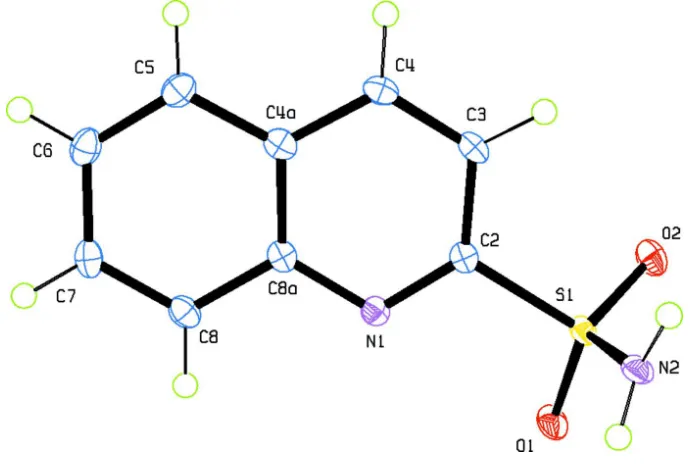
2-quinoline-sulfonamide (I) (Fig.1) is the N1—C2—S1—N2 torsion angle of -95.88 (14)°. The geometry of the 2-quinoline-sulfonamide group
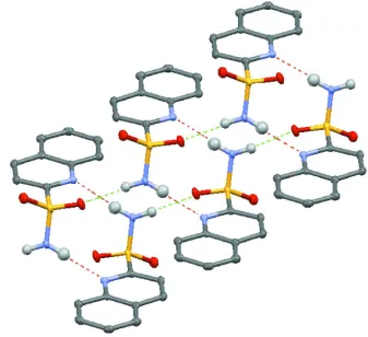
does not allow for intramolecular hydrogen-bond ring formation. In the crystal structure, the molecules form dimers
through N2—H1···N1 intermolecular hydrogen bonds [graph set R22(10)] which are extended into one-dimensional chains
[graph set C(4)] along the b axis, through the sulfonamide N2—H2···O1 hydrogen bonds (Table 1; Fig. 2). Thus the
first-level graph set is N1= C(4) R22(10).
The second-level graph-set notation (for combinations of two hydrogen bonds: N2—H2···O1 and N2—H1···N1) was
determined to be as follows N2= R44(14)C44(16) R44(16) R44(18) R66(22) R66(24) R66(26).
The crystal packing is further stabilized by week intermolecular hydrogen bonds C3—H3···O2 and C4—H4···O2 which
generate R21(5) ring and C6—H6···O1 hydrogen bond which generates D motif.
The molecular structure of (I) in the gas phase was optimized by the density-functional theory at the B3LYP/6–31
G(d,p) level, using the computer program GAUSSIAN09 (Parr & Yang, 1989; Frisch et al., 2009). Calculations were
performed using the X-ray coordinates as the input structure. The calculated geometry of the sulfonamide group allows
for intramolecular hydrogen-bond five-membered ring formation (H1···N1 = 2.618 Å; N2—H1···N1 = 100.3°; N1—C2—
S1—N2 = -35.2°) (Fig. 3). In the crystal structure of quinoline-2-sulfonamide (I) an intermolecular hydrogen bond is
more difficult to break than comparable intramolecular hydrogen bond formed between the the sulfamoyl NH2 group and
state. Intermolecular interactions, and hydrogen bonding in particular, might strongly influence the conformations
adopted in the solid state, whereas intramolacular interactions and and hydrogen bonding will dominate the gas-phase
conformation.
The interaction energies for 2-quinolinesulfonamide (I) were investigated by adding successive units, in their
crystallographic positions, once for the chain [graph set C(4)], once for the centrosymmetric dimer [graph set R22(10)].
These energies were then compared with the energy for the same number of independent asymmetric units to determine
the stabilization energy for the interaction between units. The results are presented in table 2.
The energy difference as a function of the number of bridging interactions is nearly linear and independent of choice of
generated cavity. The resulting interaction energy is ~4.4 kcal/mol/bridge for C(4) chain and ~5.9 kcal/mol/bridge for
R22(10) dimer. The small increase in stabilization energy per unit as the number of formula units suggests that there are
negligible contributions from extended molecular orbitals, supporting the original assumption that the significant
interactions result from hydrogen bonding contributions.
S2. Experimental
The title compound was prepared by the reaction of hydrochloric acid solution of quinoline-2-sulfochloride with an
excess ammonia at -10 °C according to the procedure reported by Maślankiewicz et al. (2007). Single crystals of the title
compound suitable for X-ray structure determination were obtained by recrystallization from an ethanolic solution.
S3. Refinement
The hydrogen atoms participating in hydrogen bonding were located in a difference Fourier map and freely refined. The
twin matrix, 1 0 0 0 - 1 0 - 0.344 0 - 1, has been determined with the ROTAX program (Cooper et al., 2002). For the
further refinement, the reflection data file in HKLF 5 format was prepared using "Make HKLF5" function of the WinGX
[image:4.610.133.481.432.664.2]program (Farrugia, 2012).
Figure 1
The molecular structure of the title compound with the atom labeling and displacement ellipsoids drawn at the 50%
supporting information
sup-3
[image:5.610.139.476.71.379.2]Acta Cryst. (2013). E69, o1357–o1358 Figure 2
Intermolecular N-H···N and N-H···O hydrogen bonds (dashed lines) in the title compound
Figure 3
Intramolecular hydrogen bond in 2-quinolinesulfonamide (I) (calculated) and 8-quinolinesulfonamide (II)
(experimentally determined).
(I)
Crystal data
C9H8N2O2S
Mr = 208.23
Monoclinic, P21/c Hall symbol: -P 2ybc
a = 8.5907 (1) Å
b = 5.1716 (1) Å
c = 20.0375 (3) Å
β = 94.230 (1)°
V = 887.79 (2) Å3
[image:5.610.132.472.424.553.2]Dx = 1.558 Mg m−3 Melting point: 441.2 K
Mo Kα radiation, λ = 0.71073 Å Cell parameters from 14635 reflections
µ = 0.34 mm−1
T = 100 K Plate, colorless 0.27 × 0.23 × 0.05 mm
Data collection
Agilent SuperNova
diffractometer with an Atlas detector Radiation source: SuperNova (Mo) X-ray
Source
Mirror monochromator
Detector resolution: 10.4498 pixels mm-1
ω scans
Absorption correction: multi-scan (CrysAlis PRO; Agilent, 2011)
Tmin = 0.919, Tmax = 1.000 27311 measured reflections 1552 independent reflections 1530 reflections with I > 2σ(I)
Rint = 0.024
θmax = 25.1°, θmin = 2.0°
h = −10→10
k = −6→6
l = −23→23
Refinement
Refinement on F2 Least-squares matrix: full
R[F2 > 2σ(F2)] = 0.030
wR(F2) = 0.072
S = 1.12 1552 reflections 160 parameters 0 restraints
Primary atom site location: structure-invariant direct methods
Secondary atom site location: difference Fourier map
Hydrogen site location: inferred from neighbouring sites
All H-atom parameters refined
w = 1/[σ2(F
o2) + (0.0262P)2 + 0.9337P] where P = (Fo2 + 2Fc2)/3
(Δ/σ)max < 0.001 Δρmax = 0.35 e Å−3 Δρmin = −0.32 e Å−3
Special details
Geometry. All e.s.d.'s (except the e.s.d. in the dihedral angle between two l.s. planes) are estimated using the full
covariance matrix. The cell e.s.d.'s are taken into account individually in the estimation of e.s.d.'s in distances, angles and torsion angles; correlations between e.s.d.'s in cell parameters are only used when they are defined by crystal symmetry. An approximate (isotropic) treatment of cell e.s.d.'s is used for estimating e.s.d.'s involving l.s. planes.
Refinement. Refinement of F2 against ALL reflections. The weighted R-factor wR and goodness of fit S are based on F2,
conventional R-factors R are based on F, with F set to zero for negative F2. The threshold expression of F2 > σ(F2) is used only for calculating R-factors(gt) etc. and is not relevant to the choice of reflections for refinement. R-factors based on F2 are statistically about twice as large as those based on F, and R- factors based on ALL data will be even larger.
Fractional atomic coordinates and isotropic or equivalent isotropic displacement parameters (Å2)
x y z Uiso*/Ueq
S1 0.24131 (5) 0.61370 (8) 0.04453 (2) 0.01307 (14)
O1 0.18557 (15) 0.3614 (2) 0.06117 (6) 0.0193 (3)
O2 0.38294 (14) 0.7120 (3) 0.07708 (6) 0.0188 (3)
N1 0.19733 (16) 0.4337 (3) −0.07942 (7) 0.0132 (3)
N2 0.10504 (19) 0.8125 (3) 0.05580 (8) 0.0164 (3)
C2 0.26951 (19) 0.6149 (3) −0.04339 (8) 0.0129 (4)
C3 0.3627 (2) 0.8112 (4) −0.06810 (9) 0.0153 (4)
C4 0.3835 (2) 0.8125 (4) −0.13486 (9) 0.0156 (4)
C4A 0.31105 (19) 0.6202 (4) −0.17656 (9) 0.0147 (4)
C5 0.3263 (2) 0.6090 (4) −0.24669 (9) 0.0169 (4)
supporting information
sup-5
Acta Cryst. (2013). E69, o1357–o1358
C7 0.1578 (2) 0.2367 (4) −0.25488 (9) 0.0175 (4)
C8 0.1389 (2) 0.2425 (4) −0.18758 (9) 0.0159 (4)
C8A 0.21642 (19) 0.4342 (3) −0.14675 (8) 0.0129 (4)
H2N2 0.126 (3) 0.971 (5) 0.0528 (11) 0.027 (6)*
H1N2 0.019 (3) 0.751 (5) 0.0549 (12) 0.035 (7)*
H3 0.409 (2) 0.940 (4) −0.0389 (11) 0.020 (5)*
H4 0.444 (2) 0.945 (4) −0.1538 (10) 0.021 (5)*
H5 0.391 (2) 0.744 (4) −0.2674 (9) 0.012 (5)*
H6 0.265 (3) 0.415 (4) −0.3299 (12) 0.026 (6)*
H7 0.107 (2) 0.103 (4) −0.2823 (11) 0.023 (6)*
H8 0.073 (2) 0.117 (4) −0.1661 (11) 0.023 (6)*
Atomic displacement parameters (Å2)
U11 U22 U33 U12 U13 U23
S1 0.0168 (2) 0.0096 (2) 0.0124 (2) −0.00169 (17) −0.00137 (16) −0.00008 (16)
O1 0.0290 (7) 0.0112 (6) 0.0172 (6) −0.0037 (6) −0.0005 (5) 0.0013 (5)
O2 0.0188 (6) 0.0195 (7) 0.0172 (6) −0.0019 (6) −0.0046 (5) −0.0012 (5)
N1 0.0128 (7) 0.0115 (7) 0.0152 (7) 0.0013 (6) −0.0002 (6) −0.0011 (6)
N2 0.0166 (8) 0.0106 (8) 0.0223 (8) −0.0043 (7) 0.0030 (6) −0.0020 (6)
C2 0.0114 (8) 0.0123 (9) 0.0146 (8) 0.0028 (7) −0.0011 (6) −0.0010 (7)
C3 0.0142 (8) 0.0135 (9) 0.0176 (9) −0.0015 (7) −0.0023 (7) −0.0015 (7)
C4 0.0119 (8) 0.0144 (9) 0.0203 (9) −0.0007 (7) 0.0007 (7) 0.0014 (7)
C4A 0.0116 (8) 0.0149 (9) 0.0176 (9) 0.0023 (7) 0.0002 (7) 0.0005 (7)
C5 0.0153 (8) 0.0187 (9) 0.0169 (9) 0.0017 (8) 0.0029 (7) 0.0015 (7)
C6 0.0164 (9) 0.0225 (10) 0.0149 (9) 0.0048 (8) 0.0006 (7) −0.0015 (8)
C7 0.0167 (9) 0.0177 (9) 0.0173 (9) 0.0031 (8) −0.0032 (7) −0.0046 (7)
C8 0.0138 (8) 0.0146 (9) 0.0192 (9) 0.0006 (7) −0.0005 (7) −0.0010 (7)
C8A 0.0112 (8) 0.0116 (8) 0.0157 (9) 0.0033 (7) −0.0010 (6) −0.0004 (7)
Geometric parameters (Å, º)
S1—O2 1.4308 (13) C4—H4 0.95 (2)
S1—O1 1.4376 (13) C4A—C8A 1.419 (2)
S1—N2 1.5866 (16) C4A—C5 1.422 (2)
S1—C2 1.7959 (18) C5—C6 1.361 (3)
N1—C2 1.311 (2) C5—H5 1.00 (2)
N1—C8A 1.371 (2) C6—C7 1.409 (3)
N2—H2N2 0.84 (3) C6—H6 0.93 (2)
N2—H1N2 0.80 (3) C7—C8 1.370 (3)
C2—C3 1.406 (3) C7—H7 0.97 (2)
C3—C4 1.363 (3) C8—C8A 1.419 (2)
C3—H3 0.96 (2) C8—H8 0.98 (2)
C4—C4A 1.414 (3)
O2—S1—O1 120.23 (8) C4—C4A—C8A 117.98 (16)
O2—S1—N2 108.44 (8) C4—C4A—C5 122.96 (17)
O1—S1—C2 107.59 (8) C6—C5—H5 121.3 (11)
N2—S1—C2 106.96 (8) C4A—C5—H5 118.4 (11)
C2—N1—C8A 117.05 (15) C5—C6—C7 120.69 (17)
S1—N2—H2N2 117.0 (16) C5—C6—H6 118.9 (14)
S1—N2—H1N2 115.3 (19) C7—C6—H6 120.4 (14)
H2N2—N2—H1N2 126 (3) C8—C7—C6 120.79 (17)
N1—C2—C3 125.52 (17) C8—C7—H7 119.6 (13)
N1—C2—S1 116.44 (13) C6—C7—H7 119.6 (13)
C3—C2—S1 118.02 (13) C7—C8—C8A 119.87 (17)
C4—C3—C2 117.95 (17) C7—C8—H8 122.0 (13)
C4—C3—H3 121.2 (13) C8A—C8—H8 118.1 (13)
C2—C3—H3 120.9 (13) N1—C8A—C8 118.75 (16)
C3—C4—C4A 119.54 (17) N1—C8A—C4A 121.94 (16)
C3—C4—H4 120.5 (13) C8—C8A—C4A 119.31 (16)
C4A—C4—H4 119.9 (13)
C8A—N1—C2—C3 1.0 (3) C4—C4A—C5—C6 −179.17 (17)
C8A—N1—C2—S1 179.28 (12) C8A—C4A—C5—C6 −0.5 (3)
O2—S1—C2—N1 148.59 (13) C4A—C5—C6—C7 0.7 (3)
O1—S1—C2—N1 18.84 (15) C5—C6—C7—C8 −0.1 (3)
N2—S1—C2—N1 −95.88 (14) C6—C7—C8—C8A −0.7 (3)
O2—S1—C2—C3 −32.97 (16) C2—N1—C8A—C8 −179.41 (15)
O1—S1—C2—C3 −162.72 (13) C2—N1—C8A—C4A 0.6 (2)
N2—S1—C2—C3 82.55 (15) C7—C8—C8A—N1 −179.13 (16)
N1—C2—C3—C4 −1.5 (3) C7—C8—C8A—C4A 0.9 (3)
S1—C2—C3—C4 −179.75 (13) C4—C4A—C8A—N1 −1.6 (2)
C2—C3—C4—C4A 0.4 (3) C5—C4A—C8A—N1 179.74 (15)
C3—C4—C4A—C8A 1.0 (3) C4—C4A—C8A—C8 178.43 (16)
C3—C4—C4A—C5 179.67 (17) C5—C4A—C8A—C8 −0.3 (2)
Hydrogen-bond geometry (Å, º)
D—H···A D—H H···A D···A D—H···A
N2—H2N2···O1i 0.84 (3) 2.09 (3) 2.922 (2) 171 (2)
N2—H1N2···N1ii 0.80 (3) 2.18 (3) 2.962 (2) 165 (2)
C3—H3···O2iii 0.96 (2) 2.68 (2) 3.308 (2) 123.5 (16)
C4—H4···O2iii 0.95 (2) 2.72 (2) 3.327 (2) 122.3 (15)
C6—H6···O1iv 0.93 (2) 2.66 (2) 3.431 (2) 141.5 (18)
[image:8.610.57.533.69.480.2]Symmetry codes: (i) x, y+1, z; (ii) −x, −y+1, −z; (iii) −x+1, −y+2, −z; (iv) x, −y+1/2, z−1/2.
Table 2. Calculation of stabilization energies for quinoline-2-sulfonamide (kcal mol-1)
B3LYP/6-31G(d,p)
Energy ΔE
Asymmetric unit -631075.7
2 units N1 = C(4) -1262155.9 -4.4
supporting information
sup-7
Acta Cryst. (2013). E69, o1357–o1358
3 units N1 = C(4)R22(10) -1893243.7 -16.6