organic papers
o1170
Hayneset al. C23H16O6 doi:10.1107/S1600536806005812 Acta Cryst.(2006). E62, o1170–o1172 Acta Crystallographica Section E
Structure Reports Online
ISSN 1600-5368
Pamoic acid determined from powder
diffraction data
Delia A. Haynes,aJacco Van de Streek,bJonathan C. Burley,a* William Jonesaand W. D. Sam Motherwellb
aUniversity Chemical Laboratory, Lensfield
Road, Cambridge CB2 1EW, England, and bCambridge Crystallographic Data Centre, 12
Union Road, Cambridge CB2 1EZ, England
Correspondence e-mail: [email protected]
Key indicators
Powder X-ray study T= 298 K
Mean(C–C) = 0.005 A˚ Rfactor = 0.030 wRfactor = 0.039
Data-to-parameter ratio = 3.69
For details of how these key indicators were automatically derived from the article, see http://journals.iucr.org/e.
Received 22 December 2005 Accepted 16 February 2006
#2006 International Union of Crystallography All rights reserved
The title compound [systematic name: 4,40
-methylenebis(3-hydroxy-2-naphthoic acid)], C23H16O6, has one half-molecule in the asymmetric unit. The molecular twofold rotational axis about the central C atom is preserved on crystallization. A chain formed by R2
2
(8) hydrogen bonds runs along thec axis
and an intramolecular O—H O C—OH hydrogen bond is
also formed. The crystal structure was solved by simulated annealing from laboratory X-ray powder diffraction data, with data collected at room temperature. Rietveld refinement of this model led to a final Rwp value of 0.0391 at 1.39 A˚ resolution.
Comment
The title compound, (I), is used in the pharmaceutical industry as a counter-ion to obtain long-acting formulations of certain basic drugs (Jorgensen, 1998), and is also of fundamental interest because of the possibility of extensive hydrogen bonding in the solid state. Attempts have been made to grow single crystals of pamoic acid for structure determination (Blackburn et al., 1996, Haynes et al., 2005) but these have proved unsuccessful; thus we have employed X-ray powder diffraction to solve and refine the crystal structure, which is reported here for the first time.
The compound crystallizes in the space groupC2/cwith one half-molecule of pamoic acid in the asymmetric unit (Fig. 1). A twofold rotation axis passes through C1. The –OH and –CO2H groups lie in the plane of the attached ring.
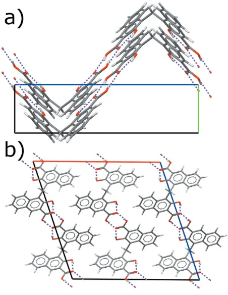
A corrugated chain of molecules running along thecaxis is formed by intermolecularR2
2(8) hydrogen bonds (Fig. 2a). The hydrogen bonds themselves are oriented at approximately 45
to the chain propagation vector (Fig. 2b). Other than hydrogen bonding, the only short intermolecular contact (less than the sum of the van der Waals radii) is an aromatic C— H interaction (symmetry code:1
2x, 1 2+y,
3
Database (CSD; Version 5.26 of November 2005; Allen, 2002), only 624 (1.3%) are as short or shorter; the median is 2.837 A˚ . The bis-pyridinium salt of pamoic acid (CSD refcode TABMAK; Blackburnet al., 1996) exhibits a similar C—H
distance of 2.646 A˚ , although between a different pair of atoms. This type of contact may therefore play a role in stabilizing the observed crystal structures in pamoic acid
derivatives; however, further work will be necessary to confirm this.
Experimental
Pamoic acid (97%+) was obtained from Sigma and used without further purification. No impurities were detected by X-ray powder diffraction. The sample was lightly ground and loaded into a 0.7 mm-diameter Lindemann glass capillary. Data were collected in Debye– Scherrer geometry employing CoK1radiation.
Crystal data
C23H16O6 Mr= 388.37
Monoclinic,C2=c a= 19.7348 (7) A˚
b= 4.78768 (12) A˚
c= 19.2544 (4) A˚
= 108.9622 (17) V= 1720.5 (1) A˚3
Z= 4
Dx= 1.499 Mg m
3
CoK1radiation
= 2.19 mm1 T= 298 K
Specimen shape: cylinder 120.70.7 mm Specimen prepared at 298 K
Data collection
Stoe linear PSD diffractometer Specimen mounting: 0.7 mm
Lindemann glass capillary Specimen mounted in transmission
mode
Scan method: step Absorption correction: none 2min= 2.0, 2max= 80.0
Increment in 2=0.0
Refinement
Refinement onInet Rp= 0.030 Rwp= 0.039 Rexp= 0.035 S= 1.20
Profile function: pseudo Voigt (Thompsonet al., 1987) with the asymmetry correction (Fingeret al., 1994)
362 reflections 98 parameters
H-atom parameters constrained Weighting scheme based on
measured s.u.’sw= 1/(Yobs)2
(/)max= 1.61
Preferred orientation correction: none
organic papers
Acta Cryst.(2006). E62, o1170–o1172 Hayneset al. C
[image:2.610.316.567.70.250.2]23H16O6
o1171
Figure 1View of (I), with the atom-numbering scheme. The unlabelled atoms are related to the labelled ones by the symmetry operation (1x,y,3
[image:2.610.47.294.71.247.2]2z).
Figure 2
The crystal packing viewed (a) along theaaxis and (b) along thebaxis. The O—H O hydrogen bonds are indicated by dashed lines.
Figure 3
Final observed (points), calculated (line), difference [(yobsycalc)] and
weighted difference [(yobsycalc)/] profiles for the Rietveld refinement
[image:2.610.57.283.309.601.2]Table 1
Hydrogen-bond geometry (A˚ ,).
D—H A D—H H A D A D—H A
O2—H2A O3i
0.82 1.85 2.643 (4) 166 O1—H1A O3 0.90 1.72 2.529 (4) 147
Symmetry code: (i)xþ1;y1;zþ1.
As noted in _pd_proc_ls_special_details, the refinement was characterised by a relatively shallow minimum with respect to the lattice parameters and zero point which were refined for the final cycle to generate s.u.’s..
Two powder diffraction patterns were collected. Initially a pattern was collected using a Philips Xpert diffractometer operating in Bragg–Brentano mode with a flat-plate sample. No monochromation or collimation was employed. A full pattern was collected in under five minutes; this was used to solve the crystal structure. Subsequent attempts to use this pattern in a Rietveld analysis were not successful owing to difficulties in modelling the diffraction peak shapes from the high-intensity operation mode. Thus a second pattern was collected for approximately 24 h using a monochromatic Stoe Stadi-P instru-ment operating in Debye–Scherrer geometry with the sample contained in a glass capillary. The resulting pattern proved suitable for a full Rietveld analysis.
The initial X-ray powder diffraction pattern was used for structure determination with the program DASH(David et al., 2004). The powder pattern was truncated to 45.32(CuK), corresponding to
a real-space resolution of 2.0 A˚ . The background was subtracted with a Bayesian high-pass filter (David & Sivia, 2001). Peak positions for indexing were obtained by fitting with an asymmetry-corrected Voigt function, followed by indexing with the programDICVOL(Boultif & Louer, 1991). Pawley refinement was used to extract integrated intensities and their correlations, from which the space group was determined using Bayesian statistical analysis (Markvardsen et al., 2001). Possible space groups wereCcorC2/c, the latter implying that the molecule sits on a special position; both space groups were tried. Simulated annealing was used to solve the crystal structure from the powder pattern in direct space. The starting molecular geometry was taken from entry TABMAK (Blackburnet al., 1996) from the CSD. When choosingCcas the space group, the asymmetric unit consists of a full pamoic acid molecule, which then has four independent flexible torsions, one for each of the two carboxylic acid groups and two across the central C atom. All these four torsion angles were left fully flexible during the simulated annealing, which, combined with three translational and three rotational degrees of freedom, gives a total of ten degrees of freedom. When choosingC2/cas the space group, the pamoic acid molecule is constrained by symmetry to sit on a twofold rotation axis through the central C atom, C1. Suitable constraints for atom C1 were therefore included in the simulated annealing runs for that space group, namely fixing itsx coordinate at 1/2, fixing its z
coordinate at 3/4 and setting its occupancy to 0.5 to account for site multiplicity. Imposing these constraints reduces the number of degrees of freedom from ten to five. The background subtraction, peak fitting, indexing, Pawley refinement, space-group determination and simulated annealing algorithms used are as implemented in the program DASH. With the default settings for the simulated annealing, ten simulated annealing runs for each of both space groups readily yielded ten identical crystal structures. The two space groups CcandC2/cgave identical crystal structures with comparable figures of merit, indicating that the higher-symmetry space group was the correct one, and Rietveld refinement was carried out inC2/conly. Suitable constraints were imposed on bond lengths, angles and planar groups, including bonds to H atoms. The CH and CH2C—H distances
were constrained to be 0.93 and 0.97 A˚ respectively, with C—C and C—O contraints taken from CSD entry TABMAK (Blackburnet al., 1996). The refinement (Fig. 3), using theGSASsoftware suite (Larson & Von Dreele, 2000), converged readily to yield acceptable figures of merit (2= 1.425,R
p= 0.0298 andRwp= 0.0392) and a chemically
reasonable structural model. An overall isotropic displacement parameter was employed to model the entire molecule. Standard deviations are taken from the program employed and represent statistical uncertainties rather than estimates of the absolute error, which are likely to be considerably greater.
Data collection: WinXPow (Stoe & Cie, 1999); cell refinement: GSAS (Larson & Von Dreele, 2000); program(s) used to solve structure: DASH (David et al., 2004); program(s) used to refine structure:GSAS; molecular graphics:PLATON(Spek, 2003).
DH, WJ and WDSM thank the Pfizer Institute for Phar-maceutical Materials Science for funding. JB thanks Jesus College, Cambridge, for the award of a Junior Research Fellowship.
References
Allen, F. H. (2002).Acta Cryst.B58, 380–388.
Blackburn, A. C., Dobson, A. J. & Gerkin, R. E. (1996).Acta Cryst.C52, 1269– 1272.
Boultif, A. & Louer, D. (1991).J. Appl. Cryst.24, 987–993.
David, W. I. F., Shankland, K., Van de Streek, J., Pidcock, E. & Motherwell, S. (2004). DASH. Version 3.0. Cambridge Crystallographic Data Centre, England.
David, W. I. F. & Sivia, D. S. (2001).J. Appl. Cryst.34, 318–324.
Haynes, D. A., Jones, W. & Motherwell, W. D. S. (2005).CrystEngComm,7, 538–543.
Jorgensen, M. (1998).J. Chromatogr. B Biomed. Sci. Appl.716, 315–323. Larson, A. C. & Von Dreele, R. B. (2000).General Structure Analysis System
(GSAS). (2000). Report LAUR 86-748, Los Alamos National Laboratory, New Mexico, USA.
Markvardsen, A. J., David, W. I. F., Johnson, J. C. & Shankland, K. (2001).Acta Cryst.A57, 47–54.
Spek, A. L. (2003).J. Appl. Cryst.36, 7–13.
Stoe & Cie (1999).WinXPow. Version 1.06. Stoe & Cie, Darmstadt, Germany.
organic papers
o1172
Hayneset al. Csupporting information
sup-1
Acta Cryst. (2006). E62, o1170–o1172
supporting information
Acta Cryst.
(2006). E
62
, o1170–o1172 [https://doi.org/10.1107/S1600536806005812]
Pamoic acid determined from powder diffraction data
Delia A. Haynes, Jacco Van de Streek, Jonathan C. Burley, William Jones and W. D. Sam
Motherwell
4,4
′
-methylenebis(3-hydroxy-2-naphthoic acid)
Crystal data
C
23H
16O
6Mr
= 388.37
Monoclinic,
C
2/
c
Hall symbol: -C 2yc
a
= 19.7348 (7) Å
b
= 4.78768 (12) Å
c
= 19.2544 (4) Å
β
= 108.9622 (17)°
V
= 1720.5 (1) Å
3Z
= 4
F
(000) = 808.0
D
x= 1.499 Mg m
−3Co
Kα
1radiation,
λ
= 1.78892 Å
µ
= 2.19 mm
−1T
= 298 K
yellow
cylinder, 12 × 0.7 mm
Specimen preparation: Prepared at 298 K
Data collection
Stoe linear PSD
diffractometer
Radiation source: sealed X-ray tube, Stoe
STADI-P
Primary focussing, Ge 111 monochromator
Specimen mounting: 0.7 mm Lindemann glass
capillary
Data collection mode: transmission
Scan method: step
2
θ
min= 2.0°, 2
θ
max= 79.99°, 2
θ
step= 0.01°
Refinement
Refinement on
I
netLeast-squares matrix: selected elements only
R
p= 0.030
R
wp= 0.039
R
exp= 0.035
R
(
F
2) = 0.07174
7199 data points
Profile function: pseudo_Voigt (Thompson et
al., 1987) with the asymmetry correction
(Finger et al., 1994)
98 parameters
82 restraints
H-atom parameters constrained
Weighting scheme based on measured s.u.'s
1/
σ
(Y
obs)
2(Δ/
σ
)
max= 1.61
Background function: Chebyshev polynomial
Preferred orientation correction: none
Special details
supporting information
sup-2
Acta Cryst. (2006). E62, o1170–o1172
Fractional atomic coordinates and isotropic or equivalent isotropic displacement parameters (Å
2)
x
y
z
U
iso*/
U
eqC1
0.5
0.4911 (9)
0.75
0.1191 (10)*
H1
0.52716
0.6087
0.72753
0.1191 (10)*
C2
0.44887 (7)
0.3128 (8)
0.69098 (14)
0.1191 (10)*
C3
0.47616 (10)
0.1354 (10)
0.6498 (3)
0.1191 (10)*
C4
0.42915 (11)
−0.0317 (10)
0.5937 (2)
0.1191 (10)*
C5
0.45887 (14)
−0.2268 (11)
0.5504 (3)
0.1191 (10)*
C6
0.35639 (12)
0.0016 (10)
0.5744 (2)
0.1191 (10)*
H6
0.32608
−0.1053
0.5367
0.1191 (10)*
C7
0.32741 (10)
0.1893 (9)
0.6134 (2)
0.1191 (10)*
C8
0.25262 (11)
0.2114 (10)
0.5963 (2)
0.1191 (10)*
H8
0.22249
0.1056
0.5582
0.1191 (10)*
C9
0.22445 (14)
0.3951 (11)
0.6325 (3)
0.1191 (10)*
H9
0.17496
0.4089
0.6205
0.1191 (10)*
C10
0.26911 (15)
0.5591 (10)
0.6896 (3)
0.1191 (10)*
H10
0.24890
0.6825
0.7146
0.1191 (10)*
C11
0.34234 (13)
0.5319 (8)
0.7105 (2)
0.1191 (10)*
H11
0.37152
0.6486
0.7466
0.1191 (10)*
C12
0.37302 (8)
0.3591 (8)
0.6683 (2)
0.1191 (10)*
O1
0.54800 (13)
0.1241 (11)
0.6627 (3)
0.1191 (10)*
H1A
0.5579
−0.024
0.6387
0.1191 (10)*
O2
0.41729 (19)
−0.3766 (9)
0.5024 (3)
0.1191 (10)*
H2A
0.4407
−0.479
0.4852
0.1191 (10)*
O3
0.5278 (2)
−0.2538 (10)
0.5663 (3)
0.1191 (10)*
Geometric parameters (Å, º)
C1—H1
0.97
C7—C8
1.4078 (12)
C1—H1
i0.97
C7—C12
1.4035 (11)
C2—C3
1.3845 (11)
C8—H8
0.93
C2—C12
1.4337 (11)
C8—C9
1.3494 (12)
C3—C4
1.4201 (11)
C9—H9
0.93
C3—O1
1.3587 (12)
C9—C10
1.4046 (12)
C4—C5
1.4930 (12)
C10—H10
0.93
C4—C6
1.3707 (12)
C10—C11
1.3744 (12)
C5—O2
1.244 (4)
C11—H11
0.93
C5—O3
1.299 (4)
C11—C12
1.4253 (12)
C6—H6
0.93
O1—H1A
0.90
C6—C7
1.4063 (11)
O2—H2A
0.82
H1—C1—H1
i109
C7—C8—H8
120
C3—C2—C12
118.84 (8)
C7—C8—C9
120.28 (12)
C2—C3—C4
120.13 (14)
H8—C8—C9
120
C2—C3—O1
119.80 (11)
C8—C9—H9
120
C4—C3—O1
120.07 (13)
C8—C9—C10
120.66 (14)
supporting information
sup-3
Acta Cryst. (2006). E62, o1170–o1172
C3—C4—C6
120.54 (9)
C9—C10—H10
120
C5—C4—C6
119.25 (9)
C9—C10—C11
120.60 (11)
C4—C5—O2
119.4 (2)
H10—C10—C11
120
C4—C5—O3
120.09 (12)
C10—C11—H11
120
O2—C5—O3
120.31 (14)
C10—C11—C12
119.26 (10)
C4—C6—H6
120
H11—C11—C12
120
C4—C6—C7
120.12 (9)
C2—C12—C7
119.36 (9)
H6—C6—C7
120
C2—C12—C11
120.75 (9)
C6—C7—C8
119.99 (10)
C7—C12—C11
118.57 (14)
C6—C7—C12
119.98 (13)
C3—O1—H1A
109
C8—C7—C12
120.01 (9)
C5—O2—H2A
109
Symmetry code: (i) −x+1, y, −z+3/2.
Hydrogen-bond geometry (Å, º)
D
—H···
A
D
—H
H···
A
D
···
A
D
—H···
A
O2—H2A···O3
ii0.82
1.85
2.643 (4)
166
O1—H1A···O3
0.90
1.72
2.529 (4)
147