Original Article
High plasma long non-coding RNA MALAT1 expression
predicts a poor prognosis of cervical cancer
Yuan Li1, Haitao Ren2
1Department of Ultrasound, Women’s Hospital, School of Medicine, Zhejiang University, Hangzhou 310006, China; 2Department of Burns and Wound Care Center, The Second Affiliated Hospital, School of Medicine, Zheji-ang University, HZheji-angzhou 310009, China
Received December 14, 2016; Accepted January 20, 2017; Epub April 1, 2017; Published April 15, 2017
Abstract: Aims: There is a great need and urgency in searching new and less invasive biomarkers to improve the detection and prognostic outcome of cervical cancer. This study aimed to investigate diagnostic and prognostic value of plasma MALAT1 expression in cervical cancer. Methods: In this research, a total of 122 cervical cancer patients and 61 age-matched healthy participants were included. Total RNA was isolated from serum using TRIzol reagent and MALAT1 expression were quantified by reverse transcription-PCR. Area under the ROC curve (AUC) was generated to assess the diagnostic values of the LncRNA MALAT1. Kaplan-Meier method to analyze the correlation between MALAT1 expression and survival. Hazard risks were calculated by Cox proportional hazards model. Results: Plasma MALAT1 expression was significantly increased in cervical cancer than in normal participants (P<0.001). The AUC (95% CI) of plasma MALAT1 in diagnosing cervical cancer patients was 0.788 (0.709-0.867). Cox propor-tional hazards model indicated that tumor size, FIGO stage and MALAT1 expression were independent prognostic factors for overall survival, with HRs of 1.97 (1.01-3.82, P=0.046), 2.46 (1.23-4.80, P=0.008), 2.46 (1.11-5.45, P=0.027), respectively. No significant interaction between MALAT1 expression and other risk factors was observed in predicting prognosis of cervical cancer. Conclusions: Plasma MALAT1 might be an ideal marker of prognosis in cervical cancer.
Keywords: Plasma MALAT1, cervical cancer, prognosis
Introduction
Cervical cancer is the second most common cancer in females worldwide, with 500,000 new patients and 300,000 deaths due to this cancer reported globally each year [1]. Among cervical cancer cases, 80% occur in developing countries and about 70% are identified as advanced cancer [2, 3]. Increasing evidences showed that early detection by testing for high-risk human papillomavirus (HPV) and cervical papilloma smears have reduced cervical can-cer mortality. However, these methods do not detected the development of cervical cancer directly. Therefore, there was a great need and urgency in searching new and less invasive bio-markers to improve the detection and prognos-tic outcome of cervical cancer.
Deep sequencing recently facilitated the dis-covery of thousands of novel transcripts, now
with some other solid tumors [11]. It was an interesting target for anti-metastatic therapy in cancer [12]. Guo etal has declared that descending MALAT1 level reduced cell migra-tion ability and lessened the tumor growth of cervical cancer in vivo, indicating that MALAT1 was related to the metastasis process in cervi-cal cancer [13].
In the current study, we aimed to investigate the clinical significance of plasma MALAT1 by assessing its diagnostic values in cervical can-cer screening and prognosis ability in survival of cervical cancer patients.
Materials and methods
Subjects
The research was conducted in strict accor-dance with the protocol approved by the Ethics Committee of Women’s Hospital of Zhejiang University, and a written informed consent was obtained from each subject before their partici-pation in the study. A total of 122 patients with cervical cancer were recruited from the hospi-tal from Jan 2008 to Jan 2010 and 61 age-matched normal subjects from Medical Examination Center in the same period were included. Each enrolled patient has to meet the following criteria: 1) patients have no severe infection, active clinical comorbidities, or a his-tory of any other malignancy; 2) all of the patients never received preoperative radiother-apy and/or chemotherradiother-apy before this study. All patients with cervical cancer were diagnosed as infiltrating carcinoma by pathology. The assessment was performed according to the International Federation of Gynecology and Obstetrics (FIGO) staging system for cervical cancer. The tumor stage and differentiation was examined by two experienced gynecologi-cal oncologists without authorship in this study. 5 mL of whole blood were collected from each participant in an EDTA gel tube. The separation procedure was performed within 2 h of sample collection. Blood samples were centrifuged at 1000 g for 10 min at 4°C to separate the blood cells. The supernatant was then centrifuged at 13000 g for 10 min at 4°C to completely remove cellular contaminants. Then plasma were aliquoted into microcentrifuge tubes, marked and stored at -80°C until use.
RNA extraction
Total RNA was isolated from 0.2 ml plasma using TRIzol reagent according to the manufac-turer’s protocol (Invitrogen). Briefly, 1 ml TRIzol was added to 0.2 ml of sample plasma, and then homogenated completely by a power homogenizer. 0.2 mL isopropanol was added to the mixture and incubate at room temperature for 10 minutes. Then the sample was centri-fuged at 12,000× g for 10 minutes at 4°C. Wash the pellet, with 1 mL of 75% ethanol and centrifuge the tube at 7500× g for 5 minutes at 4°C. Finally, the RNA pellet was resuspended with RNase-free water. The concentration and purity of the RNA solution was measured by detecting its absorbance at 260/280 and 260/230 nm with NanoDrop 1000A spectro-photometer. (NanoDrop Technologies, Wilming- ton, DE). All the purified RNA samples were stored at -80°C for further processing.
Reverse transcription and quantitative real-time PCR (q-RTPCR)
mocycler (Applied Biosystems). Relative gene expression level of each LncRNA was analyzed using the 2-∆∆Ct method. Each blood sample was performed in duplicate wells and repeated 3 times.
Statistical analysis
All the statistical analyses were performed with R software version 2.8 (R Development Core Team 2013). P<0.05 (two-sided) was consid-ered as statistical significance. MALAT1 expres-sion was divided into two groups according the middle of MALAT1. Plasma MALAT1 expression between cervical cancer patients and healthy participants were compared using unpaired
Results
Clinical characteristics of study population
Plasma samples were acquired from 183 sub-jects (122 cervical cancer patients, 61 healthy participants). As presented in Table 1, no sig-nificant difference was observed in age bet- ween cervical cancer patients and healthy par-ticipants (57.7±7.6 vs. 56.9±6.7, P=0.469). Of 122 cervical cancer patients, 54 patients were older than 45 years, 44 patients had tumor size larger than 4 cm, 39 patients had high FIGO stage and 38 patients had poor differentia- tion.
MALAT1 level and cervical cancer susceptibil-ity
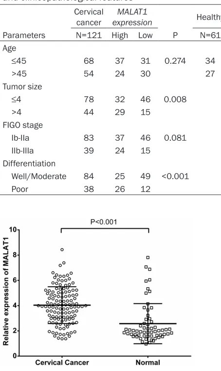
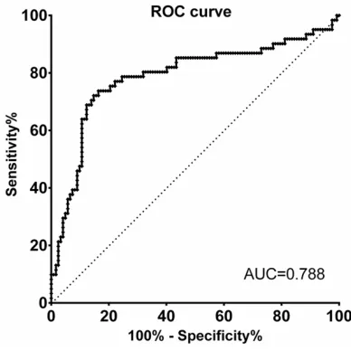
[image:3.612.90.317.92.473.2]To identify plasma MALAT1 expression in cervi-cal cancer and healthy cases, we firstly quanti-fied its expression in 122 cervical cancer cases and 61 age-matched healthy controls by qRT-PCT. Plasma relative level of MALAT1 was plot-ted in the form of scatter dots in Figure 1. As it shown, relative expression of MALAT1 in cervi-cal cancer patients was 3.97 (2.97, 5.07), while that in healthy participants was 1.98 (1.61, 2.81). Statistically significant difference can be observed between cervical cancer patients and healthy participants (P<0.001). We further eval-uated the diagnostic value of MALAT1 in distin-guishing cervical cancer and normal people by ROC curves and AUC values. Figure 2 shows Table 1. The association between MALAT1 expression
and clinicopathological features Cervical
cancer expressionMALAT1 Healthy
Parameters N=121 High Low P N=61
Age
≤45 68 37 31 0.274 34
>45 54 24 30 27
Tumor size
≤4 78 32 46 0.008
>4 44 29 15
FIGO stage
Ib-IIa 83 37 46 0.081
IIb-IIIa 39 24 15
Differentiation
Well/Moderate 84 25 49 <0.001
Poor 38 26 12
Mann-Whitney test, while the relationship between MALAT1 expression and clinico-pathological characteristics was assessed using Pearson’s χ2 test. Area under the ROC curve (AUC) was generated to assess the diagnostic values of the LncRNA MALAT1. For survival analyzes, we used the Kaplan-Meier method to analyze the correlation between MALAT1 expression and overall actual survival, and the log-rank test to compare survival curves. The Cox proportional hazards model was used to make multivariate survival analysis for all of the significant parameters observed in the univariate analysis and transaction-al antransaction-alysis was performed to investigate the interaction between clinicopathologi-cal characteristics, MALAT1 expression and survival.
the ability of MALAT1 to diagnose cervical can-cer patients. The AUC was 0.788 (0.709-0.867). We got the best cuff off value of 2.51, with sen-sitivity of 72.13% (59.17%-82.85%) and speci-ficity of 85.25% (77.69%-91.02%).
To assess to association between plasma MALAT1 expression and the clinicopathological parameters, the 122 cervical cancer patients were divided into high MALAT1 expression (higher than the median level, n=61) and low MALAT1 expression (lower than the median level, n=61) groups according to the median level of MALAT1 expression (3.97). Comparisons were performed between the two groups (Table 1). The results showed that high MALAT1 expression was significantly correlated with
tumor size (P=0.008) and differentiation (P<0.001), however age (P=0.274) and FIGO stage (P=0.081) did not show a significant association with MALAT1 expression.
High MALAT1 predicts a poor prognosis of cervical cancer
Overall survival curves in the high MALAT1 group and low MALAT1 group were given in Figure 3. Cervical cancer patients with high MALAT1 expression had significantly poorer overall survival (P<0.001) than that with low MALAT1 expression. As shown in Table 2, uni-variate analysis showed that HRs (95% CI, P
value) of age (>45 years vs. ≤45 years), tumor size (>4 cm vs. ≤4 cm), FIGO stage (IIb-IIIa vs. Ib-IIa), differentiation(poor vs. well/moderate) and MALAT1 expression (high vs. low) were 1.01 (0.52-1.92, P=0.994), 2.32 (1.21-4.31, P=0.011), 2.94 (1.54-5.63, P=0.001), 2.96 (1.51-5.62, P=0.001), 3.52 (1.66-7.47, P= 0.001), respectively. Then we applied multivari-ate analysis using the Cox proportional hazards model including these parameters and found that tumor size, FIGO stage and MALAT1 expres-sion were independent prognostic factors for overall survival, with HRs of 1.97 (1.01-3.82, P=0.046), 2.46 (1.23-4.80, P=0.008), 2.46 (1.11-5.45, P=0.027), respectively.
Interaction between MALAT1 and other risk factors
[image:4.612.91.285.72.266.2]Table 3 shows the interaction between MALAT1 and other parameters. Compared with refer-ence, univariate analysis showed that HRs of Age & MALAT1 (>45 years & high), Tumor size & MALAT1 (>4 cm & high), FIGO stage & MALAT1 (IIb-IIIa & high), Differentiation & MALAT1 (Poor & high) were 3.67 (1.31-10.33), 6.34 (2.32-17.33), 11.64 (3.34-40.56), 8.53 (2.80-25.94), respectively (all P<0.05). However, we only ob- served significant interaction between Diffe- rentiation & MALAT1, with P value of 0.035. Then multivariate analysis were performed to calculate adjusted HRs. Compared with refer-ence, HRs of Age & MALAT1 (>45 years & high), Tumor size & MALAT1 (>4 cm & high), FIGO stage & MALAT1 (IIb-IIIa& high), Differentiation & MALAT1 (Poor & high) were 1.95 (0.64-5.97, P=0.244), 4.74 (1.64-13.70, P=0.004), 8.83 (2.42-32.26, P=0.001), 5.82 (1.85-18.25, P= 0.003). No significant interaction between MALAT1 expression and other risk factors was Figure 2. Receiver operating characteristic curve
[image:4.612.90.288.325.458.2]analysis of plasma MALAT1 in cervical cancer pa-tients and healthy participants.
observed, suggesting MALAT1 may be a stable prognosis factor for overall survival of cervical cancer patients.
Discussion
In this study, we confirmed that LncRNA MALAT1 expression is significantly increased in plasma of cervical cancer patients than that of normal participants. In addition, we also found high MALAT1 expression predicts a poor pr- ognosis of cervical cancer. Furthermore, we showed that MALAT1 expression is correlated with tumor size, FIGO stage, and differentiation. More importantly, we demonstrated that there is no significant interaction between MALAT1 expression and other risk factors, suggesting MALAT1 may be a stable prognosis factor for overall survival.
In recent years, with the emergence of sequenc-ing technique, widespread existence of lncRNAs in cervical cancer has been confirmed as well as their action mechanisms and important bio-logical functions have gradually been elucidat-ed. Hox transcript antisense intergeniclncRNA, named HOTAIR, has been proven to make criti-cal effect on the most biologicriti-cal process of cer-vical cancer, via upregulating VEGF, MMP-9 and EMT-related genes and would be a potential new target in predicting recurrence and progno-sis [14-16]. Additional studies have indicated
LAT1 as a noncoding transcript enriched in the nucleus of human primary fibroblasts or trans-formed lymphoblasts [23, 24]. Since its discov-ery a decade ago, a large amount of data have accumulated that link MALAT1 to other cancer types or diseases and provided insights into its biogenesis, interaction partners and cellular, as well as molecular functions [11]. In cervical cancer, reduction of MALAT1 in CaSki cervical cancer cells affects cervical cancer cell growth, cell cycle progression, and invasion through the regulation of gene expression, such as cas-pase-3, -8, Bax, Bcl-2, and BclxL [13]. Recently, a study showed that MALAT1 expression was memorably increased in tumorous tissue in comparison with adjacent tissue and was cor-related with the size, FIGO stage, vessel inva-sion, and lymphatic diffusion acting indepen-dently as a predictive factor on prognosis in cervical cancer [25]. Due to the features of lncRNAs, such as long fragments (>200 nt), easy degradation in plasma, and extremely low concentration of total RNA in plasma, the current detection of plasma lncRNA as tumor markers becomes extremely challenging. How- ever, some studies demonstrated that certain fragments in the plasma are highly stable and abundant [26, 27]. Chen et al. [28] indicated a specific stable MALAT1 fragment existed in plasma, which was identified by a previous study, thus making circulating lncRNA expres-sion detection available [29].
Table 2. Univariate and multivariate analysis of risk factors for overall survival
Univariate analysis Multivariate analysis
Parameters HR (95% CI) P HR (95% CI) P
Age
≤45 Ref Ref
>45 1.01 (0.52-1.92) 0.994 0.98 (0.50-1.94) 0.962 Tumor size
≤4 Ref Ref
>4 2.32 (1.21-4.31) 0.011 1.97 (1.01-3.82) 0.046 FIGO stage
Ib-IIa Ref Ref
IIb-IIIa 2.94 (1.54-5.63) 0.001 2.46 (1.23-4.80) 0.008 Differentiation
Well/Moderate Ref Ref
Poor 2.96 (1.51-5.62) 0.001 1.69 (0.83-3.43) 0.148 MALAT1
Low Ref Ref
High 3.52 (1.66-7.47) 0.001 2.46 (1.11-5.45) 0.027
Previous studies showed that MALAT1 expres-sion was increased in tumorous tissue and was correlated with the size, FIGO stage, vessel invasion, and lymphatic diffusion. In present study, we firstly assessed the relevance of plas-ma MALAT1 to survival of cervical cancer and demonstrated that high plasma MALAT1 ex- pression would also predict a poor prognosis of cervical cancer. Additionally, interaction analy-sis was performed between plasma MALAT1 expression and other clinical factors in predict-ing the prognosis of cervical cancer. Result showed no significant interaction between MALAT1 expression and other factors by multi-variate analysis, indicating that plasma MALAT1 would be a stable and noninvasive biomarker for cervical cancer prognosis.
Obviously, the study on circulating LncRNA profiles offer an exciting expectation. However, some limitations and merit comments in this study should be mentioned. The population of enrolled patients and healthy participants were relatively small. Further study on a larger sam-ple and longer follow-up is needed to confirm our results. Secondly, personal information
were relatively insufficient. Lifestyle and dietary factors, which may be helpful, should be record-ed. Finally, the plasma have been stored at -80°C until use. Immediate analysis should be taken after blood samples are collected for a better reflection of real condition.
In conclusion, above all, our results extend the findings of previous studies about MALAT1 in cervical cancer patients. Our data provide plas-ma MALAT1 was significantly higher in cervical cancer compared to normal participants, and demonstrated the ideal prognosis value to pre-dict the survival of patients with cervical cancer.
Acknowledgements
The study was supported by the Health Department of the Zhejiang Province (No. 201345919).
Disclosure of conflict of interest
None.
Address correspondence to: Haitao Ren, Depart- ment of Burns and Wound Care Center, The Second Table 3. The interaction between plasma MALAT1 and other risk factors in predicting prognosis of cervical cancer patients
Parameters N Unadjusted HR (95% CI) P interactionP for Adjusted HR (95% CI) P interactionP for Age & MALAT1
≤45 & low 31 Ref 0.343 Ref 0.279
≤45 & high 37 2.53 (0.69-6.98) 0.074 1.61 (0.56-4.61) 0.372 >45 & low 30 0.70 (0.19-2.62) 0.594 0.52 (0.14-1.99) 0.340 >45 & high 24 3.67 (1.31-10.33) 0.014 1.95 (0.64-5.97) 0.244
Tumor size & MALAT1 0.757
≤4 & low 46 Ref Ref 0.912
≤4 & high 32 3.51 (1.23-9.96) 0.019 2.36 (0.79-7.06) 0.123 >4 & low 15 2.30 (0.62-8.57) 0.215 1.84 (0.48-7.06) 0.374 >4 & high 29 6.34 (2.32-17.33) <0.001 4.74 (1.64-13.70) 0.004
FIGO stage & MALAT1 0.092
Ib-IIa & low 46 Ref
Ib-IIa & high 37 6.34 (1.82-22.1) 0.004 4.80 (1.33-17.29) 0.017 0.131 IIb-IIIa & low 15 6.88 (1.71-29.62) 0.007 6.05 (1.50-24.45) 0.012
IIb-IIIa & high 24 11.64 (3.34-40.56) <0.001 8.83 (2.42-32.26) 0.001 Differentiation & MALAT1
Well/Moderate & low 49 0.035 Ref 0.068
Affiliated Hospital, School of Medicine, Zhejiang University, Hangzhou 310009, China. E-mail: [email protected]
References
[1] Jemal A, Bray F, Center MM, Ferlay J, Ward E and Forman D. Global cancer statistics. CA Cancer J Clin 2011; 61: 69-90.
[2] Parkin DM, Bray F, Ferlay J and Pisani P. Global cancer statistics, 2002. CA Cancer J Clin 2005; 55: 74-108.
[3] Tewari KS, Sill MW, Long HJ 3rd, Penson RT, Huang H, Ramondetta LM, Landrum LM, Oaknin A, Reid TJ, Leitao MM, Michael HE and Monk BJ. Improved survival with bevacizumab in advanced cervical cancer. N Engl J Med 2014; 370: 734-743.
[4] Schmitz SU, Grote P and Herrmann BG. Mech-anisms of long noncoding RNA function in de-velopment and disease. Cell Mol Life Sci 2016; 73: 2491-2509.
[5] He JH, Han ZP and Li YG. Association between long non-coding RNA and human rare diseas-es (Review). Biomed Rep 2014; 2: 19-23. [6] Ernst C and Morton CC. Identification and
func-tion of long non-coding RNA. Front Cell Neuro-sci 2013; 7: 168.
[7] Lorenzen JM and Thum T. Long noncoding RNAs in kidney and cardiovascular diseases. Nat Rev Nephrol 2016; 12: 360-373.
[8] Jiang C, Li X, Zhao H and Liu H. Long non-cod-ing RNAs: potential new biomarkers for pre-dicting tumor invasion and metastasis. Mol Cancer 2016; 15: 62.
[9] Evans JR, Feng FY and Chinnaiyan AM. The bright side of dark matter: lncRNAs in cancer. J Clin Invest 2016; 126: 2775-2782.
[10] Schmitt AM and Chang HY. Long Noncoding RNAs in cancer pathways. Cancer Cell 2016; 29: 452-463.
[11] Gutschner T, Hammerle M and Diederichs S. MALAT1--a paradigm for long noncoding RNA function in cancer. J Mol Med (Berl) 2013; 91: 791-801.
[12] Peng L, Yuan X, Jiang B, Tang Z and Li GC. Ln-cRNAs: key players and novel insights into cer-vical cancer. Tumour Biol 2016; 37: 2779-2788.
[13] Guo F, Li Y, Liu Y, Wang J, Li Y and Li G. Inhibi-tion of metastasis-associated lung adenocarci-noma transcript 1 in CaSki human cervical cancer cells suppresses cell proliferation and invasion. Acta Biochim Biophys Sin (Shanghai) 2010; 42: 224-229.
[14] Wu Y, Zhang L, Wang Y, Li H, Ren X, Wei F, Yu W, Wang X, Zhang L, Yu J and Hao X. Long noncod-ing RNA HOTAIR involvement in cancer. Tumour Biol 2014; 35: 9531-9538.
[15] Li J, Wang Y, Yu J, Dong R and Qiu H. A high level of circulating HOTAIR is associated with progression and poor prognosis of cervical cancer. Tumour Biol 2015; 36: 1661-1665. [16] Kim HJ, Lee DW, Yim GW, Nam EJ, Kim S, Kim
SW and Kim YT. Long non-coding RNA HOTAIR is associated with human cervical cancer pro-gression. Int J Oncol 2015; 46: 521-530. [17] Sun NX, Ye C, Zhao Q, Zhang Q, Xu C, Wang SB,
Jin ZJ, Sun SH, Wang F and Li W. Long noncod-ing RNA-EBIC promotes tumor cell invasion by binding to EZH2 and repressing E-cadherin in cervical cancer. PLoS One 2014; 9: e100340. [18] Naemura M, Murasaki C, Inoue Y, Okamoto H
and Kotake Y. Long noncoding RNA ANRIL reg-ulates proliferation of non-small cell lung can-cer and can-cervical cancan-cer cells. Anticancan-cer Res 2015; 35: 5377-5382.
[19] Yang M, Zhai X, Xia B, Wang Y and Lou G. Long noncoding RNA CCHE1 promotes cervical can-cer cell proliferation via upregulating PCNA. Tumour Biol 2015; 36: 7615-7622.
[20] Cao S, Liu W, Li F, Zhao W and Qin C. Decreased expression of lncRNA GAS5 predicts a poor prognosis in cervical cancer. Int J Clin Exp Pathol 2014; 7: 6776-6783.
[21] Liao LM, Sun XY, Liu AW, Wu JB, Cheng XL, Lin JX, Zheng M and Huang L. Low expression of long noncoding XLOC_010588 indicates a poor prognosis and promotes proliferation through upregulation of c-Myc in cervical can-cer. Gynecol Oncol 2014; 133: 616-623. [22] Zhang J, Yao T, Wang Y, Yu J, Liu Y and Lin Z.
Long noncoding RNA MEG3 is downregulated in cervical cancer and affects cell proliferation and apoptosis by regulating miR-21. Cancer Biol Ther 2016; 17: 104-113.
[23] Ji P, Diederichs S, Wang W, Boing S, Metzger R, Schneider PM, Tidow N, Brandt B, Buerger H, Bulk E, Thomas M, Berdel WE, Serve H and Muller-Tidow C. MALAT-1, a novel noncoding RNA, and thymosin beta4 predict metastasis and survival in early-stage non-small cell lung cancer. Oncogene 2003; 22: 8031-8041. [24] Hutchinson JN, Ensminger AW, Clemson CM,
Lynch CR, Lawrence JB and Chess A. A screen for nuclear transcripts identifies two linked noncoding RNAs associated with SC35 splic-ing domains. BMC Genomics 2007; 8: 39. [25] Yang L, Bai HS, Deng Y and Fan L. High MALAT1
expression predicts a poor prognosis of cervi-cal cancer and promotes cancer cell growth and invasion. Eur Rev Med Pharmacol Sci 2015; 19: 3187-3193.
plas-ma-based biomarker for diagnosing prostate cancer. Eur J Cancer 2013; 49: 2949-2959. [27] Ng EK, Tsui NB, Lau TK, Leung TN, Chiu RW,
Panesar NS, Lit LC, Chan KW and Lo YM. mRNA of placental origin is readily detectable in maternal plasma. Proc Natl Acad Sci U S A 2003; 100: 4748-4753.
[28] Chen Q, Su Y, He X, Zhao W, Wu C, Zhang W, Si X, Dong B, Zhao L, Gao Y, Yang X, Chen J, Lu J, Qiao X and Zhang Y. Plasma long non-coding RNA MALAT1 is associated with distant metas-tasis in patients with epithelial ovarian cancer. Oncol Lett 2016; 12: 1361-1366.