Original Article
Programmed cell death ligand 1 (PD-L1) expression on
gastric cancer and its relationship with
clinicopathologic factors
Lin Zhang1*, Miaozhen Qiu2,3*, Ying Jin2*, Jiao Ji4, Baoxia Li4, Xueping Wang1, Shumei Yan5, Ruihua Xu2, Dajun Yang4
Departments of 1Clinical Laboratory, 2Medical Oncology, 4Experimental Research, 5Pathology, Sun Yat-Sen University Cancer Center, State Key Laboratory of Oncology in South China, Collaborative Innovation Center for Cancer Medicine, Guangzhou 510060, China; 3Department of Oncology, The Sidney Kimmel Comprehensive Cancer Center, The Johns Hopkins University School of Medicine, Baltimore, MD 21231, USA. *Equal contributors. Received July 27, 2015; Accepted August 28, 2015; Epub September 1, 2015; Published September 15, 2015
Abstract: Background: Targeting the immune checkpoints in solid tumors becomes hot recently. Programmed cell death ligand 1 (PD-L1) is ligand for programmed death 1 (PD-1), which is known to negatively regulate T-cell activa-tion. In the present study, we investigated the expression of PD-L1 in tumor specimens of gastric cancer and its relationships with clinicopathological variables and survival. Methods: The expression of PD-L1 in 132 surgically resected specimens of stage II and III gastric cancer was evaluated by immunohistochemistry in microarray tissue. Results: Expression of PD-L1 was observed in 50.8% (67/132) of gastric cancer tumor specimens. Patients whose tumor size over 5cm had a higher positive rate of PD-L1 expression. There was no relationship between the expres-sion of PD-L1 and other clinicopathological variables including age, gender, clinical stage, location as well as
histo-logical differentiation. PD-L1 positive patients had significantly poorer survival than negative patients. The 5-year
survival rates was 83.1% in those with PD-L1 negative patients and 50.7% for PD-L1 positive patients (P<0.001). The multivariate analysis indicated that both PD-L1 positive and Tumor-node-metastasis stage were independent prognostic factors in gastric cancer patients (P=0.001 and 0.025, respectively). Conclusions: The expression of PD-L1 was found in half of stages II and III gastric cancer patients. Positive of PD-PD-L1 expression indicated poor survival in Chinese stages II and III gastric adenocarcinoma patients. These results may provide the clue for immunotherapy in the adjuvant treatment setting of gastric cancer patients.
Keywords: Clinicopathologic features, gastric cancer, PD-L1, prognosis
Introduction
Gastric cancer is the second leading cause of cancer-related death worldwide [1]. The
inci-dence of gastric carcinoma varies significantly
from one part of the world to another and it is particularly common in Eastern Asia, especially in China [2]. In our previous report, we found out that at the time of diagnosis, stage II and III gastric cancer patients accounted for about 28% and 33% of the whole population
respec-tively [3]. The five-year survival rate for the
stage II and III gastric cancer patients was around 70% and 40% respectively [3, 4]. Stages II and III gastric cancer patients are a special group of patients. They have the potential to be cured, however, they are also under threaten to develop disease recurrent or distant
metasta-sis [5]. To figure out the prognostic factors for
this group of patients would be helpful to pick up patients who need close follow up.
In normal condition, PD-L1 is expressed on the endothelium in the thymus, heart, and placenta in both human and mice [9, 13, 14] in addition to lymphoid cells, such as activated T cells, B cells, macrophages, and dendritic cells on the protein level. PD-L1 is also abundant on tumor tissues, including lung carcinomas, ovarian car-cinomas, breast carcar-cinomas, glioblastoma, and squamous cell carcinoma of the head and neck [13-16]. In 2015 American Society of Clinical Oncology Gastrointestinal Cancers Symposium there was a poster about the relationship between PD-L1 expression and clinical out-comes in patients with advanced gastric can-cer treated with the PD-1 monoclonal anti-body Pembrolizumab. In this study, they found that 40% of the advanced gastric cancer patients had PD-L1 positive tumors. They enrolled 39 patients to receive the monoclonal antibody treatment and the response rate was 22.2%. They also observed a trend toward improved outcomes with higher levels of PD-L1
patients: 94 males and 38 females with a median age of 62 years (range 24-80 years). The histological types of the patients were all adenocarcinoma, including 25 signet ring cells, 59 low differentiated, 42 moderate differenti-ated and 6 high differentidifferenti-ated. Staging was performed according to the
tumor-node-metas-tasis (TNM) classification of the American Joint
Committee on Cancer (AJCC, sixth edition,
2002). Twenty-one of them were classified as stage II tumors and the rest 111 were classified
as stage III patients. No patient underwent radi-ation or chemotherapy before surgery. 63 (47.7%) patients received adjuvant chemother-apy. Forty (30.3%) patients received D1 resec-tion and the rest 92 patients received D2 resection. The median follow-up was 66.0 months (range 3.0-153.0 months).
[image:2.612.91.318.98.401.2]Tissue specimens were obtained from the his-topathology archive in the Department of Pathology, Cancer Center of Sun Yat-sen
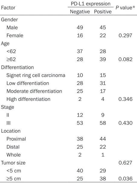
Table 1. Distribution of 132 patients according to PD-L1 expression status
Factor PD-L1 expression P value* Negative Positive
Gender
Male 49 45
Female 16 22 0.297
Age
<62 37 28
≥62 28 39 0.082
Differentiation
Signet ring cell carcinoma 10 15 Low differentiation 28 31 Moderate differentiation 25 17
High differentiation 2 4 0.346 Stage
II 12 9
III 53 58 0.430
Location
Proximal 38 44
Distal 25 22
Whole 2 1
Tumor size 0.627
<5 cm 40 29
≥5 cm 25 38 0.036
*The fisher’s exact test was used to evaluate the correlation be -tween PD-L1 expression and the clinicopathologic characteristics.
P<0.05 indicates statistical significance. PD-L1: Programmed cell death ligand 1.
expression. However, there is no data about the prognostic value of PD-L1 in gastric can-cer. To better understand the role of PD-L1 in gastric cancer, we started this study. In the present study, using immunohistochem-istry, we investigated the expression of PD-L1 in tumor specimens of gastric can-cer, and we analyzed the relationship between the expression and clinic patho-logical variables as well as the postopera-tive survival.
Materials and methods
Patients
Between January 1996 and December 2004, the medical records of pathology-proven gastric adenocarcinoma patients who were diagnosed and received radical resection in the Cancer Center of Sun Yat-Sen University were retrospectively ana-lyzed. We excluded patients who received neoadjuvant chemotherapy, or had a histo-ry of other primahisto-ry cancer. All patients received gastrectomy and D1/2
lymphade-nectomy. We identified 200 patients with
gastric adenocarcinoma in our institution but excluded 32 patients because of miss-ing baseline characteristics, 31 patients because of incomplete follow-up and 5 patients who died of non-malignant related reasons (car accidents and coronary heart
University. Histological examinations were car-ried out on tissue preparations with hematoxy-lin and eosin (H&E) staining. The best tissue sections that suited for immunohistochemistry were selected by the pathologist and the
satis-fying formalin-fixed paraffin-embedded resec -tion specimens were obtained.
Tissue microarray construction
Formalin-fixed, paraffin-embedded tumor sam -ples were taken from the selected cases. Tissue microarray construction was prepared as described by Kononen et al [17]. Three 1.0-mm tumor tissue cores were taken from each tumor sample. The arraying machine was from Beecher Instruments (Sun Prairie, WI). The
array blocks were cut to produce 4-μm
sections.
Immunohistochemistry
Slides were dewaxed and rehydrated in xylene and graded ethanol solutions for antigen retrieval. Slides were then blocked with 3% H2O2, goat serum, avidin solution, and biotin solution. Primary PD-L1 [(ab58810) Rabbit polyclonal, Abcam, 1:50] was added and then probed with biotinylated rabbit secondary anti-body (Vector Laboratories) and high-sensitivity streptavidin-HRP conjugate. To visualize
stain-ing, slides were incubated in 3,3’-diaminobenzi
-dine in 0.1% H2O2 in Tris-HCl buffer, and subse-quently counterstained with Hematoxylin QS (Vector Laboratories). PDL1-positive samples
were defined as those showing cytoplasmic
staining pattern of tumor tissue. PD-L1 staining intensity was graded into four groups: no stain-ing (0), weak stainstain-ing (1+), moderate stainstain-ing (2+), and intense staining (3+). Tumors without
staining and weak staining were classified as
negative while tumors which were moderate or
intense staining were classified as positive. The
immunostained slide was evaluated under the microscope. The staining intensity of cells showing positive membrane and cytoplasmic staining for PDL1 was calculated by reviewing the entire spot.
Analysis of immunohistochemistry was carried out by two independent observers (Shumei Yan and Lin Zhang) who were blinded to any prior information on clinic pathological features of
the patients’ samples. If there was difference
between these two observers, these slides were reinvestigated by both investigators using a multiheaded microscope.
Statistical analysis
[image:3.612.89.523.72.280.2]All statistical analyses were performed by Statistical Package of Social Sciences 16.0 software for Windows (SPSS, Inc., Chicago, IL). P value <0.05 was considered to be statistically
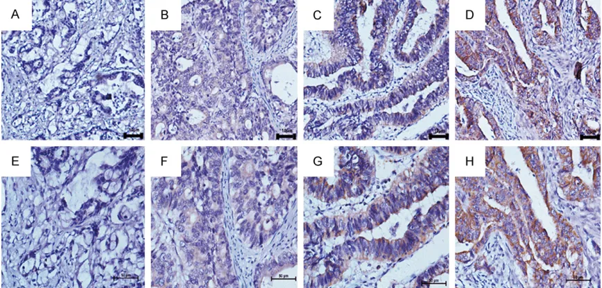
Figure 1. The expression of PD-L1 in gastric adenocarcinoma tissues by IHC. (A) Negative expression of PD-L1 protein (100×). (B) Weak expression of PD-L1 protein (100×), (C) Moderate expression of PD-L1 protein (100×), (D)
Intense expression of PD-L1 protein (100×). (E-H) demonstrate the higher magnification (400×) from the area of the
significant. All tests were two-tailed. The
Kaplan-Meier method was used to estimate the 5-year overall survival. For patients who remained alive, data were censored at the date
[image:4.612.90.359.71.310.2]expression. Patients whose tumor size over 5 cm had a higher positive rate of PD-L1 expres-sion (P=0.036). There was no statistical differ-ence between the expression of PD-L1 and
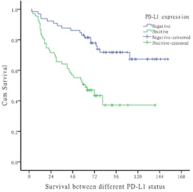
Figure 2. The overall survival of the whole population.
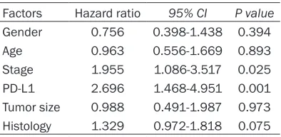
Figure 3. The survival of gastric cancer patients according to the expres-sion of PD-L1.
of the last contact. Kaplan-Meier analysis with log-rank testing was used for univariate analysis. Variables showing a trend for association with survival (P<
0.05) were selected in the final
multivariate Cox proportional hazards model. The Chi-squared test and t test were used to com-pare the relationship between PD-L1 expression and the vari-ous clinicopathological charac-teristics of the patients.
Ethics, consent and permissions
All patients provided written informed consent. Study approv-al was obtained from indepen-dent ethics committees at Cancer Center of Sun Yat-Sen University. The study was under-taken in accordance with the ethical standards of the World Medical Association Declaration of Helsinki.
Results
The characteristics of the 132 gastric adenocarcinoma patients are shown in Table 1. Until December 2014, there were 55 patients died from the disease. Expression of PD-L1 in surgically resected specimens
In immunohistochemical stain-ing of PD-L1, the protein expres-sion in gastric adenocarcinoma patients was 50.8% (67/132) (Figure 1 and Table 1).
PD-L1 expression and clinic pathological features
For the correlation with clinico-pathological features, Table 1
[image:4.612.91.362.361.631.2]age, gender, disease stage, histological grade and tumor location.
Effect of PD-L1 expression on survival
Till December 2014, 55 patients had died of
cancer. The 1, 3, 5, 7-year disease specific sur -vival rate of the entire cohort was 93.9%, 77.3%, 70.5%, 66.7% respectively. Figure 2
showed the survival curve of the whole group of patients.
There was significantly difference between
PD-L1 positive and negative patients. (5-year survival rates: 83.1% in those with PD-L1 nega-tive patients and 50.7% in those with PD-L1 positive patients, P<0.001, Figure 3).
Univariate analysis showed that tumor stages (P<0.001), smaller tumor size (P=0.003), high tumor differentiation (P=0.016) and PD-L1 neg-ative expression (P<0.001) were all significant -ly associated with longer survival.
To test the independent prognostic effect of
these clinical factors, Cox’s proportional hazard
model was applied. The results revealed that tumor stage (P=0.025) and PD-L1 expression (P=0.001) remained independent prognostic variables for survival when age, gender, tumor size, tumor stage, PD-L1 and differentiation were all included for test (Table 2).
Discussion
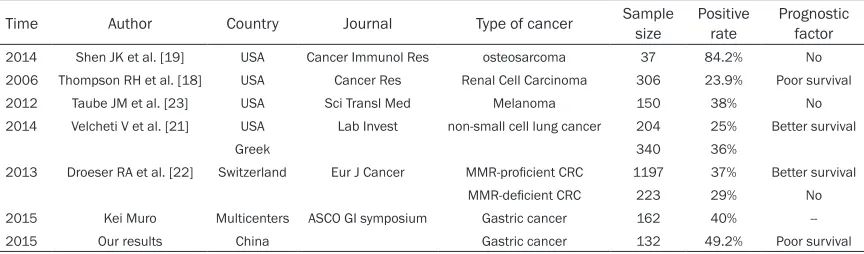
We have demonstrated the expression of PD-L1 in surgically resected specimens of gastric can-cer. The expression of PD-L1 is found in cyto-plasm of cancer cells. The expression pattern of PD-L1 was consistent with previous reports, which examined their expression in human tumor tissues [13-16]. The positive rate of PD-L1 in cancer patients varied in different
cancer types. Table 3 listed some of the results using the method of immunohistochemistry to detect the expression of PD-L1 in human tumor tissues. It ranged from 23.9%-84.2%. In 2015 American Society of Clinical Oncology Gas- trointestinal Cancers Symposium there was a poster about the relationship between PD-L1 expression and clinical outcomes in patients with advanced gastric cancer treated with the anti-PD-1 monoclonal antibody Pembrolizumab. They found that 40% of the advanced gastric cancer patients had PD-L1 positive tumors. The present study also examined the relation-ship between the expression of PD-L1 and clini-copathological variables as well as the progno-sis of gastric cancer patients. In our analyses, there was no correlation of PD-L1 expression with clinicopathologic features except for tumor size. Thompson RH et al. found that renal cell carcinoma patients with PD-L1 expression were more likely to exhibit adverse pathologic features including advanced tumor stage, tumor size over 5 cm, nuclear grade 3 or 4, and coagulative tumor necrosis [18]. However, in the disease of osteosarcoma and non-small cell lung cancer, the expression of PD-L1 had no relationship with the clinicopathologic vari-able [19, 20].
As for the prognostic value of PD-L1 in the malignant disease, several previous studies
had demonstrated conflicting results varying
from positive correlation of prognosis in non-small cell lung cancer [21] and mismatch repair
(MMR)-proficient colorectal cancer [22], to
observing no correlation with survival in
osteo-sarcoma, Melanoma as well as MMR-deficient
Colorectal cancer [19, 22, 23], to an inverse relationship in which high PD-L1 expression was associated with poor survival in renal cell carcinoma [18, 24]. To our knowledge, this is
the first study to analyze the prognostic value of
[image:5.612.91.288.95.191.2]PD-L1 in gastric cancer patients. We found that PD-L1 positive was associated with poor prog-nosis. The multivariate analysis indicated that PD-L1 positive was an independent prognostic factor in stage II and III gastric cancer patients. Immunotherapy has made some progress in several cancers, such as melanoma and non-small cell lung cancer [25]. Recently, immuno-therapy got its indication in the lung squamous cancer. In the multicenter phase 1 trial, anti-PD-L1 antibody was given to several kinds of cancer including gastric cancer (seven pati-
Table 2. Multivariate analysis of overall sur-vival in gastric cancer
Factors Hazard ratio 95% CI P value Gender 0.756 0.398-1.438 0.394 Age 0.963 0.556-1.669 0.893 Stage 1.955 1.086-3.517 0.025 PD-L1 2.696 1.468-4.951 0.001 Tumor size 0.988 0.491-1.987 0.973 Histology 1.329 0.972-1.818 0.075
ents), but no clinical benefit was found in the
gastric cancer patients [25]. Till now most of the target therapy agent failed in the gastric cancer except for trastuzumab and ramucirum-ab [26, 27]. We still have no idea whether anti
PD-L1 antibody would bring survival benefit to
the advanced gastric cancer patients. At least basing on our result, we found out that PD-L1 was an independent prognostic factor for stage II and III gastric cancer patients. Patients who were PD-L1 positive deserved closer follow up. There were some limitations of our study. Firstly, we only collected patients of stage II and III. It would be a good idea to explore the patient population to stages I to IV patients and ana-lyzed the relationship of PD-L1 expression and tumor stage. Secondly, the sample size of our study was small. Even for the limitations here,
this is the first study to evaluate the prognostic
value of PD-L1 in Chinese gastric adenocarci-noma patients. We found that about half of the patients had PD-L1 positive expression and PD-L1 positive indicated a poor survival. In conclusion, The authors are not aware of any previous studies which address the clinicopath-ological characteristics and prognostic impact of PD-L1 with resectable gastric cancer in China. In this retrospective study conducted with 132 patients with gastric adenocarcinoma we came to the following conclusions: 1) we demonstrated the positive rate of PD-L1 in gas-tric cancer patients. 2) The expression of PD-L1 was associated with larger tumor size. 3) PD-L1 positive expression was associated with poor survival.
Acknowledgements
We gratefully thank our colleagues in the department of medical oncology. This work was
supported by: 1. The third outstanding young talents training plan of Sun Yat-Sen University
cancer center. 2. Medical Scientific Research of Guangdong province B2014161. 3. Scientific
and Technological projects Guangdong Esoph- ageal Cancer Institute Q201408.
Disclosure of conflict of interest
None.
Address correspondence to: Dr. Ruihua Xu, De- partment of Medical Oncology, Sun Yat-Sen Uni- versity Cancer Center, 651 Dong Feng Road East, Guangzhou 510060, China. Tel: +86-20-87343333; Fax: +86-20-8734 3295; E-mail: [email protected]. cn; Dr. Dajun Yang, Department of Experimental Research, Sun Yat-Sen University Cancer Center, 651 Dong Feng Road East, Guangzhou 510060, China. Tel: 87342285; Fax: +86-20-87342285; E-mail: [email protected]
References
[1] Jemal A, Bray F, Center MM, Ferlay J, Ward E and Forman D. Global cancer statistics. CA Cancer J Clin 2011; 61: 69-90.
[2] Moore MA, Eser S, Igisinov N, Igisinov S, Mohagheghi MA, Mousavi-Jarrahi A, Ozenturk G, Soipova M, Tuncer M and Sobue T. Cancer epidemiology and control in North-Western and Central Asia - past, present and future. Asian Pac J Cancer Prev 2010; 11 Suppl 2: 17-32.
[3] Qiu MZ, Wang ZQ, Zhang DS, Liu Q, Luo HY, Zhou ZW, Li YH, Jiang WQ and Xu RH. Comparison of 6th and 7th AJCC TNM staging
classification for carcinoma of the stomach in
China. Ann Surg Oncol 2011; 18: 1869-1876. [4] Park JM, Kim JH, Park SS, Kim SJ, Mok YJ and
[image:6.612.90.522.85.212.2]Kim CS. Prognostic factors and availability of D2 lymph node dissection for the patients with stage II gastric cancer: comparative analysis of
Table 3. The expression of PD-L1 in different kinds of tumors
Time Author Country Journal Type of cancer Sample size Positive rate Prognostic factor
2014 Shen JK et al. [19] USA Cancer Immunol Res osteosarcoma 37 84.2% No
2006 Thompson RH et al. [18] USA Cancer Res Renal Cell Carcinoma 306 23.9% Poor survival
2012 Taube JM et al. [23] USA Sci Transl Med Melanoma 150 38% No
2014 Velcheti V et al. [21] USA LabInvest non-small cell lung cancer 204 25% Better survival
Greek 340 36%
2013 Droeser RA et al. [22] Switzerland Eur J Cancer MMR-proficient CRC 1197 37% Better survival
MMR-deficient CRC 223 29% No
2015 Kei Muro Multicenters ASCO GI symposium Gastric cancer 162 40%
subgroups in stage II. World J Surg 2008; 32: 1037-1044.
[5] Jin Y, Qiu MZ, Wang DS, Zhang DS, Ren C, Bai L, Luo HY, Wang ZQ, Wang FH, Li YH and Xu RH. Adjuvant chemotherapy for elderly patients with gastric cancer after D2 gastrectomy. PLoS One 2013; 8: e53149.
[6] Dong H, Zhu G, Tamada K and Chen L. B7-H1, a third member of the B7 family, co-stimulates T-cell proliferation and interleukin-10 secre-tion. Nat Med 1999; 5: 1365-1369.
[7] Latchman Y, Wood CR, Chernova T, Chaudhary D, Borde M, Chernova I, Iwai Y, Long AJ, Brown
JA, Nunes R, Greenfield EA, Bourque K,
Boussiotis VA, Carter LL, Carreno BM, Malenkovich N, Nishimura H, Okazaki T, Honjo T, Sharpe AH and Freeman GJ. PD-L2 is a sec-ond ligand for PD-1 and inhibits T cell activa-tion. Nat Immunol 2001; 2: 261-268.
[8] Carter L, Fouser LA, Jussif J, Fitz L, Deng B, Wood CR, Collins M, Honjo T, Freeman GJ and Carreno BM. PD-1:PD-L inhibitory pathway af-fects both CD4(+) and CD8(+) T cells and is overcome by IL-2. Eur J Immunol 2002; 32: 634-643.
[9] Liang SC, Latchman YE, Buhlmann JE, Tomczak MF, Horwitz BH, Freeman GJ and Sharpe AH. Regulation of PD-1, PD-L1, and PD-L2 expres-sion during normal and autoimmune respons-es. Eur J Immunol 2003; 33: 2706-2716. [10] Mozaffarian N, Wiedeman AE and Stevens AM.
Active systemic lupus erythematosus is associ-ated with failure of antigen-presenting cells to express programmed death ligand-1. Rheu- matology (Oxford) 2008; 47: 1335-1341. [11] Nishimura H and Honjo T. PD-1: an inhibitory
immunoreceptor involved in peripheral toler-ance. Trends Immunol 2001; 22: 265-268. [12] Nishimura H, Nose M, Hiai H, Minato N and
Honjo T. Development of lupus-like autoim-mune diseases by disruption of the PD-1 gene encoding an ITIM motif-carrying immunorecep-tor. Immunity 1999; 11: 141-151.
[13] Brown JA, Dorfman DM, Ma FR, Sullivan EL,
Munoz O, Wood CR, Greenfield EA and Freeman
GJ. Blockade of programmed death-1 ligands on dendritic cells enhances T cell activation and cytokine production. J Immunol 2003; 170: 1257-1266.
[14] Dong H, Strome SE, Salomao DR, Tamura H, Hirano F, Flies DB, Roche PC, Lu J, Zhu G, Tamada K, Lennon VA, Celis E and Chen L. Tumor-associated B7-H1 promotes T-cell apop-tosis: a potential mechanism of immune eva-sion. Nat Med 2002; 8: 793-800.
[15] Strome SE, Dong H, Tamura H, Voss SG, Flies DB, Tamada K, Salomao D, Cheville J, Hirano F, Lin W, Kasperbauer JL, Ballman KV and Chen L. B7-H1 blockade augments adoptive T-cell
immunotherapy for squamous cell carcinoma. Cancer Res 2003; 63: 6501-6505.
[16] Wintterle S, Schreiner B, Mitsdoerffer M, Schneider D, Chen L, Meyermann R, Weller M and Wiendl H. Expression of the B7-related molecule B7-H1 by glioma cells: a potential mechanism of immune paralysis. Cancer Res 2003; 63: 7462-7467.
[17] Kallioniemi OP, Kononen J and Sauter G. Introducing tissue microarrays to molecular pathology. Clin Chem 2012; 58: 1717-1718. [18] Thompson RH, Kuntz SM, Leibovich BC, Dong
H, Lohse CM, Webster WS, Sengupta S, Frank I, Parker AS, Zincke H, Blute ML, Sebo TJ, Cheville JC and Kwon ED. Tumor B7-H1 is as-sociated with poor prognosis in renal cell carci-noma patients with long-term follow-up. Cancer Res 2006; 66: 3381-3385.
[19] Shen JK, Cote GM, Choy E, Yang P, Harmon D, Schwab J, Nielsen GP, Chebib I, Ferrone S, Wang X, Wang Y, Mankin H, Hornicek FJ and Duan Z. Programmed cell death ligand 1 ex-pression in osteosarcoma. Cancer Immunol Res 2014; 2: 690-698.
[20] Konishi J, Yamazaki K, Azuma M, Kinoshita I, Dosaka-Akita H and Nishimura M. B7-H1 ex-pression on non-small cell lung cancer cells
and its relationship with tumor-infiltrating lym -phocytes and their PD-1 expression. Clin Cancer Res 2004; 10: 5094-5100.
[21] Velcheti V, Schalper KA, Carvajal DE, Anagnostou VK, Syrigos KN, Sznol M, Herbst RS, Gettinger SN, Chen L and Rimm DL. Programmed death ligand-1 expression in non-small cell lung cancer. Lab Invest 2014; 94: 107-116.
[22] Droeser RA, Hirt C, Viehl CT, Frey DM, Nebiker C, Huber X, Zlobec I, Eppenberger-Castori S, Tzankov A, Rosso R, Zuber M, Muraro MG, Amicarella F, Cremonesi E, Heberer M, Iezzi G, Lugli A, Terracciano L, Sconocchia G, Oertli D, Spagnoli GC and Tornillo L. Clinical impact of programmed cell death ligand 1 expression in colorectal cancer. Eur J Cancer 2013; 49: 2233-2242.
[23] Taube JM, Anders RA, Young GD, Xu H, Sharma R, McMiller TL, Chen S, Klein AP, Pardoll DM, Topalian SL and Chen L. Colocalization of
in-flammatory response with B7-h1 expression in
human melanocytic lesions supports an adap-tive resistance mechanism of immune escape. Sci Transl Med 2012; 4: 127ra137.
[25] Brahmer JR, Tykodi SS, Chow LQ, Hwu WJ, Topalian SL, Hwu P, Drake CG, Camacho LH, Kauh J, Odunsi K, Pitot HC, Hamid O, Bhatia S, Martins R, Eaton K, Chen S, Salay TM, Alaparthy S, Grosso JF, Korman AJ, Parker SM, Agrawal S, Goldberg SM, Pardoll DM, Gupta A and Wigginton JM. Safety and activity of anti-PD-L1 antibody in patients with advanced can-cer. N Engl J Med 2012; 366: 2455-2465. [26] Bang YJ, Van Cutsem E, Feyereislova A, Chung
HC, Shen L, Sawaki A, Lordick F, Ohtsu A, Omu-ro Y, Satoh T, Aprile G, Kulikov E, Hill J, Lehle M, Ruschoff J and Kang YK. Trastuzumab in com-bination with chemotherapy versus chemo-therapy alone for treatment of HER2-positive advanced gastric or gastro-oesophageal junc-tion cancer (ToGA): a phase 3, open-label, ran-domised controlled trial. Lancet 2010; 376: 687-697.