Original Article
Compromised natural killer
cells in pulmonary embolism
Xiaoyu Zhang1*, Qiang Wang2*, Yuqin Shen2, Haoming Song2, Zhu Gong2, Lemin Wang2
1Department of Rheumatology, Tongji Hospital of Tongji University, Shanghai 200065, China; 2Department of Cardiology, Tongji Hospital of Tongji University, Shanghai 200065, China. *Equal contributors.
Received April 28, 2015; Accepted June 22, 2015; Epub July 1, 2015; Published July 15, 2015
Abstract: Objective: The high morbidity, mortality and misdiagnosis rate render pulmonary embolism (PE) as a worldwide health problem. However, the etiology and pathogenesis of this disease have not been well character-ized. Increasing studies indicate infection and immunity play a crucial role in PE. Natural killer (NK) cells act as a bridge between the innate immune and acquired immune. This study aimed to investigate the possible function of NK cells in PE. Methods: Human cDNA microarray analysis was employed to detect genes associated with NK cells in peripheral blood mononuclear cells (PBMCs). Random variance model corrected t-test was used for statisti-cal analysis of differential gene expression. Flow cytometry was performed to detect the CD16+CD56+ NK cells. Results: In the present study, based on gene expression microarray analysis, we showed four inhibitory receptors (KLRB1, KLRD1, KLRF1, KLRG1) and four activating receptors (KLRC1, KLRC3, KLRK1 and NCR1) on NK cells were remarkably down-regulated and the cytological experiment demonstrated the proportion of CD16+CD56+ NK cells
among PBMCs decreased in the PE group. Conclusions: We confirmed the presence of reduced expression of critical
activating as well as inhibitory NK cell receptors and low proportion of CD16+CD56+ NK cells in PE. The consistence between genomic and cytological examination suggests compromised NK cells may contribute to the pathogenesis of PE.
Keywords: Pulmonary embolism, gene, natural killer cell
Introduction
Venous thromboembolism (VTE) represents a spectrum of conditions that include pulmonary embolism (PE), chronic thromboembolic pulmo-nary hypertension (CTEPH) and deep venous thrombosis (DVT). The high morbidity, mortality and misdiagnosis rate render VTE as a world-wide health problem [1]. The American College of Chest Physicians has recommended nine editions of the guideline for the diagnosis and prevention of VTE since 1995 and also posed the concept of risk stratification for pro-phylaxis of VTE [2]. However, the incidence of symptomatic VTE is increasing gradually ins- tead of reducing, partly because the etiology and pathogenesis of this disease have not been well characterized [3].
It has been demonstrated that genetic and environmental factors are involved in the devel-opment of VTE and increasing studies indicate that infection and immunity play a crucial role
sys-tem. What’s more, numerous experiments have worked to demonstrate their ability to readily adjust to the immediate environment and for-mulate antigen-specific immunological memo-ry, fundamental for responding to secondary infections with the same antigen. On the basis of previous findings, this study was undertaken to detect the mRNA expression levels of NK cells in PBMCs isolated from PE patients and controls by the whole human genome microar-ray and Gene Ontology analysis. In addition, flow cytometry was performed to investigate the changes in NK cells in PE patients, which aimed to validate the results from genome analysis.
Materials and methods
Subjects
All subjects were from Tongji Hospital of Tongji University. The diagnostic criteria for pulmo-nary embolism were any two of the following: (1) selective pulmonary angiography showing pulmonary artery occlusion or filling defect; (2) pulmonary ventilation-perfusion scan showing single or multiple perfusion defect, normal or abnormal ventilation, and ventilation-perfusion mismatch; (3) clinical diagnosis: risk factors for PE are present and other cardiovascular dis-eases may be excluded by clinical performance, electrocardiogram and chest film; arterial blood gas analysis, D-dimer detection, echocardiog-raphy and/or chest computed tomogechocardiog-raphy sup-port PE diagnosis. The diagnosis of DVT was based on the criteria previously reported [11] and the criteria for CTEPH was used in accor-dance with the diagnosis of CTEPH [12]. Mali- gnancies, use of immunosuppresants or auto-immune diseases were excluded in all subjects. Healthy controls underwent physical examina-tion before they were recruited from the outpa-tient clinic. The present study was approved by the Ethics Committee of Tongji Hospital and informed consent was obtained before study.
Genomic study
Twenty PE inpatients and 20 controls were randomly selected in Cardiology Department, Tongji Hospital of Tongji University. In the PE group, there were 11 males and 9 females, with a mean age of 70±14 years (44~89 years). There were 13 patients with acute PE and 7 with CTEPH. The pulmonary artery pressure
was 50-108 mmHg in CTEPH patients. In the control group, 20 patients (11 males and 9 females) with a mean age of 72±14 years (44~91 years) were enrolled during the same period. No significant difference in age was found between PE patients and controls (P> 0.05).
Cellular immunology
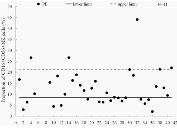
A total of 41 clinically proven VTE patients (31 with APE, 9 with DVT and 10 with CTEPH) aged 67±12 years (31~88 years) were recruited from Tongji Hospital, including 18 males and 23 females. The proportion of CD16+CD56+ NK cells in VTE patients were all compared with the detection intervals of normal population (8.6- 21.1%).
Extraction of total RNA
A total of 5 ml of venous EDTA anti-coagulated blood was obtained from patients of both groups and mononuclear cells were isolated by density gradient centrifugation. Red blood cell lysis buffer (Qiagen, Hilden, Germany) was used to isolate mononuclear cells and total RNA was extracted from mononuclear cells with TRIzol (Invitrogen, Carlsbad, USA) followed by purifica-tion with RNeasy column (QIAGEN). Treatment with DNase was performed to avoid the influ-ence by genomic DNA. Quantification of extract-ed RNA was performextract-ed with Nanodrop ND- 1000 spectrophotometer (Nanodrop Techno- logy, Cambridge, UK).
Gene expression profiling
Agilent G4112A Whole Human Genome Oligo Microarrays were purchased from Agilent (USA). A microarray is composed of 44,290 spots including 41675 genes or transcripts, 314 neg-ative control spots, 1924 positive control spots and 359 blank spots. The functions of more than 70% of genes in the microarray have been known. All patients of both groups were sub-jected to microarray analysis.
Sample marking and hybridization
Kit (Agilent, Pal alto, USA) and modified UTP [aaUTP, 5-(3-aminoally1)-UTP] was used to replace UTP. The integrated aaUTP can interact with Cy3 NHS ester forming fluorescent prod-ucts which are then used for hybridization. The integration rate of fluorescence can be deter-mined with a NanodropND-1000 spectropho-tometer. Then, hybridization mixture was pre-pared with Agilent oligonucleotide microarray in situ hybridization plus kit. About 750 ng of
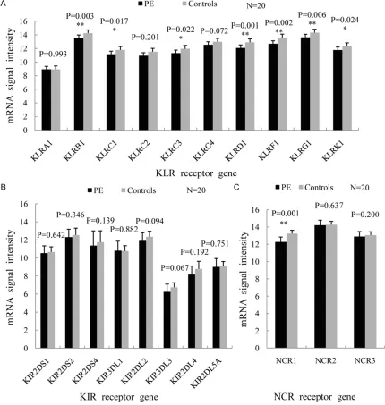
fluo-rescent products were fragmented at 60°C and hybridization was conducted in Human Whole-Genome 60-mer oligo-chips (G4112F, Agilent Technologies) at 60°C for 17 h at 10 rpm. After hybridization, the chips were washed with Agilent Gene Expression Wash Buffer accord-ing to manufacturer’s instructions. Original sig-nals were obtained Agilent scanner and Feature Extraction software. The standardization of original signals was carried out with RMA stan-Figure 1.Microarray results of NK cell surface receptors. A. KLR receptors were down-regulated totally in PE group.
The difference of KLRC1, KLRC3 and KLRK1 showed statistical significance (*P<0.05), and KLRB1, KLRD1, KLRF1 and KLRG1 were remarkably down-regulated (**P<0.01). B. The mRNA expression of KIR receptors was lower
than that of controls consistently, but the difference had no statistical significance. C. NCR receptors were
[image:3.612.91.521.73.526.2]dardized method and standardized signal val-ues were used for screening of differentially expressed genes.
RT-PCR
The spots in the microarray were randomly selected and their expressions were confirmed by RT-PCR. Among genes with differential expressions, 3 genes were randomly selected and these genes and house-keeping gene (GAPDH) were subjected to RT-PCR. The rela-tive expressions were expressed as the expres-sions of target genes normalized by that of GAPDH (2-ΔΔCt). Melting curve and 2-ΔΔCt method were used to compare the difference in the expressions between control group and PE group. Results from RT-PCR were consistent with microarray analysis.
Significant differential gene expression analy-sis
Agilent Feature extraction software was used to collect original data from microarray followed by analysis with robust multichip average (RMA). Gene intensity data between PE group and control group were compared with t test after calibration with a stochastic variance
detect the differentiation antigens on NK cells with BECKMANCOULTER EPICS XL-II flow cytom-eter. The NK cell marker CD16+CD56+ was detected in 41PE patients.
Results
mRNA expressions of NK cells related genes
NK cells related surface receptors, including killer cell lectin-like receptor (KLR), killer cell immunoglobulin-like receptor (KIR) and natural cytotoxicity receptor (NCR) were uniformly down-regulated. In the PE group, four inhibitory receptors (KLRB1, KLRD1, KLRF1, KLRG1) and four activating receptors (KLRC1, KLRC3, NCR1 and KLRK1) which positively correlate with NK cytotoxicity, were remarkably down-regulated compared with control group (P<0.05), while the difference of KIR receptors, KLRC2, KLRC4, NCR2 and NCR3 between two groups had no statistical significance (Figures 1, 2).
Flow cytometry analysis of CD16+CD56+ NK cells
[image:5.612.90.373.121.325.2]Flow cytometry was performed to detect periph-eral blood NK cell surface marker CD16+CD56+. The CD16+CD56+ NK cells were decreased in Figure 2. Microarray results of remarkably down-regulated receptors of NK cells. Crucial receptors including KLRB1
(A), KLRC1 (B), KLRC3(C), KLRD1 (D), KLRF1 (E), KLRG1 (F), KLRK1 (G) and NCR1 (H) were significantly
down-regulated.
Figure 3.Analysis of peripheral CD16+CD56+ NK cells proportion by flow cy -tometry. The CD16+CD56+ NK cells were decreased in 16 out of 41 patients compared with the detection intervals of normal population (8.6-21.1%), in-dicating that the proportion of activated NK cells declined.
model. Differentially express- ed genes were identified from whole genomes. Independent-Samples T Test was used to compare mRNA levels in sam-ples from PE patients and con- trols. Statistical tests were performed using SPSS 17.0, and P values <0.05 were con-sidered significant. Before t test, test for equality of ances was performed, if vari-ances were not equal, t test result would be corrected.
Detection of differentiation antigens on NK cells
16 out of 41 patients (39.02%), suggesting that the proportion of activated NK cells decreased (Figure 3).
Discussion
Based on gene expression microarray analysis, we have shown in the present study four inhibi-tory receptors (KLRB1, KLRD1, KLRF1, KLRG1) and four activating receptors (KLRC1, KLRC3, KLRK1 and NCR1) on NK cells were remarkably down-regulated and the cytological experiment demonstrated the proportion of CD16+CD56+ NK cells among PBMCs reduced in the PE group. The activating and inhibitory receptors of NK cells are key mediators in controlling the immune reaction to viral infection [13]; thus, it is likely that both the low gene expression of NK cell receptors and the reduced proportion of activated NK cells may lead to impairment of NK cell function in this disease. On the other hand, our results showed no significant differ-ences in the expression of KLRA1, KLRC2, KLRC4, NCR2, NCR3 and KIR receptors in this study. Considered together, while it was unknown if they were correlated with each other, these findings indicated that the reduced receptors might play a role at least in part, among the molecules related to NK cell dys-function in the development of PE.
It is well known that individuals with defective NK cells are susceptible to virus infection and cancer formation. In this study, malignant tumors or use of immunosuppressive drugs were all excluded, so we speculated compro-mised NK cells in PE patients might be associ-ated with virus infection. Successive scholars demonstrate that individuals who are infected special virus, such as influenza virus[14], her-pes simplex virus[15], Epstein-Barr virus [16], cytomegalovirus [17], human immunodeficien-cy virus[18], have high tendency to VTE this decade. Zhu et al [19] conducted a case-con-trol study involving 1,454 adults enrolled in 11 French centers to investigate whether influenza vaccination reduced the risk of VTE. They con-cluded that influenza vaccination was associ-ated with a reduced risk of VTE. Viral infections associated with decreased NK cell activity in other human diseases have also been report-ed. Nakata S et al pointed that the presence of reduced NK activating receptor gene expres-sion and low proportion of NK cells was involved in fulminant type 1 diabetes [20]. Since 2004,
we have been engaged in basic and clinical investigation of VTE. In 2010, we reported virus-like microorganisms observed in cyto-plasm and intercellular substance of lympho-cytes from peripheral venous blood in VTE patients with pulmonary hypertension and T cell immune dysfunction/disorder [6]. We also observed rod-shaped bacteria like microorgan-isms in apoptotic phagocytes from peripheral venous blood in patients with repeated PE/DVT and T cell immune dysfunction/disorder [7]. Besides, we also reported VTE in multiple or-gans of a patient who died of SARS, suggesting viral infection is closely related with VTE [8]. All these findings indicate that viral infections associated with decreased NK cell activity con-tribute to the onset of VTE. Yet most of the molecular mechanisms whereby compromised NK cells interact with virus to promote the occurrence of VTE remain largely undefined. It was reported previously that certain viruses have developed strategies that interfere with NK cell cytotoxicity. For example, through mod-ulation of NK cell receptor ligands on the sur-face of infected cells or down-regulation of cer-tain activating NK cell receptors to reduce in their cytolytic effectiveness with consequent immune evasion [13]. On basis of our previous findings, we suppose that the impaired NK cells immune function may be the internal cause of susceptibility to acquired VTE, and infection acts as a triggering factor. When the pathogens invade the subjects with NK cell dysfunction, the pathogens cannot be completely removed by the immune system. Thus, patients with compromised NK cell function are susceptible to infection, especially viral infection, which is a high risk factor for VTE. The current study pro-vided a new insight into a novel role for NK cells in the pathogenesis of venous thrombosis from an immune perspective. However, the method-ology of this study is relatively simple and understanding the cellular mechanisms where-by NK cells dysfunction contribute to VTE is still an important challenge of biomedical research. Furthermore, Long term follow-up is needed as regards the restoration of pulmonary perfusion, the rate of survival, and the occurrence of new diseases such as cancer in future.
Acknowledgements
Disclosure of conflict of interest
None.
Address correspondence to: Dr.Lemin Wang, De- partment of Cardiology, Tongji Hospital of Tongji University, 389 Xincun Road, Shanghai 200065, P. R. China. Tel: 86-21-66111291; Fax: 86-21-560- 50502; E-mail: [email protected]
References
[1] Cardiovascular Disease Educational and Research Trust; European Venous Forum; Nor- th American Thrombosis Forum; International Union of Angiology; Union Internationale du Phlebologie. Prevention and treatment of ve-nous thromboembolism: international consen-sus statement (guidelines according to
scien-tific evidence). Clin Appl Thromb Hemost 2013;
19: 116-118.
[2] Kahn SR, Lim W, Dunn AS, Cushman M, Dentali F, Akl EA, Cook DJ, Balekian AA, Klein RC, Le H, Schulman S and Murad MH. Prevention of VTE in nonsurgical patients: Antithrombotic The- rapy and Prevention of Thrombosis, 9th edi-tion. American College of Chest Physicians Evidence-Based Clinical Practice Guidelines. Chest 2012; 141 Suppl: e195S-226S.
[3] Shackford SR, Rogers FB, Terrien CM, Bouchard P, Ratliff J and Zubis R. A 10-year analysis of venous thromboembolism on the surgical service: the effect of practice guide-lines for prophylaxis. Surgery 2008; 144: 3-11. [4] Smeeth L, Cook C, Thomas S, Hall AJ, Hubbard
R and Vallance P. Risk of deep vein thrombosis and pulmonary embolism after acute infection in a community setting. Lancet 2006; 367: 1075-1079.
[5] Schmidt M, Horvath-Puho E, Thomsen RW, Smeeth L and Sørensen HT. Acute infections and venous thromboembolism. J Intern Med 2012; 271: 608-618.
[6] Wang L, Gong Z, Liang A, Xie Y, Liu SL, Yu Z, Wang L and Wang Y. Compromised T-cell im-munity and virus-like structure in a patient with pulmonary hypertension. Am J Respir Crit Care Med 2010; 182: 434-435.
[7] Wang L, Zhang X, Duan Q, Lv W, Gong Z, Xie Y, Liang A and Wang Y. Rod-like bacteria and re-current venous thromboembolism. Am J Respir Crit Care Med 2012; 186: 696.
[8] Xiang-Hua Y, Le-Min W, Ai-Bin L, Zhu G, Riquan L, Xu-You Z, Wei-Wei R and Ye-Nan W. Severe acute respiratory syndrome and venous throm-boembolism in multiple organs. Am J Respir Crit Care Med 2010; 182: 436-437.
[9] Wang L, Song H, Gong Z, Duan Q and Liang A. Acute pulmonary embolism and dysfunction of CD3+ CD8+ T cell immunity. Am J Respir Crit Care Med 2011; 184: 1315.
[10] Lv W, Wang L, Duan Q, Gong Z, Yang F, Song H and Song Y. Characteristics of the complement
system gene expression deficiency in patients
with symptomatic pulmonary embolism. Thro- mb Res 2013; 132: e54-7.
[11] Qaseem A, Snow V, Barry P, Hornbake ER, Rodnick JE, Tobolic T, Ireland B, Segal JB, Bass EB, Weiss KB, Green L and Owens DK. Current diagnosis of venous thromboembolism in pri-mary care: a clinical practice guideline from the American Academy of Family Physicians and the American College of Physicians. Ann Intern Med 2007; 146: 454-458.
[12] McLaughlin VV, Archer SL, Badesch DB, Barst RJ, Farber HW, Lindner JR, Mathier MA, McGoon MD, Park MH, Rosenson RS, Rubin LJ, Tapson VF and Varga J. ACCF/AHA 2009 expert consensus document on pulmonary hyperten-sion: a report of the American College of Cardiology Foundation Task Force on Expert Consensus Documents and the American Heart Association: developed in collaboration with the American College of Chest Physici-ans, American Thoracic Society, Inc., and the Pulmonary Hypertension Association. Circula- tion 2009; 119: 2250-2294.
[13] Vivier E, Raulet DH, Moretta A, Caligiuri MA, Zitvogel L, Lanier LL, Yokoyama WM and Ugolini S. Innate or adaptive immunity? The ex-ample of natural killer cells. Science 2011; 331: 44-49.
[14] Ohrui T, Takahashi H, Ebihara S, Matsui T,
Nakayama K and Sasaki H. Influenza A virus
infection and pulmonary microthromboembo-lism. Tohoku J Exp Med 2000; 192: 81-86. [15] Görek A, Akçay S, Ibiş OA, Atar I and Eyüboğlu
FO. Herpes simplex virus infection, massive pulmonary thromboembolism, and right atrial thrombi in a single patient: case report. Heart Lung 2007; 36: 148-153.
[16] Mashav N, Saar N, Chundadze T, Steinvil A and Justo D. Epstein-Barr virus-associated venous thromboembolism: a case report and review of the literature. Thromb Res 2008; 122: 570-571.
[17] Justo D, Finn T, Atzmony L, Guy N and Steinvil A. Thrombosis associated with acute cytomeg-alovirus infection: a meta-analysis. Eur J Intern Med 2011; 22: 195-199.
[18] Bibas M, Biava G and Antinori A. HIV-Associated Venous Thromboembolism. Mediterr J Hematol Infect Dis 2011; 3: e2011030.
[19] Zhu T, Carcaillon L, Martinez I, Cambou JP, Kyndt X, Guillot K, Vergnes MC, Scarabin PY
-cination with reduced risk of venous throm- boembolism. Thromb Haemost 2009; 102: 1259-1264.
[20] Nakata S, Imagawa A, Miyata Y, Yoshikawa A, Kozawa J, Okita K, Funahashi T, Nakamura S,