ASPECTS OF COlV1PARATIVE VEGETATIVE MORPHOLOGY
AS AN AID TO
ACTINIDIA
TAXONOMY
A thesis submitted in partial fulfilment of the requirements for the
degree of Doctor of Philosophy in Plant and Microbial Sciences
in the
University of Canterbury
by
James M. Condon
;:::
THESIS
/ \ .
(I,: ABSTRACT
This study explores the practical value of comparative morphology as an aid to Actinidia taxonomy, using vegetative characters derived from spring and
summer shoots of 20-30 taxa, selected from the "NZ D.S.LR. Actinidia germplasm
collection" at Auckland and Te Puke during 1988-1989. Data are collected from field-based obselVations and from samples processed for light and electron microscopy; these are supplemented with obselVations derived from herbarium specimens collected in China. The taxonomic potential of clfaracters is further tested using multivariate and other statistical methods.
Actinidia are morphologically variable vines which, nevertheless, express
genetically-determined form in: their manner of climbing, the types and growth characteristics of shoots and in the ontogenetic expression of shoot form. There is however a strong "opportunistic component" in the realisation of plant form. Some more conservative characters include leaf venation pattern, trichome morphology, arrangement of sclerenchyma fibres and the complement of ergastic crystals associat.ed with vascular bundles of the leaf. Microscopic examination of abaxial foliar trichomes, currently used to demarcate sections of the genus, reveal branched hair types in M aculatae and Strigosae, which are supposed to be absent
(Dunn 1911, Liang 1984) from these sections of the genus. Re-examination of these groups and the characters delimiting them is recommended.
Comparative morphological studies of Actinidia in the gerrnplasm
collection show that many of the characters of winter-dormant shoots are genotypic in nature. Vines of Leiocarpae and Stellatae may be identified below
the species level by their bud-form characteristics. Discriminant analysis shows the value of bud height and ostiole size in separating major taxonomic and geographic groups. Detailed analysis of bud characters is justified as poor or uneven budbreak currently limits the productivity of commercial cultivars.
Taxonomists need to be more aware of the spatial and temporal potential afforded by vegetative morphological characters in this genus.
The discovery of "water-excreting glands" (= hydathodes) in all Actinidia
seen, culminating in a combination of "water spending" characters in A. deliciosa,
has important implications for water-relations in these plants. Hydathodes in A. deliciosa are well supplied by craspedodromous venation and the ultimate
tracheids terminate in a spatially diffuse but metabolically active environment, in the apices of these glands. The fate and functioning of hydathodes in Actillidia
needs further research.
The results from this exploratory study are intended to contribute to the programme of genetic and taxonomic studies of Actinidia; currently being
TABLE OF CONTENTS
ABSTRACT
CHAPTER 1 : General Introduction
1.1 The klwifmit industry
1.2 Actinidia in taxonomy and horticulture
1.3
1.4
Actinidia taxonomy-past and present
Horticultural potential of some Actinidia species
1.5 Reproductive botany of Actinidia
1.6
1.7
1.8
Vegetative structure in Actinidia
Development of topic
Aims of investigation
Addendum Chapter 1 : Name Changes
CHAPTER 2 : Comparative (vegetative) morphology of "summer-shoots" in some Actinidia
2.1 Introduction
2.2 Materials and Methods
2.2.1 Plant collection
2.2.2 Descriptive terminology
2.2.3 Leaf form measurements
2.2.4 Light microscopy of leaf surfaces
2.2.5 Scanning electron microscopy of leaf surfaces
2.2.6 Internal organisation of leaves
2.2.6.1 Light microscopy
2.2.6.2 Dehydration (chemical)
2.2.6.3 Infiltration
2.2.6.4 Polymerisation
2.2.6.5 Microtomy for light microscopy (L.M.)
2.2.6.6 Staining for L.M.
2.2.6.7 Photomicrography for L.M.
2.2.7 Internal structure of leaves, S.E.M. methods
2.2.7.1 Preparation of fractured leaves for S.E.M.
2.2.7.2 Photomicroscopy
2.2.8 Internal structure of leaves T.E.M. methods
Page
1
2
6
9
10
11
12
14
15
16
17
19
20
11
2.2.9
Cluster analysis of winter shoot characters2.2.9.1 Characters
22
2.2.9.2 Similarity coefficients
23
2.2.9.3 Cluster methods
2.2.9.4 Cophenetic correlation coefficient
24
2.2.10
Discriminant analysis (D.A) of summer shoot characters2.3
Results25
2.3.1
Comparative morphology of summer shoots in some Actinidia under cultivation2.3.1.1 Stem characters
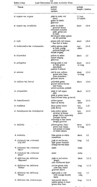
2.3.1.2 Leaf characters
29
2.3.1.3 Juvenile shoots
38
2.3.2
Leaf venation in some Actinidia46
2.3.2.1 Orders of leaf venation 2.3.2.2 Pectinal venation
2.3.2.3 Primary venation
47
2.3.2.4 Secondary venation
2.3.2.5 Tertiary venation
48
2.3.2.6 Basal venation in Actinidia leaves
2.3.2.7 Areolation and ultimate venation
52
2.3.2.8 Leaf venation and margins
72
2.3.2.9 Marginal hydathodes in Actinidia leaves
2.3.2.9.1 Histology of hydathodes
73
2.3.2.9.2 Electron microscopy ofhydathodes
2.3.3
Surfaces of mature leaves78
2.3.3.1 Adaxial (uppermost) surface
2.3.3.2 Abaxial (lowermost) surface
79
2.3.3.3 Surfaces of juvenile leaves 88
2.3.4
Anatomy of some Actinidia leaves92
2.3.4.1 Common trends
2.3.4.2 Taxonomic sectional trends in leaf
94
anatomy2.3.5
Cluster analysis of summer shoot characters 106 2.3.5.1 Clustering by average-linkage method2.3.5.2 Clustering by single-linkage Jnetizod
2.3.6 Discriminant analysis of summer shoot 111
characters
2.3.6.1 Validation of "a priori" groups
2.4 Discussion
2.4.1 Nature of plant growth form
2.4.2 Architecture of Actinidia vines
2.4.3 Types of shoots on Actinidia vines
2.4.4 Stress-induced morphology
2.4.5 Plant size and vigour
2.4.6 Phenology
2.4.7 Juvenile shoot characters
2.4.8 Determinants of three-dimensional form of leaves
2.4.9 Upper (Adaxial) leaf surface
2.4.10 Lower (Abaxial) leaf surface
2.4.11 Types of leaf hairs (trichomes) 2.4.11.1 Leiocarpae
2.4.11.2 Maculatae 2.4.11.3 Strigosae 2.4.11.4 Stellatae
2.4.12 Leaf venation
2.4.13 Hydathodes
2.4.14 Leaf anatomy
2.4.15 Systematic affinities of Actinidia
2.4.16 Cluster analysis of v1etative
characters derive from summer shoots
2.4.17 Discriminant analysis of some characters from summer shoots
2.5 Conclusions
CHAPTER 3 : Comparative (vegetative) morphology of "winter-shoots" in some Actinidia .
3.1 Introduction
3.3
Materials and Methods
3.2.1
3.2.2 3.2.33.2.4
3.2.5
ResultsCollection -of plant material List of plants studied
Descriptions of plant form Bud photography
Selection of variables obtained from winter shoots
3.3.1 Descriptions of winter shoots in some Actinidia
3.3.1.1 Stem surface
3.3.1.2 Bud form
3.3.1.3 Bud emergence
3.3.2 Cluster analysis of winter shoot characters
3.3.2.1 Clustering by average-linkage method
3.3.2.2 Clustering by single-linkage method
3.3.2.3 Cluster analysis of a reduced
number of taxa using average-linkage 3.3.2.4 Cluster analysis of the reduced data
set using single-linkage
IV
3.3.3 Discriminant analysis of winter shoot characters
3.3.3.1 Validation of the 'a priori' groups 3.3.3.2 Discriminant effiacacy of winter
shoot characters
3.3.4 Internal bud structures of some Actinidia 3.3.4.1 Types of bud structures
3.3.4.2 Bud structure complements of selected Actinidia
3.3.5 Infraspecific variation in selected genotypes of A. chinensis var. chinensis
3.3.5.1 Descriptive trends in genotypes 3.3.5.2 Remarks on specific genotypes
3.4 Discussion
3.5
3.4.1 Taxonomy
3.4.1.1 Stem surface characters 3.4.1.2 Stem texture
3.4.1.3 Bud shape
3.4.2 Cluster analysis of winter shoot characters 3.4.3 Bud form in relation to temperature and
geography
3.4.4 Emergence from bud dormancy
3.4.5 Bud morphology and breeding potential
of Actinidia
3.4.5.1 Cold tolerance
3.4.5.2 Budbreak and flowering time 3.4.5.3 Bud productivity
Conclusions
CHAPTER 4 : General Discussion
4.1 Context of problem
4.2 Cluster analysis of the combined data set
4.3 Defining taxonomic groups
4.3.1 4.3.2 4.3.3 4.3.4 Taxonomic "identity" Taxonomic "range" Use of characters Evolution of the genus
Addendum Chapter 4
Taxonomy
Molecular systematics
1. Restriction fragment length polymorphisms 2. Genomic (D.N.A repeat) sequences
References (Addendum)
AC~~O~DGE~ffiNTS REFERENCES
v
Page
Vl
LIST OF TABLES
Page
CHAPTER 1
Table
1.1
History of A ctin idia taxonomy5
CHAPTER 2
2,1
Vines of Actinidia Undl. examined18
Stem characters in some Actinidia taxa
26-28
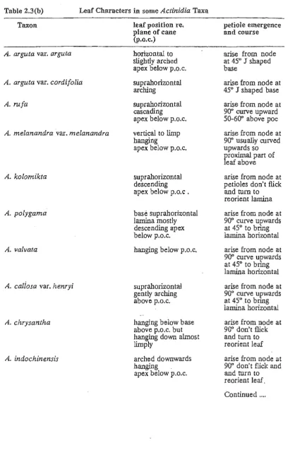
Leaf characters in some Actinidia taxa
32-34
Leaf shape characters in mature leaves of some Actinidia taxa
Juvenile shoot characters in some Actinidia taxa
2,6
Criteria used for classifying leaf venation2.7
Comparison of selected leaf venation characters50-51
2.8
Comparative form of areolation in leaves of someActinidia
69-70
2.9
Leaf venation and margins of some Actinidia71a,b
10
Adaxial leaf surface characters in mature leaves80-81
of Actinidia
11 Abaxial leaf surface characterse in mature leaves of Actinidia
Abaxial surfaces of outer leaves in A. deliciosa
85-86
some related taxa
Hair dimensions of some Actinidia
87
2.14
Stomatal sizes of some Actinidia93
Leaf anatomy of some Actinidia
100-105
16
Linkage coefficients (average-linkage)107
Linikage coefficients (single-linkage)
109
18
Statistics summarising "discriminant efficacy"112
vii
Page
CHAPTER 3 Table
3,1
list of plants studied134
3.2
Genotypes of A. chinensis var. chinensis135
3.3
Cane surfaces of some Actinidia142-145
3.4
Form of lenticels on some Actinidia canes146-149
Bud shape in some Actinidia
150-157
3.6
Bud emergence characters of some Actinidia158-161
3.7
linkage coefficients (U.F.GM.A)177
3.8
linkage coefficients (single)179
3.9
Linkage coefficients reduced matrix (average)181
3.10
Linkage coefficients reduced matrix (single)183
3.11
Group validation by D.A185
3.12
Statistics summarising discriminant efficacy of189
winter shoot characters
3.13
Bud descriptions of some A. chinensis vaT. chinensis genotypes198-199
3.14
Bud variables (ranges) of some A. chinensz's200
genotypes
CHAPTER 4
4.1
linkage coefficients "combined data set"219
(winter and summer shoot characters) (average-linkage)
viii
LIST OF FIGURES
Page CHAPTER 2
Figure
2.1 Leaf measurements 19
Drawings of leaf venation and margins of
2.2 A. arguta var. cordifolia 53
2.3 A. valvata (Adaxial) 54
2.4 A. valvata (Abaxial) 55
2.5 A. chrysantha (Abaxial) 56
2.6 A. melliana (Abaxial) 57
2.7 A., hemsleyana (Abaxial) 58
2.8 A. latifolia (Abaxial) 59
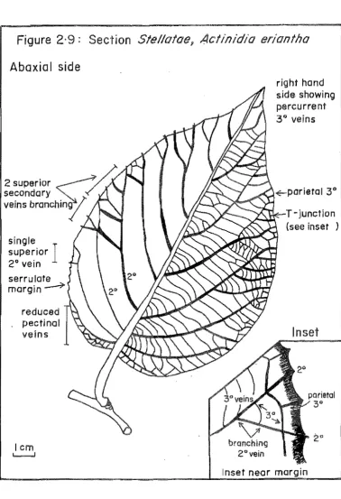
2.9 A. eriantha (Abaxial) 60
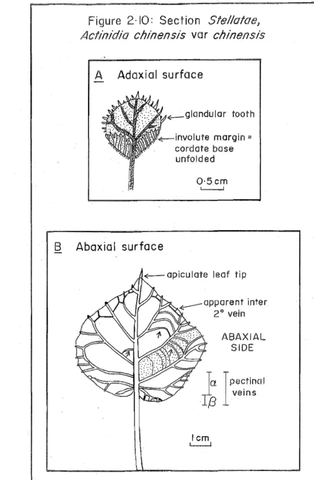
2.10 A. chinensis var. chinensis juvenile leaves 61
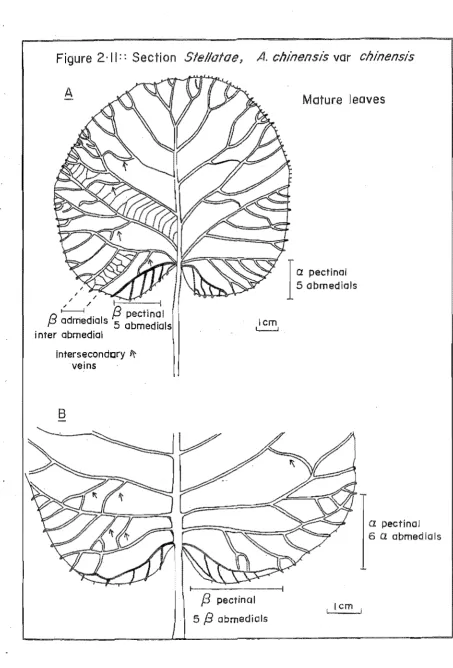
2.11 A. chinensis var. chinensis mature leaves 62
2.12 A. deliciosa var. deliciosa cv. Hayward 63
2.13 Pectinal venation in cv. Hayward leaves 64
2.14 Leaf margin (and adaxial surface) cv.'Hayward 65
2.15 A. deliciosa var. chlorocarpa 66.
2.16 Cluster analysis (average-linkage) dendrogram 108
2.17 Cluster analysis (single-linkage) dendrogram 110
CHAPTER)
3.1 Map of Eastern Asia (after Krussmann 1984) 136
3.2 External parts of an A. deliciosa bud soon 138
after bud-burst
Page
3.4 Graphs of means ± 95% confidence limits 167
3.4.1 Lentice1 density on cane (em -2) Longest lenticel cane means
3.4.3 Bud-length means 168
3.4.4 Bud-height means Bud-width means
3.4.6 BUd-le?~h to Bud-height (Bud-case shape 169 ratIO means
3.4.7 B.T.O.H. (= Base of bud to height vertical 170 point) means
3.4.8 Depth of petiole scar means
3.4.9 Ostiole WIdth means 171
3.4.10 Cane diameter means
3.4.11 Height of visible bud-structures means 172
3.4.12 Number of visible bud-structures means 3.4.13 L.P.D (Length of 12etiole diameter) means
3.4.14 Ratio of Bud-case width to ostiole width 173 means
3.5 Cluster analysis (average-linkage = U.P.G.M.A) 178 3.6
dendrogram
Cluster analysis (single-linkage) dendrogram 180
3.7 Cluster analysis reduced m~trix (average-linkage) 182 3.8 Cluster analysis reduced matrix (single-linkage) 184 3.9 Plot of "canonical variate 1" against 186
"canonical variate 2" for the four speCIes groups
3.10 Means (±: S.B.) of selected variables for the 188 four groups
CHAPTER 4
x
LIST OF PLATES
Page
Plate
1.1 Fruit of selected Actinidia taxa 4
CHAPTER 2
2.1 Habit shots of selected Actinidia taxa 30
2.2 Distal (apical) shoots of Actinidia with juvenile 40
characters
2.3 "Semi-cleared" leaf of A. valvata revealing 68
areolation
2.4 Hydathode morphology 74
2.5 U1trastructure of hydathodes in A. de lie iosa cv.
Hayward
77
2.6 Abaxial leaf hairs of A. callosa var. henryi and 89
A. hemsleyana
2.7 Abaxial leaf surfaces and fractures of some Stellatae 90
2.8 Abaxial leaf surfaces of mise Actinidia 91
2.9 Leaf anatomy cjf Ac tinidia I 97
2.10 Leaf anatomy of Actinidia II 99
CHAPTER 3
3.1 External morphology of Leiocarpae buds 163
3.2 External morphology of M aculatae and Strigosae buds 164
3.3 External morphology of buds (Stellatae I) 165
3.4 External morphology of buds (Stellatae II) 166
3.5
Types of bud-structure in Actinidia buds 1913.6 Bud structure complements of some Actinidia 193
3.7 External morphology of some A. chinensis var. 196
chinensis genotypes
3.8 Dissected buds of some A. chinensis var. chinensis 197
PERSONAL CO:Ml\l1UNICATIONS
1. Ferguson, AR. (Dr),
D.S.I.It Fruit andTrees, Mt Albert Research Centre, D.s.I.R.,
AUCKLAND.
2. Morley-Bunker, M. (Mr),
Department of Horticulture, Parks and Recreation, Lincoln University,
CHRISTCHURCH.
3. Breitwieser, I. (Dr)
Department of Plant and Microbial Sciences, Canterbury University,
CHRISTCHURCH.
4. Fineran, B.A (Dr),
Department of Plant and Microbial Sciences, Canterbury University,
Xli
LIST OF APPENDICES
Table
A2.1.1 Leaf form variables (outer leaves) ± S.E.
for different Actinidia taxa
2 pp
A2.1.2 Leaf form variables (inner leaves) ± S.E. 2 pp
for different Actinidza taxa
. A2.1.3 Ranges of leaf size variables (Condon 1911) A2.1.4 Ranges of leaf size variables derived from
Chinese Actinidia (Liang 1984)
A2.1.S Leaf form measurements of some N.Z. cultivars of Actinidia (Adapted from Zhang and Thorp 1986)
A2.2.1 Summer shoot characters used in cluster analysis
4 pp
A2.2.2 Table of Gower's coefficients
A2.3 List of herbarium specimens consulted (Botany 2 pp Division D.S.I.~., Dec. 1990)
CHAPTER 3
A3.1 List of winter shoot characters used in 2 pp
cluster analysis
A3.2 (Table of Gower's coefficients 3 pp
A3.3 Data used in calculation of Gower's 6 pp
similarity coefficients
CHAPTER 4
A4.1 Table of Gower's coefficients 2pp
A4.2 Pearson correlation matrix 14 pp
A4.3 Key to character codes in correlation matrix 3 pp
Calculation of Gower similarity coefficient
APPENDIX 5
CHAPTER ONE
INTRODUCTION
This chapter is a general introduction to aspects of the taxonomy, domestication and morphology of Actinidia.
1.1 THE KIWIFRUIT INDUSTRY
Kiwifruit is an important horticultural crop developed in New Zealand in
recent years and is now also being established in other countries. Current world production of the crop is estimated at 0.53 million tonnes with 0.3 million tonnes of this being produced in N.Z. (Warrington 1990). Export revenue from kiwifruit production in N.Z. in 1988 was $450 millionl. Kiwifruit berries are prized for their attractive colour, flavour and high nutritional qualities and their longevity in storage and shipping. The kiwifruit industry is based largely on the vegetative propagation of a single female cultivar 'Hayward', selected by Hayward Wright in
1930, and derived in the first instance from a single importation of Chinese seed in 1904. Domestication of the crop and its history has been reviewed by Ferguson and Bollard (1990) and Yerex and Haines (1983).
1.2 ACTINIDIA IN TAXONOMY AND HORTICULTURE
The kiwifruit [:::: A. deliciosa var. deliciosa cv. Hayward] represents only one cultivar of one species of the 50 or so species in the genus Actinidia. A. deliciosa now recognised as a separate species (Uang and Ferguson 1984, 1986)
was formerly considered to be a variety of the polymorphic species A. chinensis. It
differs from the latter however, in having "stiff hairs", more elongate fruit and a higher chromosome number (McNeilage and Considine 1989). A. deliciosa is a
hexaploid (2n ::: 6x :::: 174), whereas, A. chinensis is·a diploid (2n :::: 2x ::::: 58).
Both of these species provide a limited indication of the range of fruit forms found within the genus.
The number of fruits per inflorescence varies from 1-3 in cv. Hayward, which is consistent with the "reduced form" of this pseudodichasial inflorescence. Some taxa such as A. latifolia may have up to 20-30 flowers per inflorescence
(Plate LID).
2
Fruit shape in the genus may be spherical (e.g. A. chinensis var. chinensis, Plate 1.1E and A. eriantha Plate 1.1B) ar avaid (e.g. A. chrysantha (Plate 1.lA) A. deliciosa cv. Bruna, Plate 1.1C). The shape af the fruit cantributes to. its
aesthetic appeal, ease af packing and mechanical processing, e.g. canning. The familiar fruit af 'Hayward' is shawn in Plate 1.1P.
1.3 ACTINIDIA TAXONOMY - PAST AND PRESENT
Lindley (1836) defined the genus Actinidia an the basis af plants with a climbing habit and flawers with radiating styles. [Actinidia fram the Greek ward aktis meaning ray].
Siebald and Zuccarini (1843) assigned five climbing species fram Japan to. the Ternstromiaceae and gave them the generic name Trichostigma.
Planchan (1847) described A. chinensis fram ane af Fartune's specimens callected in China and reunited Actinidia and Trichostigma under the genus Actinidia which had priarity af namenclature.
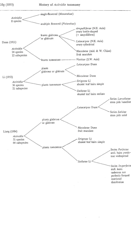
Gilg (1893) emphlliiised the number af flawers per cyme when he subdivided the graup into. single (Monanthae) and many-flawered (Pleianthae) types. Dunn (1911) perceived the graup as a series af geagraphically differentiated subunits, which he separated an the basis af abundance af shaat hairs, avary shape and the presence or absence af lenticels (= "spats"). Li (1952) subdivided Dunn's graups using the marphalagy camplexity af faliar tric~ames an the underside af the leaf. Sectian Stellatae were characterised by stellate abaxial hairs, whereas, all ather sectians af the genus had simple abaxial hairs. Liang (1984) extended the work af Li and he recognised many new taxa belaw the species level. He subdivided the Leiocarpae into. twa series based an the salidity or lamellatian af the pith; the Stellatae were alSo. subdivided into. series based an the persistence, marphalagical cansistency and camplexity af the stellate hairs. The taxanamic history af the genus is summarised in Table 1.1.
The current taxanamic revisian af Actinidia by Liang Chau-Fen (1984) cantains ca. 50 species and as many infraspecific taxa. Actinidia are differentiated fram related genera by their lianaid habit, diaecy and flawers with numeraus styles. Seeds af Actinidia are nan-arillate with capiaus quantities af albumen (Dunn 1911).
PLATE 1.1
Fruit of selected Actinidia taxa.
A Single ovoid, spotted (= lenticellate) fruit of A. chrysantha (Maculatae).
B Single ovoid, furry fruit of A. eriantha (Stellatae).
C Single elongate, hispid fruit of A. deliciosa var. deliciosa cv. Bruno (Stel/atae).
D Single infructescence comprising ten glaucous fruit of A. latifolia (Stel/atae).
E Single spherical fruit (hairs caducous) of A. chinensis var. chinensis (= soft-haired mihoutao) (Stellatae).
F Single elongate fruit (hairs persistent; hispid) of A. deliciosa var. deliciosa cv. Hayward (= stiff-haired mihoutao, the cultivar term 'kiwifruit') (Stellatae).
5
Table 1.1 Gilg (1893) History of Actinidia taxonomy
<
single-flowered (MO/zallthae) Actinidia8 species
multiple flowered (P/eiantlzae)
Du= (1911) A ctillidia 24 species
12 subs pecies
Ampulliferae (N.E. Asia) ovary bottle-shaped leaves glabrou.s (= ampulliform) or glabrate
Leiocarpae (N.E. Asia) ovary cylindrical
Macll/alae (mid. & W. China)
fruit maculate leaves tomentose - - - - -Vestitae (S.W. Asia)
Leiocarpae Dunn plants
<
Z
glabrous or glabrateLi (1952) MaCll/alae Dunn
AcLinidia < S t r i g o s a e Li
36 species plants tomenlose abaxial le:rt hairs simple 21 subspecies
Liang (1984) Actinidia 51 species 64 subs pecies
plants glabrou.s or glabrate
plants tomentose
Stellatae Li
abaxial leaf hairs stellate
Leiocarpae Dunn
Mawlalae Dunn fruit maculate
<
Series Lanzellatae stem pith iameilateSeries So/idae stem pilh solid
/ Strigosae Li
/ abaxial leaf hairs simple
~
Series PerfecraeZ
stell. hairs ;cersis-tent widespread Stellatae Li6
characters such as caducous hairs, or indeed vegetative and floral characters themselves, since most Actinidia are deciduous plants. The identification of winter plants and some hybridising fonns is problematical, particularly among widespread polymorphic taxa such as A. arguta, which may still be in the process of actively evolving.
For the time being, I accept the composition of the Actinidiaceae as defined by Liang (1984). I am not able to comment on this as I have not examined other genera, e.g. Clematacletlzra, Saurauia or Sladenia. It should be noted however that the composition of this family is systematically contentious. Importance has been attached to the following characters in Actinidia and related genera: plant habit, presence of raphides, number, arrangement and degree of fusion of floral parts, pattern of embryonic development, number of integuments and the presence of arils.
1.4 HORTICULTURAL POTENTIAL OF SOME ACTINIDIA SPECIES
The genus Actinidia is centred in the mountains of S.W. China between the Pearl and Yang-tze rivers, where the greatest diversity of taxa occurs (Liang 1983). The ecological range of Actinidia is considerable with some taxa extending as far north as Manchuria and others as far south as Indonesia.
Other species besides A. deliciasa and A. chinensis offer potential for improvement of the crop. Some taxa have very high levels of vitamin C (e.g. A.
eriantlza, A. lati/alia); others have very different coloured fruits (e.g. red-fleshed
A. arguta x A. melanandra, Seal and McNeilage 1988), or fruits with superior flavour qualities (e.g. A. chinensis). Some taxa produce hairless fruit (e.g. A.
arguta var. arguta; A. clzrysantha), while the fruit of others, (e.g. A. chinensis) matures earlier than that of existing cultivars, (e.g. Hayward). The ecological range of Actinidia cultivars might be extended by hybridisation with: cold-tolerant Siberian species (e.g. A. kalamikta), or with tropical plants (e.g. A. lati/alia;
A. indachinensis). Mindful of this potential for improving kiwifruit, the D.S.I.R. established an Actinidia germplasm collection in 1971 (Ferguson pers. comm.).
1.5 REPRODUCTIVE BOTANY OF ACTINIDIA
7
ensure adequate fruit set in the orchard (Sale 1985). Efforts are being directed toward the development of fruiting males (Ferguson pers. comm.) as dioecy is not absolute in this genus. A well-managed ldwifruit vine in a commercial orchard may produce 1500-2000 fruit, equivalent to a yield of 25 tonnes per hectare (Ferguson and Davison 1986). The development of artificial spray-pollination has resulted in substantial improvements in fruit-set (Hopping 1985). The fruit is harvested when ripe once it has attained a level of 6.2% soluble solids (Harman 1981). The fruit of
A. deliciosa cv. Hayward is a large, sweet many-seeded berry with a brown hairy
exocarp and a fleshy green pericarp.
Reproductive anatomy is documented in several Actinidia sp and is
reviewed in Ferguson (1984).
Vijayaraghavan (1965) describes the development of the anther. Sporogenous tissues originate from divisions of archesporial mother cells beneath Young anthers have two plates of hypodermal, multicelled archesporial tissue. The central layer of cells becomes sterile in each plate, so that 4 layers of archesporial cells remain. Each plate divides periclinally producing parietal and archesporial cells, respectively. Parietal tissues undergo further cell divisions producing epidermal, endothecial, middle and tapetal layers. Only the epidermal and endothecial cells persist; the middle and tapetal layers eventually degenerate and come absorbed. Microspore mother cells undergo meiosis, during which the cell walls round off and become enclosed by a gelatinous wall. Microspore mother cells undergo cytokinesis simultaneously and their cell walls differentiate into a thick exine and thin intine layer. Division of the microspore nucleus produces a large generative and a small vegetative nucleus and pollen grains are released at the 2-celled stage. In pistillate flowers the microspore nucleus does not divide, the protoplast degenerates and the anther dehisces. In staminate vines the microspore nucleus undergoes normal cell division producing a large vegetative cell and a small generative celL Pollen of staminate vines is tricolpate with crassimarginate colpi and the grains are prolate to prolate spheroidal in form. Pollen grains are binucleate (Lechner 1915, Schmid 1978) and the pollen is shed in dry clumps (Palmer-lones and Clinch 1975). According to Corbet et al. (1988), Actinidia
anthers dehisce at the resonant frequency at which bees "buzz". Fertilisation and pollen tube growth in Actinidia are described by Harvey et al. (1987).
Ovules are axile, anatropous, unitegmic and tenuinucellate (Van Tieghem 1899, Lechner 1915, Vijayaraghavan 1965, Harvey and Fraser 1988). AU but one of the megaspores degenerates to form the archesporium (An et al. 1983, Harvey and
7a
According to Harvey and Fraser (1988), the polar nuclei fuse to form a uninucleate cell. However, it is generally held (e.g. Esau 1976) that the triploid state of the endosperm in angiosperms originates in one of two ways: either by fusion of one of the sperm nuclei with two haploid nuclei, or by fusion of a single sperm nucleus with a diploid "secondary" nucleus (e.g. Esau 1976). Vijayaraghavan (1965) indicates that the two polar nuclei in A. polygmna fuse to form a
secondary nucleus, but the author does not specify the ploidy of the latter. Further work is needed to clarify the details of endosperm formation in Actinidia. Harvey
and Freaser lac. cit. further note that the primary endosperm nucleus begins to
divided 96-180 hrs after fertilisation in A. deliciosa and A. chinensis. The zygote
divides 8 wks after fertilisation and develops through the globular to the heart-shaped stage 12 wks after fertilisation. The embryo is fully formed 16 wks after fertilisation.
The general phenology of Actinidia from budbreak to fruiting is
8
Phenology of A. deliciosa var. deliciosa 'Hayward'
Adapted from Brundell (197Sb); Ferguson and Davison (1986)
.
Shoot Bud Development Flower Bud Developm~nt
undifferentiated pr10rdium
New Zealand Calendar
bud dormant
bud swell
bud-burst
open cluster
20 I
30
r
40
I
lO
60
formation of floral primordia
initiation of carpels formation of gynoecial
plateau
anther and filament initiation
I
ovule initiation
I
!:alyx split
I
FUlL BLOOl\tl
New buds are thought to be initiated at time of bud-burst.
January to
September
late tptember
to October
I
NovemberDecember
January
9
1.6 VEGETATIVE STRUCTURE Jl\T ACTINIDIA
Aspects of the morphology of Actinidia have been described in the course
of taxonomic revisions of the genus (e.g. Dunn 1911, Li 1952, Liang 1984). These authors have noted the morphological plasticity and variability of vegetative organs in these plants. Whereas the reproductive biology of Actinidia has been
well-researched (e.g. Anon 1979, Brundell 1975b, Corbet et al. 1988, Harvey et al.
1987, Harvey and Fraser 1988, Liang 1984, Schmidt 1978, White 1986 a,b) vegetative structures have received much less attention.
There have been some morphological descriptions of buds (Brothers 1988, Brundell 1975a, Lionakis and Schwabe 1984a), internodes (BrundeU loc. cit.
Ferguson 1984, 1990e) and the root systems of the vine (Clothier et al. 1986, 1988,
Lemon 1988), but these have referred to A. deliciosa. Recent observations by Gui
(1981), Ferguson (1984, 1990e) and Zhang and Thorp (1986), indicate that vegetative characters might be used to identify Actinidia from winter-dormant
shoots, given the perennial deciduous nature of most of these vines.
Anatomical studies of stems have largely been exploratory and these have been confined to only a few species, i.e. A. polygama; A. deliciosa (e.g.
Hitzemann 1886, Metcalfe and Chalk 1950). The size and distribution of vessels in
kiwifruit stems have been examined in relation to water transport (McAneney and Judd 1983).
Work on leaf anatomy has been more extensive and here the systematic studies of Lechner (1915), Gao (1988) are discussed in Chapter Two.
The physiological responsiveness of plant form to light quality and quantity was assessed by Morgan-et al. (1985) in A. deliciosa. Low red: far-red light (=
simulated shade) increased petiole elongation, but vegetative growth was not markedly affected. Low-light quantity (::: low PPFD) did not reduce floral initiation, but development of floral primordia was severely diminished, particularly when the daily integral of PPFD (::: photosynthetic photon flux density) was less than or equal to 30% of the incident radiation. Seedlings were shown to be more light-sensitive than adult vines of A. deliciosa (Zhu 1983). Actinidia behaved as as a warm-temperate plant with increased biomass
accumulation under "long-day" conditions. Shoot tips showed increased twisting under "short-days" and petioles were unexpectedly succulent (Lionakis and Schwabe 1984b). Actinidia vines grew rapidly in response to water (Ferguson
10
Actinidia ill the wild are straggly, rambling or climbing vines forming
impenetrable thickets on occasion. They prefer well-lit humid and sheltered conditions and are often found on margins of forests or near the banks of streams (Kolbasina 1963).
Kiwifruit vines are established as grafted plants in sheltered and intensively managed orchards. They are arranged in an orderly manner on T -bar or pergola frames (Sale 1985), where a permanent trunk supports a perpendicular pair of "leader" branches. The crop is raised 1-2 rn above the ground on fruiting arms, which emerge from these leaders at regularly spaced intervals. Fruiting arms consist of 2 yr old stems termed canes, which are pruned in the winter, leaving only the basal 5 .. 10 internodes. The following spring flowers are borne in the axils of the current season's shoots, which arise from buds on the fruiting arms. Summer pruning is used to control excessive vigour of shoots and to increase the penetration of light for photosynthesis and flowering. Vines are pruned (in winter) after flowering, when new fruiting arms are tied down for the next season and. arms which have flowered are removed. Under a system of replacement pruning, fruiting arms are renewed every 2-3 years.
Actinidia vines in the germplasm collection are all grown under common conditions in an "experimental garden" (Clausen, Keck and Riesey 1940) and plants are managed as if they were commercial kiwifruit vines. Thus genotypic variation emerges in a horticultural context.
1.7 DEVELOPMENT OF TOPIC
The thesis began in 1987 with the aim to explore stem structure in relation to transport, with reference to A. deliciosa var. deliciosa cv. Hayward. Results of some of this work were presented at the 1991 Kiwifruit symposium (see Appendix 5 for conference abstracts). Further examination of plants in the D.S.I.R. germplasm collection and the literature, however, revealed that little was lmown of the basic morphology of many of these plants. Emphasis was therefore redirected (in July 1988) in studying the taxonomic potential of vegetative morphology in some Actinidia. Given the difficulty of obtaining plant material from China, attention was directed towards those taxa of economic potential, currently grown
11
1.8 AIMS OF INVESTIGATION
The aims of this thesis are as follows :
(1) To evaluate the genetic and environmental components of plant form in summer shoots in Actinidia vines, in relation to taxonomy;
(2) Detailed study of the taxonomic potential of morphological characters displayed by winter-dormant shoots with some assessment of infraspecific variation in A. chinensis and A. deliciosa;
(3) Exploratory structural observations of the "water-excreting" glands (=
hydathodes) in A. deliciosa var. deliciosa cv. Hayward, with consideration
of their possible mode of operation.
12
ADDENDUM CHAPTER ONE : NAME CHANGES
Since completing the final draft of this thesis, Dr AR. Ferguson (pers. comm.) of D.S.I.R Fruit and Trees has reassessed the nomenclature of some Actinidia in the germplasm collection.
1. A. arguta complex
ARF. thinks that the N.Z. examples of A. arguta var. arguta and A. arguta var. cordifolia represent two morphological extremes within a highly variable group. He is not able to support Liang's (1984) separation of these varieties on morphological grounds, even after examination of Chinese herbarium specimens at Guilin. Further cytotaxonomic and chemical data are thus needed to settle the issue.
2. A. arguta vaL giraldii (4/1/4)
This plant differs from A. arguta vaL arguta in having glaucous leaf undersides and a diploid, rather than tetraploid genome. According to A.R.F., the petioles and leaf undersides are less "hairy" than suggested in descriptions of this plant by Diels and Liang.
Our "A. giraldii" plant may be A. hypoleuca Nakai. AR.F. had previously overlooked this species; it is confined to southern and central Japan and is therefore absent from Liang's (1984) descriptions of Actinidia. Our specimen is like A. hypoleuca with its purple, non-pointed anthers and pale green leaves which are smaller than those in A. arguta and broader than those of A. melanandra.
3. "A. rufa"
In hindsight, this is the least surprising of the name changes in the germplasm collection. A. rufa is so different from other Leiocarpae in many morphological features. According to AR.F., successive Japanese botanists and Li (1952) have treated A. rufa as a variety of A. arguta. Liang does not discuss A. rufa as his work was limited to Chinese and Taiwanese Actinidia.
Our specimen originated from a Japanese experimental station on the eastern side of Kyushu in an area where A. rufa grows naturally.
13
of /lA. rufa /I is more reminiscent of A. callosa in its: dark, rigid and thick
leaves, and orange lenticellate bark. Like A. callosa the winter buds are dome-shaped, "winged" in profile, and covered in ginger tomentum. Both AR.F. and I agree that the N.Z. plant cannot be allied with Leiocarpae. AR.F. suggests that his plant may be a variety of A. callosa, but this hypothesis needs confirmation.
4. /lA. melliana/l (36/4/3a)
This plant is quite distinct from a representative plant of the 'real'
A. melliana seen by AR.F. at Guilin. The D.S.I.R. specimen seems similar to Liang's description of A. fortunatii, but (according to Liang) the latter plant has not been collected since 1906. The identity of this plant is uncertain, and is provisionally termed "hairy hemsleyana".
In summary, Actinidia identification is still controversial. However, for this thesis, the nomenclature currently used in N.Z. has been retained. The work from this thesis indicates that considerable further research on the taxonomy of
Actinidia is warranted. Herbarium-voucher specimens of the D.S.I.R. plants are now being examined by Asian botanists for verification of their nomenclature.
References (Chapter 1 Addendum) : Not in general ref. list, pp. 230-241 Nakai, T. (1933)
C
APTER TWO
OMPA
ATIVE
(V E G E l
V E)
M
RPHO
GY OF
"S U M MER - S H 0 0
~r
Sir
14
CHAPTER lWO
2.1 INTRODUCTION
Actinidia are morphologically diverse vines (::::: lianes) (KrUssmann 1984, Li
1952), which normally adopt a rambling or climbing form (Dunn 1911, Stapf 1926), but they may become bush-like on occasion (Dunn 1908, Kolbasina 1963, Vorobiev 1939). Within the highly managed environment of the orchard (Ferguson 1990c), a range of shoot types persists, each with a characteristic potential for growth and flowering. Horticulturists produce phenotype from genotype, in order to maximise the economic gains arising from fruit production.
The current taxonomic treatment of Actinidia presented in Flora
Reipublicae Popularis Sinicae (Liang 1984), emphasises reproductive attributes
of the vines such as: number, colour shape and degree of fusion of floral parts. The size, shape and colour of fruit and seeds are also recorded by Liang loc.cit.
Diagnoses are based on vegetative characters such as solidity or lamellation of stem pith and the structural complexity and abundance of trichomes on the underside (abaxial surface) of the leaf (Dunn 1911, Li 1952; Uang 1984).
The "two-dimensional" qualities of vegetative characters have been emphasised in most taxonomic treatments of these vines, owing to a reliance on evidence obtained from herbarium specimens. However, earlier field-botanists such as Dunn (1911), Ii (1952), Rehder (1951) and Sargent (1894a) were more aware of differences in the stature, vigour and overall form of the vines. They also perceived some of the morphological changes that arose from the growth and
development of Actinidia shoots, which accounted for some anomalous
identifications in herbarium material (Dunn 1911).
Applications of shoot form in taxonomy such as those of Sargent (1894b) and Halle, Oldemann and Tomlinson (1978), suggested that the taxonomic potential of vegetative characters in Actinidia might be explored from a more
dynamic perspective, with a greater awareness of ptyxis (Davis and Heywood 1963) and its modification by growth (Hickey and Wolfe 1975) and mechanical factors (Givnish 1987).
Most drawings of Actinidia (e.g. Dunac 1899, Liang 1984) show that the
15
in Actinidia, in the light of the recent revisions of venation terminology by Hickey
(1973, 1979).
There have been several anatomical studies of leaves in Actinidia (Dunac 1899, Beauvisage 1920, Solereder 1908, Lechner 1915, and Metcalfe and Chalk 1950). The most recent of these (Gao 1988) pre-empted publication of some of the work in this chapter. Most authors have noted the occurrence of ergastic crystals, particularly rap hides, which are characteristic of this genus. Lechner lac. cit. provided anatomical diagnoses for fourteen species of Actinidia, including
nine of those described herein. Both Gao and Lechner noted the taxonomic value of variations in palisade, mesophyll structure and hair structure. Gao showed that the structure of the petiole near the leaf base was taxonomically constant in
Actinidia. Despite these studies of leaf anatomy in Actinidia, our lmowledge
remains incomplete.Furthermore, none of the above works have addressed the anatomical features of commercial Actinidia cultivars.
This chapter is an attempt to evaluate the taxonomic potential of structural characters derived from summer shoots of Actinidia. Descriptions extend existing
lmowledge of shoot structure in Actinidia. The aims of this chapter are as follows :
(1) To evaluate the genotypic components of shoot morphology as exhibited by
20-30 Actinidia vines in the D.S.I.R. germplasm collection, with
appropriate reference to plants growing elsewhere in the wild or under cultivation.
(2) To e1.'Plore experimentally, the taxonomic potential of ca. 60 vegetative
characters, using multivariate analysis to generate possible taxonomic groups and to identify the taxonomic value of selected characters.
(3) To investigate aspects of the structure, development and possible mode of action of the "water-excreting" glands in leaves of A. deliciosa var.
deliciosa cv. Hayward.
2.2 MATERIALS AND METHODS
This study is based upon plants obtained from the D.S.I.R. Actinidia
germplasm collection.
2.2.1 Plant collection
16
condition from vines in an active state of summer growth, with many juvenile shoots still present.
Shoot samples were harvested in plastic bags and refrigerated in a coLd-room at 4°C, to allow for extended observation of plants in a living state. Herbarium specimens were prepared according to Lawrence (1951).
Field data obtained from these plants were verified the following season (J an. 1990), by Dr AR. Ferguson (Tables 2.2-2.5).
2.2.2 Descriptive terminology
Descriptive terms were derived from the following sources: 1. general taxonomy
2, leaf venation
3.
hydathodes- KrUssmann (1984), Lawrence (1951) Radford et at. (1974); Stearn (1966) - Dilcher (1974), Hickey (1979); Spicer (1980).
- Esau (1976), Fahn (1982); Wilkinson (1979).
2.2.3 Leaf form measurements
Exemplars, representative individuals, were selected for each of the "taxa", as in Zhang and Thorp (1986).
SampLes were taken randomly from each of the exemplars ("taxa"), such that 20 leaves were obtained from the "outer" fully sunlit regions of the vine and a further 20 leaves were sampled from the "inner" shaded region of the vine. These regions were defined qualitatively and apply to the standard T -bar canopy system (Sale 1985).
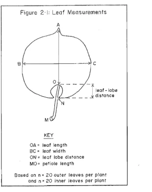
Several leaf form measurements (Figure 2.1) were made with a ruler. Biomass calculations were measured directly or generated secondarily as ratio variables. Dry weights were determined after drying leaves in a vacuum oven for 4 days at 60°C.
2.2.4 Light microscopy of leaf surfaces
17
2.2.5 Scanning electron microscopy of leaf surfaces
Detailed observations of leaf surfaces were made using a Cambridge Stereoscan 250 Mk2 scanning electron microscope, operated at 8-20 kV, equipped \\rith a Robinson backscatter detector.
Fresh leaves were affixed to aluminium stubs with copper conductive paint G.E.C. Electronics, lllinois, U.S.A), as fresh leaves (see Robards 1978, after freeze-drying from liquid nitrogen (sub cooled), or after critical-point-drying from CO2 , \\rith prior dehydration in ethanol and amyl acetate series, as described by
Fineran and Condon (1988). Specimens for S.E.M. were routinely coated \\rith a 5-10 nm layer of gold-palladium using an Edwards model coater, or a Polaron E5000 sputter coater.
2.2.6 Internal organisation of leaves
Procedures for investigating the internal structure of leaves in Actinidia largely follow those of Condon and Fineran (1989).
2.2.6.1 Light microscopy
Leaves were trimmed into pieces 0.5-1 cm2
x 1 mm thick and fixed at 20°C for 24 hrs under water vacuum in 2.5% glutaraldehyde, in a 0.075 M Na-Na phosphate buffer (pH 7.2) vehicle.
2.2.6.2 Dehydration (Chemical)
Tissue was dehydrated for 2 hr in an ethanol series for 20 min. in each of 10, 20, 40, 60; 80% grades with 2 x 30 min changes in absolute ethanoL Specimens were also re-evacuated at a lower surface tension, in 80 and 100% alcohol changes, in order to remove any oxygen adhering to hairs on the surfaces of leaves.
2.2.6.3 Infiltration
Material was infiltrated in mixtures of 25 and 75% JB4 water-miscible resin (Polysciences Inc.) dissolved in ethanol, for 2-3 weeks at each stage, initially at 4°C and subsequently at room temperature. Samples were agitated during infiltration as the resin was prone to an aerobic polymerisation as time increased.
2.2.6.4 Polymerisation
18
Table 2.1 Vines of Actlnidia Lindl. eXllmlnd. Taxa as recognised byIJ (1952) Liang (1980, 1982a,b, 1983,1984); liang and Ferguson (1984, 19861'
Introduction and source categof es : 1 = 1904, seeds, Japan; 2 1955, plants, England (Ot~inal source not known); 3 1975, seeds, China; 4 ... 1977, seeds, Japan; 5 = 1977,
see ,China; 6 = 1979, sdonwood, USSR; 7 0: 1981, seeds, China; 8 1981, scionwood,
China; 9 = unknown, selection of Pletcher and Mouat. .
Taxon Orchard Se7C Inlro
plnnt M/F nnd
number(s) Source
Genus ktlnidia Undl.. Section Leiocarpae (Dunn) Li
Series Lamellatae C.P.liang .
A. arl1uta (Sieb.et Zucc.) Planch. ex Miq.
~T,pr/l/4 P
var. arguta 2
vnr. cordifolia (Miq.) Bean T.P.4/1/16 P 2
A. rufa (Sieb. et Zucc.) Planch. ex lvliq. T.P.3/11/8 M 4 2nd vine
A. melarlGndra Pranch.
var.l11elanandra G,P'j4/1/9a M 2
A. ko[a/JIikta (Maxim. et Rupr.) Maxim. T.P.3/11/11 7' 6
Series Sa/idae c.P. Lian~
A. polygama (Sieb. et. ucc.) Maxim. [f.P.)3/12/11 M 4
A. lIa/llata Dunn K.)MlBl M 7 Section Maculatam Dunn
A. callosa Lind!.
var. Ilenryi Maxim ~T,pr/2/14b M 2
A. cllryslll1tlla C.F. Liang T.P. 36/3/15a M 7
A. indocMnensis Merr. (T.P.36/3/8b M 7 Section Strigosae IJ
A. melUana Hand.-Ml,lZz (T.P.)36/3/3a M 7
A. Jremsleyana Dunn
var. hemsleyana (T.P.)3/8/19a M 3 Section Stellatae Li
Series Perfeetae c.P. Uan~
A. latifofia (Gardn. et Camp.) Merr.
P
var.lallfolia ~K)M5[l4 7
A. eriantlln Benth. T.P.)3/7/15c P 3 Section Stella(ae Li
Series Perfectae C.P. Liang
A. chilUmsis PJanch.
ff·
p·t/
6/7a
1
var. chinel1sis F 8
T.P. 3/6/9al
P 8
T.P. 3/6/14b P 5
A. deliciosa (A Chev.) c.F. Liang et AR. Ferguson var. deliciosa
'Hayward' (K.)
destruction block P
'Bruno' [FP.~4/5/16 P 1
'Matua' T.P.4/2/16 M 9
var. ch/oroc{).rpa (C.P. Liang) .
c.P. Liang et AR. Ferguson. (K)CGM2 F 7
Footnotes: 1. Sex unknown, plant not yet fruited.
2. These genotypes of A. cllinensis var. cllinensis have aitemalive names: 3/6/7a '" 460.4
3/6/9a "' 460.9
Latter designations are used in results tables.
Abbreviations:
19
Figure 2-1: Leaf Measurements
A
Bf~---r---~
KEY
OA:: leaf length Be = leaf wid th
ON:: leaf lobe distance MO:: petiole length
leaf - lobe distance
Based on n = 20 outer leaves per plant and n:: 20 inner leaves per plant
with an intervening piece of "Parafilm". Polymerisation was completed within 2-4 hrs at 20D
e.
2.2.6.5 Microtomy for L.M.
Sections of leaves were obtained with a Reichert rotocut 2000 EX and with a Jung rotary microtome 11800 equipped with glass knives mounted in a modified knife-holder (Fineran and Johnston 1974).
2.2.6.6 Staining for L. M.
Sections were stained with one of the follo\\IDg histochemical reagents:
1. 50/50 methylene blue/toluidine Blue (aq)
2. acid fuschin (aq)/toluidine blue (acO (Feder and O'Brien 1968). After rinsing and drying slides on a hotplate for 2-3 min at 40-60D
[image:35.545.141.452.41.433.2]20
2.2.6.7 Photomicrography for L.M.
Bright-field micrographs were recorded on Ilford FP4 film using a Leitz Orthoplan photomicroscope equipped with an Orthomatt M400 camera. Films were developed in Rodinol F for 5-10 millS using a 1:20 dilution of developer to water.
2.2.7 Internal structure of leaves, S.E.M. methods
S.E.M. methods were also used to document for the chemical artifacts induced by most L.M. methods and also to document the three-dimensional form of the leaves.
2.2.7.1 Preparation of fractured leaves for S.E.M.
Leaves were prepared for S.E.M. following the freeze-fracture methods of Fineran and Condon (1988) and Condon and Fineran (1989). This involved material being immersed in subcooled liquid nitrogen ("slush"),l at -210 degrees
C. (Robards and Sleytr 1988). The leaves were subsequently broken into pieces 1-2 cm2
under the liquid and stored in small aluminium containers held in a large dewar of liquid nitrogen. For freeze-drying, the containers and their liquid nitrogen were transferred to a Bullivant-Ames device (see Fineran 1978) filled with liquid nitrogen) and placed in an Edwards vacuum coater unit for 11 hrs at a vacuum pressure of 2 x 10 Torr. The freeze-dried samples were then mounted on stubs, as described in Section 4.2.3.
2.2.7.2 Photomicroscopy
Scanning electron micrographs were recorded on l1ford FP4 developed in Microphen and rated at 200 ASA.
21
2.2.8 Internal structure of leaves T.E.M. methods
Schedule
2 hr fixation
2.5% Glutaraldehyde in 0.075M Na-Na phosphate buffer$ pH ::::: 7.2 at 20°C
1
post-fixation buffering*
1
2-3 hr post-fixation
1 % OS04 in O.02SM Na-Na phosphate buffer pH == 7.2 at 20°C
1
post-fixation buffering*
1
Chemical dehydration
10 min in 20,40,60;80% acetone (aq) with 3 x 10 min
changes in 100% acetone
1
Infiltration
30% Spurrs resin (Spurr 1969) in acetone overnight
1
Embedment
100% Spurr's resin polymerised at 70°C
1
Ultramicrotomy
blocks trimmed and sectioned with a diamond knife for T.E.M. on an LKB Ultrotome IV
1
Post-staining of sections in uranyl acetate-lead citrate
1
Transmission electron microscopy
mOL 1200 EX Transmission electron microscope operated
at 80-100 kV. .
Indicates same buffer used during fixation and post-fixation.
2.2.9 Cluster analysis of winter shoot characters
22
overall relationships between species or individuals are assessed in "distance" (variance-covariance) or "similarity" (correlation) matrices (Sneath and Sokal
1973). The three-dimensional clusters arising from C.A are presented in two-dimensional computer-drawn diagrams termed phenograms.
Relationships between individuals (termed O.T.Us
=
Qrerational!.axonomic units) or exemplars of Actinidia taxa derived from winter shoot characters were assessed using Gower's (1971) general coefficient of similarity. Similarity values were clustered using complete, arithmetic group averages (U.P.G.M.A) and single-linkage techniques. The degree of deviation of the phenogram from its precursor similarity matrix, was assessed using the cophenetic correlation coefficient of Sokal and Rohlf (1962).
The program used for C.A was "Gower", written by Drs C.M. Frampton, G.A Findlay and 1.M. Ward, Christchurch.
2.2.9.1 Characters
Characters can be defined as attributes which taxonomists separate from whole organisms for particular purposes such as comparison or identification (Davis and Heywood 1963). Any character may be expressed (often arbitrarily) as a number of indivisible parts (henceforth termed "character states").
Variables or "characters" entered into the similarity matrix are coded as "multistate non-ordered", "discrete non-ordered" or ''binary dichotomous" in category.
"Multistate ordered" or "type 1" characters in the computer program, also termed quantitative multistate characters, are those in which the character states can be placed in an ordered sequence. These characters may include a series of measurements along a scale, (e.g. leaf width in mm), or they may include other characters, which have been subdivided into a number of discrete character states, (e.g. leaf apex form as a character with 3 character states: 1
=
acuminate, 2=
apiculate and 3=
retuse)."Discrete non-ordered" or "type 2" characters in the program, are those in which character states cannot be -placed in an ordered sequence and where both presence and absence character states are considered to be of equal importance in
assessin~ similarity. "Type 2" characters include non-ordered multistate characters, for example petal colour with 3 character states: 1
=
green, 2=
white, and 3=
yellow, and binary alternative characters such as leaf crystal form with character states: 1=
acicular and 2=
cubic.23
2.2.9.2 Similarity coefficients
Gower's general coefficient of similarity (1971) can be used with data containing different kinds of characters without the need for recoding. Gower's coefficient is a composite of three similarity coefficients. One of the three is chosen for each character in the data set. Jaccard's coefficient SJ is used with binary dichotomous characters (shared absence or negative state of the character not scored as a similarity), the simple matching coefficient Ssm is· used with "discrete non-ordered" characters (sharing of any or either state of the character scored as a similarity). With "continuous" characters Gower applies the following coefficient
where Xik is the score of OTU i for character k, Xjk is the score of OTU j for character k and Rk is the range of character k.
The simple matching coefficient of Sakal and Michener (1958) is defined as:
SSM = (Nsp
+
Nsn)/(Nsp+
Nsn+
Nu)and Jaccard's coefficient (1908) is defined as :
S
=
Nsp/(Nsp + Nu)J
where Nsp is the number of states whose presence or positive state is shared by two OTUs, Nsn is the number of shared absence or negative states in the two OTUs being compared with Nu is the number of unshared states (i.e. present/positive in one and absent/negative in the other of the two OTUs being compared). The simple matching coefficient gives equal weight to the shared presence/positive state and absence/negative state of characters, while Jaccard's coefficient ignores shared absences/negative states.
2.2.9.3 Cluster methods
The similarity values were clustered by the unweighted pair group method using arithmetic averages (UPGMA) and by the single linkage technique.
24
one cluster with all members of the other. Average-linkage provides information on average phenetic relationships. The clusters form over an intermediate range (compared with single and complete linkage clustering) and the hierarchical structure is quite clear. Average-linkage generally gives the least amount of distortion of a similarity matrix (Rohlf 1970, Sneath and Soka1 1973). However, outlying OTUs (those which are not similar to any others) may form a pair not because they are most similar to each other, but rather because their similarity to each other is higher than either one's average similarity to any existing cluster.
In single linkage clustering, an OTU has a similarity to an existing cluster which is equal to its similarity to the closest member within the cluster. The single linkage technique (Florek et al., 1951 a,b and Sneath 1957) provides information on closest phenetic relationships, and because of the criterion for entry into the fusion of clusters, it is not sensitive to cluster size.
2.2.9.4 Cophenetic correlation coefficient
Since the phenogram resulting from C.A IS a two-dimensional
representation of a multi-dimensional structure, some distortion of the relationships in the similarity matrix on which it is based is inevitable. The "degree of fit" of a phenogram to the similarity matrix from which it is derived may be measured using the cophenetic correlation coefficient proposed by Sokal and Rohlf (1962). [A matrix of cophenetic values is obtained from the phenogram by finding the similarity level that links each pair of OTUs. The cross-product correlation coefficient is then computed between the two matrices; this is the cophenetic correlation coefficient]. A value of one represents complete agreement between the two matrices.
Sections 2.2.9.2 to 2.2.9.4 were adapted, with permission, from Breitwieser (1990) with minimal modifications. Methods used in these sections of the thesis are identical to those used by Breitwieser loc. cit.
2.2.10 Discriminant analysis (D.A,) of summer shoot characters
The character set (Appendix 2.2.1) used in this chapter is large and diverse and most (binary and continuous multistate) are not suited to discriminant analysis. D.A is explained and demonstrated more comprehensively in Chapter 111ree (Section 3.3.3). Throughout this thesis D.A. is synonymous with the "standard type of discriminant analysis" (Frampton 1988) as devised by Fisher (1936). It is not to be confused with "stepwise discriminant analysisll
, which is a
25
2.3 RESULTS
2.3.1 Comparative morphology of summer shoots in some Actinidia
under cultivation
Actinidia vines in the spring and summer months are colourful and
handsome plants of extremely varied morphology. The vegetative form of these plants is described in detail in Tables 2.2 - 2.6, but the main trends are summarised in the following text.
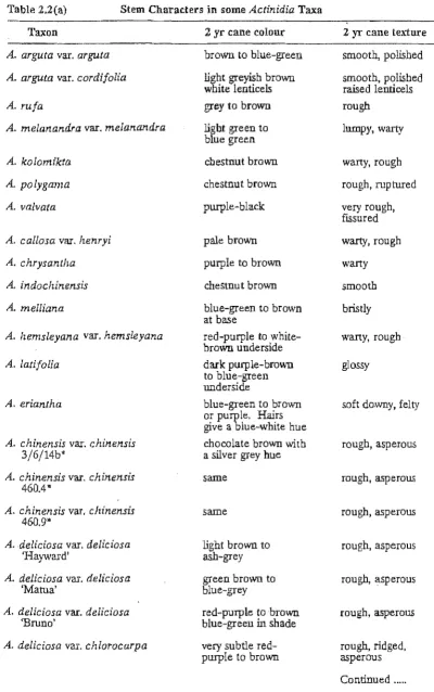
2.3.1.1 Stem characters
The colour of 2 yr canes (Table 2.2) is generally more stable than that of the current year's wood. None of the colours appear to be unique to particular sections of the genus. Two year old wood is generally brown or brown overlain with blue-green pigmentation. Red-purple colours are more common in the
Stellatae but are not restricted to this group.
Cane texture results from a combination of the underlying bark and any overlying hairs or lenticels. The wood is coarsely textured in sections Leiocarpae. M aculatae and Strigosae. The underlying bark may be lustrous with large warty
lenticels, e.g. A. polygama. Preliminary anatomical observations show that lenticel protuberance is proportional to the depth of the concave depression formed by the phellogen. The bulk of the lenticel is filled with parenchymatous "packing tissue".
Canes of the Stellatae are more finely textured, with a rough (asperous)
texture arising from a cover of stiff (hispid) hairs, which may persist as intact structures in the form of "stubblei
• or hair bases only (see Chapter Three for details).
The colour and form of lenticels (Table 2.2) can be used to identify spring and summer shoots. Most Actinidia have orange or brown, but rarely, yellow or
white lenticels. Stellatae have smaller lenticels with white edges and orange to
brown centres.
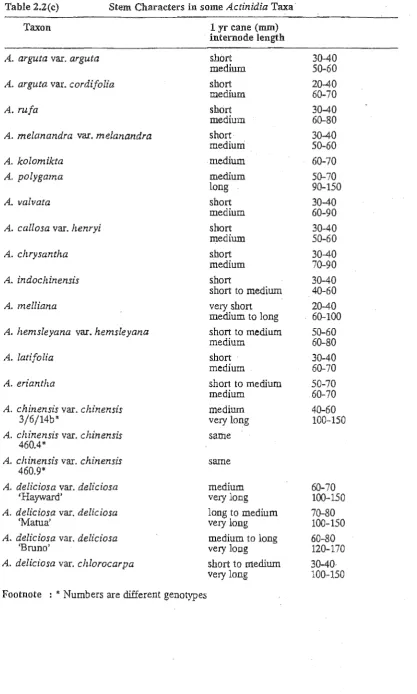
The length of internodes (Table 2.2) of the current year's canes 15
characteristic of certain sections of the genus. Leiocarpae have the shortest canes,
those of the M aculatae and Strigosae are comparable in length or longer,