ORIGINAL RESEARCH
INTERVENTIONAL
Differences in Hemodynamics and Rupture Rate of Aneurysms
at the Bifurcation of the Basilar and Internal Carotid Arteries
X R. Doddasomayajula,XB. Chung,X F. Hamzei-Sichani,X C.M. Putman, andXJ.R. Cebral
ABSTRACT
BACKGROUND AND PURPOSE:Cerebral aneurysms in the posterior circulation are known to have a higher rupture risk than those in the anterior circulation. We sought to test the hypothesis that differences in hemodynamics can explain the difference in rupture rates.
MATERIALS AND METHODS: A total of 117 aneurysms, 63 at the tip of the basilar artery (27 ruptured, 36 unruptured, rupture rate⫽43%) and 54 at the bifurcation of the internal carotid artery (11 ruptured, 43 unruptured, rupture rate⫽20%) were analyzed with image-based computational fluid dynamics. Several hemodynamic variables were compared among aneurysms at each location and between ruptured and unruptured aneurysms at each location.
RESULTS: On average, aneurysms at the basilar tip had more concentrated inflow (P⬍.001), a larger inflow rate (P⬍.001), a larger maximum oscillatory shear index (P⫽.003), more complex flows (P⫽.033), and smaller areas under low wall shear stress (P⬍.001) than aneurysms at the bifurcation of the internal carotid artery. In general, ruptured aneurysms had larger inflow concentration (P⫽.02), larger shear concentration (P⫽.02), more complex flows (P⬍.001), and smaller minimum wall shear stress (P⫽.003) than unruptured aneurysms.
CONCLUSIONS: High flow conditions, characterized by large and concentrated inflow jets, complex and oscillatory flow patterns, and wall shear stress distributions with focalized regions of high shear and large regions of low shear, are associated with aneurysm rupture, especially for basilar tip aneurysms. The higher flow conditions in basilar tip aneurysms could explain their increased rupture risk compared with internal carotid bifurcation aneurysms.
ABBREVIATIONS:BAtip⫽basilar artery tip; corelen⫽core-line length; ICAbif⫽internal carotid artery bifurcation; ICI⫽inflow concentration index; LSA⫽areas under low wall shear stress; max⫽maximum; min⫽minimum; OSI⫽oscillatory shear index; Q⫽flow rate; SCI⫽shear concentration index; VE⫽mean velocity; WSS⫽wall shear stress
I
ntracranial aneurysms are balloon-like pathologic dilations of the cerebral blood vessel walls. An estimated 2%–5% of the general population is affected by intracranial aneurysms.1 Rupture of intracranial aneurysms is an event associated with high mortality and disability rates; consequently, many physi-cians often preventively treat incidentally discovered aneu-rysms. However, the treatment risk can exceed the low natural risk of rupture of incidental aneurysms.2,3Therefore, it is im-portant to precisely distinguish high-risk aneurysms forimme-diate treatment from low-risk aneurysms, which could be con-servatively followed.
Several patient, demographic, and behavioral characteristics have been investigated as possible risk factors, including sex, age, family history, smoking, hypertension, and ethnicity. Past studies have indicated that in addition to the size of the aneurysm, loca-tion is an important risk factor for intracranial aneurysm rup-ture.4In particular, aneurysms in the posterior circulation have been shown to have an overall higher rupture risk than aneurysms in the anterior circulation. The cause of this difference in rupture risk is largely unknown.5Because aneurysm evolution is thought to be governed by a progressive degradation and weakening of the vessel wall in response to abnormal flow conditions,6it is logical to ask whether differences in aneurysm hemodynamics, induced by diverse configurations and geometries of vessels of the anterior and posterior circulations, could help explain the various rupture rates.
The purpose of this study was to test whether intrasaccular flow characteristics are different between aneurysms at the basilar
Received May 11, 2016; accepted after revision October 11.
From the Bioengineering Department (R.D., B.C., J.R.C.), Volgenau School of Engi-neering, George Mason University, Fairfax, Virginia; Department of Neurosurgery (F.H.-S.), Icahn School of Medicine at Mount Sinai, New York, New York; and Inter-ventional Neuroradiology (C.M.P.), Inova Fairfax Hospital, Falls Church, Virginia. Please address correspondence to Juan R. Cebral, PhD, Bioengineering Department, Volgenau School of Engineering, George Mason University, 4400 University Dr, MSN 2A1, Fairfax, VA 22030; e-mail: [email protected]
Indicates article with supplemental on-line tables. Indicates article with supplemental on-line photos.
artery tip (BAtip) in the posterior circulation and the internal carotid artery bifurcation (ICAbif) in the anterior circulation, as well as between ruptured and unruptured aneurysms at these lo-cations. These locations were selected because the BAtip is the most common location of intracranial aneurysm rupture in the posterior circulation7and is anatomically similar to the ICAbif in that they are both terminal bifurcations of a major feeding artery. The information generated in this study is useful for understand-ing what hemodynamic conditions predispose aneurysms to rup-ture and which mechanobiologic processes could be involved in aneurysm weakening and eventual rupture.
MATERIALS AND METHODS
Image and Patient Data
We have developed a data base of intracranial aneurysms imaged with 3D rotational angiography. This data base contains 3D rota-tional angiography images and anonymized clinical information, including rupture status, size, and location and patient sex and age of approximately 1800 intracranial aneurysms. All aneurysms from our data base located at either the BAtip or ICAbif were included in the study. A total of 117 aneurysms were studied, with the distribution of locations as follows: 63 BAtip aneurysms con-sisting of 27 ruptured and 36 unruptured with a rupture rate of 43%, and 54 ICAbif aneurysms, among which 11 were ruptured and 43 were unruptured, with a rupture rate of 20%. Among the 117 patients studied, there were 94 (80%) women and 23 (20%) men. There was no statistical difference between sexes in the BAtip and ICAbif aneurysms (P ⫽ .8163). The mean age of the ICAbif group was slightly younger (52 years) than that of the BAtip group (57 years) (P⫽.0284).
Vascular and Flow Modeling
Computational fluid dynamics models of all 117 aneurysms were constructed from the corresponding 3D rotational angiography images, by using a methodology previously developed.8Briefly, the 3D rotational angiography images were filtered to reduce noise and the vascular geometry was reconstructed with an iso-surface-deformable model.9These vascular models were subse-quently smoothed, and the vessel branches were truncated at
planes orthogonal to their axes. The ICAbif models extended proximally to the cavernous ICA, and the BAtip, at least to the origin of the basilar artery; the models included both vertebral arteries if they were adequately depicted in the 3D rotational an-giography images.
Numeric simulations based on 3D incompressible Navier-Stokes equations were performed on the 117 patient-specific an-eurysm geometries by using pulsatile flow conditions. Because patient-specific flow information was unavailable, typical flow boundary conditions for a healthy subject derived from phase-contrast MR imaging (On-line Fig 1) were scaled with the inlet cross-sectional areas to achieve a mean wall shear stress (WSS) of 15 dynes/cm2prescribed at the inlets for all the models. Fully
developed velocity profiles were prescribed at the inlets by using the Womersley solution.10Outflow boundary conditions were selected to produce flow divisions consistent with the Murray law to avoid unrealistic jumps in the wall shear stress from parent to daughter branches. Assumptions included Newtonian viscosity for blood and rigid vessel walls. The simulations had a minimum mesh resolution of 200m and a time resolution of 10 ms and were run for 2 cardiac cycles by using an in-house fully implicit finite-element solver.11,12
Flow variables such as the flow rate (Q), maximum wall shear stress (WSSmax), inflow concentration index (ICI), and the shear concentration index (SCI) were computed from the results of the second cycle to characterize the flow conditions within the aneu-rysms.13,14Geometric variables such as aneurysm size (Asize) and aspect ratio (AR) were computed from the 3D anatomic models. The definitions of vessel geometric variables are presented in On-line Fig 2. TheTablepresents a list of all the hemodynamic and geometric variables analyzed.
Data Analysis
The hemodynamic parameters listed in theTablewere used to test the following: 1) whether aneurysms at the BAtip have different hemodynamic environments than those at the ICAbif, and 2) whether ruptured and unruptured aneurysms have different flow environments at each of these 2 locations. Two-sided Wilcoxon Hemodynamic and geometric variables used in the study and the aspect of the aneurysm they characterize
Variables Metrics Characterizes
Hemodynamics
Q, ICI Inflow rate, inflow concentration index Inflow jet
VE, KE Mean velocity, kinetic energy Flow speed in aneurysm
VO, SR, VD Vorticity, shear rate, viscous dissipation Flow rotation deformation/dissipation in aneurysm
⬍WSS⬎, WSSmin, WSSmax, SCI, LSA Mean, min, and max WSS, shear concentration index, area under low WSS
WSS distribution
⬍OSI⬎, OSImax Mean, max oscillatory shear index Oscillatory flow/WSS
Corelen Length of vortex core lines Flow complexity
Podent, podenum POD entropy and number of modes Flow stability
Geometry
Aneurysym size, volume, area, SizeR Aneurysm diameter, volume, area, size ratio Aneurysm size
Neck size, area Neck diameter, area Neck size
Depth, aspect ratio Aneurysm depth, aspect ratio Aneurysm shape elongation
Artery, branch1, branch2 Diameter parent and branch arteries Artery sizes
Symmetry Ratio of branch diameters Bifurcation symmetry
⬍Taper⬎, taper1, taper2 Mean taper, branch taper Artery tapering
rank sum tests were used to compare continuous variables between these groups. Differences were considered statistically significant withP⬍.05 (95% confidence level). To test for correlations or sta-tistical dependence between the geometric and hemodynamic vari-ables, we performed a Spearman rank correlation test among all the variables. A size-based comparison was then performed to assess any size-specific differences in ruptured and unruptured aneurysms at both locations. For this purpose, aneurysms were subdivided into 3 size groups: 1) large,⬎13 mm; 2) medium, 7–13 mm; and 3, small, ⬍7 mm. Then the statistical analysis was repeated.2
RESULTS
The rupture rate among BAtip aneurysms was 43% compared with a 20% rupture rate in ICAbif aneurysms. A 2⫻2
contin-gency table analysis based on the Fisher exact test confirmed a statistically signif-icant association between the aneurysm location (BAtip or ICAbif) and rupture rate (P⫽.0107).
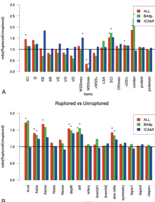
A comparison of hemodynamic and geometric variables between ruptured and unruptured aneurysms at both loca-tions (red bars), along with similar com-parisons for aneurysms at the BAtip (green bars) and at the ICAbif (blue bars), is shown inFig 1(data is provided in On-line Tables 1–3 as means and SDs for ruptured and unruptured aneurysms for all aneurysms and by location). The bars in these graphs indicate the ratio of mean values of the corresponding vari-ables of ruptured aneurysms over the mean values of the unruptured aneu-rysms. Variables that are significantly different between these groups are marked by an asterisk.
In general, irrespective of the loca-tion, ruptured aneurysms had more concentrated inflow jets (ICI,P⫽.02), more concentrated WSS distributions (SCI,P⫽.02), more complex flow pat-terns (characterized by a longer vortex core-line length [corelen],P⬍.0001), and lower minimum WSS (WSSmin, P⫽.003) than unruptured aneurysms, as indicated inFig 1A. Geometrically, ruptured aneurysms were larger (aneu-rysm volume,P⫽.006; aneurysm size, P⫽.002; aneurysm area,P⫽.005; size ratio,P⫽.003) and had more elongated shapes (depth,P ⬍ .001; aspect ratio, P⫽.001) than unruptured aneurysms (red bars inFig 1B).
Similar associations were found for aneurysms located at the BAtip (green bars in Fig 1A). Specifically, ruptured BAtip aneurysms had higher WSS con-centration (SCI,P⫽.02), more complex flow patterns (corelen, P⬍.0001), and lower minimum WSS (P⫽.02) than unruptured aneurysms at this location. Ruptured BAtip aneurysms were also larger (aneurysm size,P ⫽.02; size ratio,P⫽ .03) and more elongated (depth,P⫽.01; aspect ratio,P⫽.001) than unrup-tured BAtip aneurysms (green bars inFig 1B). In contrast, rup-tured ICAbif aneurysms had larger maximum WSS (P⫽.03) than unruptured ICAbif aneurysms (blue bars inFig 1). All other he-modynamic or geometric variables did not reach statistical significance.
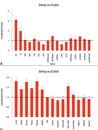
Hemodynamic and geometric characteristics of aneurysms (in-cluding both ruptured and unruptured aneurysms) located at the BAtip and the ICAbif are shown inFig 2. In general, BAtip aneu-rysms had more concentrated inflow jets (ICI,P⬍.001), larger FIG 1. Ruptured-versus-unruptured aneurysms. Ratio of mean hemodynamic (A) and geometric
[image:3.594.54.375.64.482.2]inflow rates (P ⬍.001; mean velocity [VE], P⫽.04), more oscillatory flows (maximum oscillatory shear index [OSImax], P ⫽.005; mean OSI,P⫽.04), and smaller areas under low WSS (LSA,P⬍.0001) compared with ICAbif aneurysms. Ad-ditionally, BAtip aneurysms were larger (aneurysm volume, P ⫽.0005; aneurysm size, P⫽ .0004; Area,P ⫽.0006; size ratio,P⬍.001) and had deeper domes (depth,P⫽.01) and wider necks (neck size,P⬍.0001; neck area,P⬍.0001) than ICAbif aneurysms. Most interesting, both parent and branch arteries were of a smaller caliber in the BAtip group (artery, P⫽.003; branch1,P⬍.001; branch2,P⫽.006) and had lower tapering (taper1,P⬍.001; taper2,P⫽.04; mean taper,P⫽ .0004) compared with the ICAbif group.
The differences between BAtip and ICAbif hemodynamic en-vironments were further analyzed by subdividing the aneurysms into groups based on their size (Fig 3). For large aneurysms (red
bars), there were no significant differ-ences in the hemodynamic variables. However, medium-sized BAtip aneu-rysms (green bars) had more concen-trated inflow jets and higher inflow rates (ICI,P⬍.001; Q,P⫽.003), more oscil-latory flows (OSImax, P ⫽ .03), and larger areas under low WSS (P⫽.001) compared with medium-sized ICAbif aneurysms. Similarly, small BAtip aneu-rysms had higher flow conditions char-acterized by larger inflow rates, more concentrated inflow jets, and larger in-trasaccular velocity (ICI,P⬍ .001; Q, P⫽.01; VE,P⫽.04) as well as larger areas under low WSS (P⬍.001) com-pared with small ICAbif aneurysms.
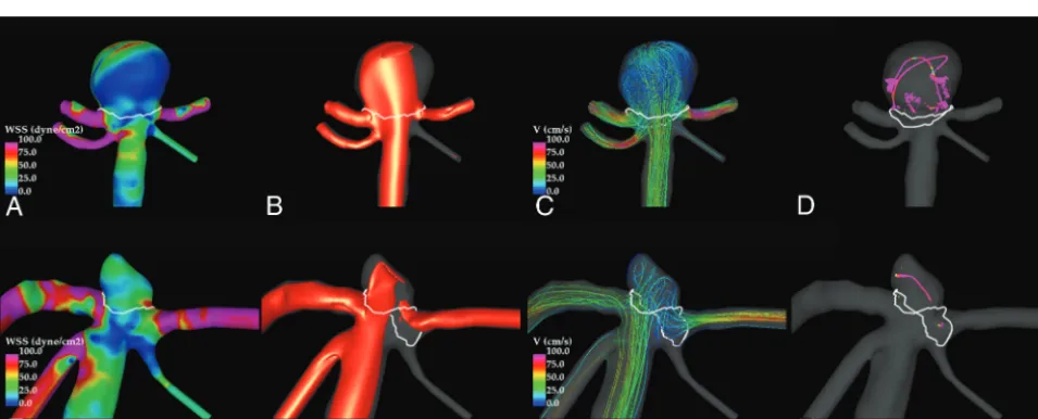
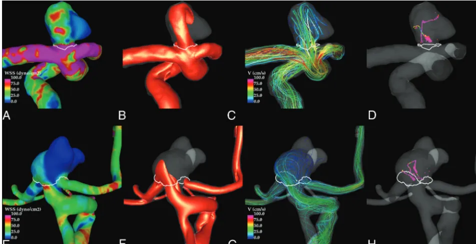
Illustrative examples of ruptured and unruptured aneurysms at the BAtip and ICAbif are shown inFigs 4and5, respec-tively. These figures show visualizations at peak systole of the WSS distribution, the inflow jets, the streamline patterns, and the flow structure by using vortex center lines. In both examples, the flow in the ruptured aneurysm (upper row) is charac-terized by a strong inflow jet impacting a concentrated area of the dome and pro-ducing a complex flow pattern within the aneurysm. In contrast, the unruptured an-eurysms (lower row) exhibit a more uni-form WSS distribution associated with a more diffuse inflow jet and simpler flow pattern.
DISCUSSION
Posterior circulation intracranial aneu-rysms are known to have higher rupture rates compared with anterior circulation intracranial aneurysms15; considering that there are 2 ICAs for every 1 basilar artery in a complete circle of Willis, the reported differences in rupture risk are probably even more substantial.5The results in our series showing a rupture rate of 43% among BAtip aneu-rysms compared with 20% among ICAbif aneuaneu-rysms (P ⫽ .0107) are consistent with those published in previous studies.5,15
The exact reasons for these differences in rupture risks or the rupture mechanisms are still relatively unknown.16Thus, several previous studies have sought to determine the effects of morpho-metric features and hemodynamic forces on the mechanobiology of the vessel wall and on the rupture risk of intracranial aneu-rysms.17-19Some studies have suggested that aneurysms located at bifurcations are at the highest risk of rupture, attributing it to the increased hemodynamic stress at the tip of the bifurcation.5,20We sought to further understand the differences between these 2 lo-cations by evaluating the intra-aneurysmal hemodynamics and FIG 2. BAtip-versus-ICAbif aneurysms. Ratio of mean hemodynamic (A) and geometric (B)
[image:4.594.57.375.53.474.2]morphometric features of analogous bifurcations representing both systems.
Can et al21suggested that the larger P1-P1 angle of the BAtip bifurcation causes greater flow divergence into the daughter posterior cerebral arteries, thus causing lower WSS and leading to aneurysmal initiation. Our results do not seem to support the idea that this same mechanism results in aneurysm pro-gression and rupture. Irrespective of whether the aneurysms were located at the BAtip or at the ICAbif, ruptured aneurysms in our series had more concentrated inflow jets, more concen-trated WSS distributions, and longer vortex core-line lengths and, at the same time, had lower WSSmin. Although not con-firmed in the ICAbif location, the BAtip location has signifi-cantly “higher flow conditions” associated with ruptured an-eurysms compared with the unruptured ones. The longer vortex core-line lengths are an indicator of more complex flows and are consistent with the findings in several previously reported studies comparing ruptured and unruptured aneu-rysms.14,22Morphologically, ruptured aneurysms in our study
were larger and more elongated than unruptured aneurysms as identified in multiple prior studies.23,24
Similarly, higher flow conditions were observed in general when comparing all BAtip and ICAbif aneurysms irrespective of rupture status. The differences observed between aneurysms at these locations were significant for small and medium-sized an-eurysms, but not for large ones. This finding may be due to in-creased inertia effects associated with larger aneurysms.
Understanding the differences in the intra-aneurysmal hemo-dynamics requires attention to both the aneurysmal morphology and the geometric characteristics of the associated arterial vascu-lar system. The BAtip aneurysms in our series were vascu-larger and had deeper domes and wider necks than ICAbif aneurysms. Larger neck sizes lead to higher intra-aneurysmal flow rates and, in com-bination with the greater depth and volume, have the possibility of greater flow complexity and instability. Why basilar tip aneu-rysms have larger sizes than ICA aneuaneu-rysms is unclear. Better understanding of the mechanisms and mechanobiology of aneu-rysmal growth may prove helpful; however, the perianeuaneu-rysmal environment may also play an impor-tant role in aneurysmal growth because contact with extravascular structures can constrain or influence the aneurysm evolution. Further studies are needed to elucidate the specific contribution of each of these factors and their combined effect on aneurysm progression.
Extending the arguments related to high flow mechanisms contributing to the initiation and progression of cere-bral aneurysms can yield insight when applied to the 2 circulations discussed in this study. We chose to compare the ICA bifurcation with the basilar bifurcation because of their analogous anatomy of a T-type bifurcation. Yet, these aneurysms have very different hemodynamics, and FIG 3. Comparison of hemodynamic variables across BAtip and ICAbif aneurysms grouped
according to their sizes as small, medium, and large. KE indicates kinetic energy; SR, shear rate; VO, vorticitiy; VD, viscous dissipation;⬍WSS⬎, mean WSS; podent, POD entropy; podenum, POD number of modes; hemo, hemodynamic.
[image:5.594.90.336.288.446.2] [image:5.594.55.533.503.696.2]a detailed assessment of the parent artery geometry reveals several distinct differences. The artery sizes (both parent and branch ar-teries) were significantly smaller in the BAtip group along with lower artery tapering compared with the ICAbif group. For a given flow rate, tapering can lead to faster flow velocities and larger pressure drops through an arterial segment, leading to in-creased WSS within that segment. If this effect occurs proximal to the site of aneurysm formation, then fewer kinetic forces are avail-able for the impaction zone of the flow division where aneurysms are typically located. The tapering of the ICA proximal to the terminus, therefore, may act to reduce the shear at the region of the terminus compared with the basilar artery. Curvature of the proximal ICA can also influence the environment of the terminus. The carotid siphon may cause a consistent deflection of the main flow stream into an impaction in the posterior wall of the supra-clinoid ICA, a region of high propensity to aneurysm formation. The curvature of the ICA distal to the posterior communicating artery origin often directs most of the flow stream within the M1 segment, avoiding a direct impaction on the terminus/origin of the A1 segment. In the basilar terminus, this outcome is less often the case with a near-perpendicular impaction and a more equally divided flow stream. Thus, the basilar artery geometry could more often lead to a much higher flow impaction and associated ele-vated WSS compared with the ICA. Variability of the bifurcation asymmetry and bifurcation angles can lead to further differences in the hemodynamic environments in an individual case. In this sense, ICAbif aneurysms seem to behave more like sidewall aneu-rysms, while BAtip aneurysms behave more like true bifurcation aneurysms.
Although a number of results reaching statistical significance were identified in this study, some limitations should be consid-ered. The sample size is small when extending into the various
subgroups, this could interfere with identifying significant differ-ences. The study was taken from a data base of all aneurysms that had undergone angiography or embolization during a 10-year period. Selection bias related to referral patterns and the indica-tions for treatment may lead to exclusion of an important subset of aneurysms. A number of assumptions were made during the modeling process, including the approximations of the vascular geometry, the inflow conditions, the Newtonian approximation, and the rigid wall assumption.
CONCLUSIONS
High flow conditions, characterized by large and concentrated inflow jets, complex and oscillatory intrasaccular flow patterns, and wall shear stress distributions with focalized regions of high WSS and large regions of low WSS are associated with aneurysm rupture in general and in basilar tip aneurysms in particular. The higher flow conditions in BAtip aneurysms could explain their increased rupture risk compared with ICAbif aneurysms. In ICAbif aneurysms, only trends toward high flow conditions were associated with aneurysm rupture, and these trends were ob-served in only smaller ICAbif aneurysms. Further studies with larger sample sizes will be needed to determine whether these trends are statistically significant.
ACKNOWLEDGMENTS
We thank Ms Alicia Suchi (George Mason University) for assis-tance in measuring vessel sizes.
Disclosures: Ravi Doddasomayajula—UNRELATED:Grants/Grants Pending: Na-tional Institutes of Health,Comments: research grant.* Christopher M. Putman—
[image:6.594.53.529.48.292.2]Grants Pending: National Institutes of Health, Philips Healthcare,Comments: re-search grants.* *Money paid to the institution.
REFERENCES
1. Rinkel GJ, Djibuti M, Algra A, et al.Prevalence and risk of rupture of
intracranial aneurysms: a systematic review.Stroke1998;29:251–56
CrossRef Medline
2. Wiebers DO, Whisnant JP, Huston J 3rd, et al; International Study of
Unruptured Intracranial Aneurysms Investigators.Unruptured
intra-cranial aneurysms: natural history, clinical outcome, and risks of sur-gical and endovascular treatment.Lancet2003;362:103–10Medline
3. Weibers DO, Piepgras DG, Meyer FB, et al.Pathogenesis, natural
history, and treatment of unruptured intracranial aneurysms. Mayo Clinic Proc2004;79:1572– 83CrossRef Medline
4. Clarke M.Systematic review of reviews of risk factors for
intracra-nial aneurysms.Neuroradiology2008;50:653– 64CrossRef Medline
5. van der Kolk NM, Algra A, Rinkel GJ.Risk of aneurysm rupture at
intracranial arterial bifurcations.Cerebrovasc Dis 2010;30:29 –35
CrossRef Medline
6. Sforza DM, Putman CM, Cebral JR.Hemodynamics of cerebral
an-eurysms.Annu Rev Fluid Mech2009;41:91–107CrossRef Medline
7. Higa T, Ujiie H, Kato K, et al. Basilar artery trunk saccular
aneurysms: morphological characteristics and management. Neu-rosurg Rev2009;32:181–91; discussion 191CrossRef Medline
8. Cebral JR, Mut F, Weir J, et al.Quantitative characterization of the
hemodynamic environment in ruptured and unruptured brain an-eurysms.AJNR Am J Neuroradiol2011;32:145–51CrossRef Medline
9. Yim PJ, Vasbinder GB, Ho VB, et al.Isosurfaces as deformable
mod-els for magnetic resonance angiography.IEEE Trans Med Imaging
2003;22:875– 81CrossRef Medline
10. Taylor CA, Hughes T Jr, Zarins CK.Finite element modeling of
blood flow in arteries.Computer Methods in Applied Mechanics and Engineering1998;158:155–96CrossRef
11. Cebral JR, Castro MA, Appanaboyina S, et al.Efficient pipeline for
image-based patient-specific analysis of cerebral aneurysm hemodynamics: technique and sensitivity.IEEE Trans Med Im-aging2005;24:457– 67CrossRef Medline
12. Mut F, Aubry R, Lo¨hner R, et al.Fast numerical solutions of
patient-specific blood flows in 3D arterial systems.Int J Numer Method Biomed Eng2010;26:73– 85CrossRef Medline
13. Mut F, Lo¨hner R, Chien A, et al.Computational hemodynamics
framework for the analysis of cerebral aneurysms.Int J Numer Method Biomed Eng2011;27:822–39CrossRef Medline
14. Byrne G, Mut F, Cebral JR.Quantifying the large-scale
hemodynam-ics of intracanial aneurysms. AJNR Am J Neuroradiol 2014;35:
333–38CrossRef Medline
15. Tykocki T, Kostkiewicz B.Aneurysms of the anterior and posterior
cerebral circulation: comparison of the morphometric features. Acta Neurochir (Wien)2014;156:1647–54CrossRef Medline
16. Wermer MJ, van der Schaaf IC, Algra A, et al.Risk of rupture of
unruptured intracranial aneurysms in relation to patient and aneu-rysm characteristics: an updated meta-analysis.Stroke 2007;38:
1404 –10CrossRef Medline
17. Chien A, Castro MA, Tateshima S, et al.Quantitative hemodynamic
analysis of brain aneurysms at different locations.AJNR Am J Neu-roradiol2009;30:1507–12CrossRef Medline
18. Robertson AM, Duan X, Aziz KM, et al.Diversity in the strength and
structure of unruptured cerebral aneurysms.Ann Biomed Eng2015;
43:1502–15CrossRef Medline
19. Cebral JR, Raschi M.Suggested connections between risk factors of
intracranial aneurysms: a review.Ann Biomed Eng2013;41:1366 – 83
CrossRef Medline
20. Meng H, Tutino VM, Xiang J, et al.High WSS or low WSS? Complex
interactions of hemodynamics with intracranial aneurysm initia-tion, growth, and rupture: toward a unifying hypothesis.AJNR Am J Neuroradiol2014;35:1254 – 62CrossRef Medline
21. Can A, Mouminah A, Ho AL, et al.Effect of vascular anatomy on the
formation of basilar tip aneurysms.Neurosurgery2015;76:62– 66;
discussion 66CrossRef Medline
22. Cebral JR, Mut F, Weir J, et al.Association of hemodynamic
charac-teristics and cerebral aneurysm rupture.AJNR Am J Neuroradiol
2011;32:264 –270CrossRef Medline
23. Dhar S, Tremmel M, Mocco J, et al.Morphology parameters for
intracranial aneurysm rupture risk assessment.Neurosurgery2008;
63:185–197; discussion 196 –97CrossRef Medline
24. Fan J, Wang Y, Liu J, et al.Morphological-hemodynamic
character-istics of intracranial bifurcation mirror aneurysms.World