warwick.ac.uk/lib-publications
A Thesis Submitted for the Degree of PhD at the University of Warwick
Permanent WRAP URL:
http://wrap.warwick.ac.uk/111600/
Copyright and reuse:
This thesis is made available online and is protected by original copyright.
Please scroll down to view the document itself.
Please refer to the repository record for this item for information to help you to cite it.
Our policy information is available from the repository home page.
L
e6EX>S M
•£•
fsfbf 6 * 0 &>fib»riGr.
N-ALKANES BY MICROORGANISMS
by
M . R . Lebens B .Sc.
A thesis submitted in fulfilment
of the requirements for the degree of Ph .D.
Department of Biological Sciences,
University of Warwick,
Coventry CV4 7AL
U.K.
Summary
Microorganisms capable of growth using n-alkanes as
sole carbon and energy source were isolated from the
environment. Subsequently selected organisms from those
isolated were subjected to morphological and biochemical
surveys that involved electron microscopy and assays of
enzymes concerned with n-alkane degradation and assimilation.
The alcohol and aldehyde dehydrogenases associated with the
growth of a pseudomonad on n-alkanes were partially purified
and shown to be NAD(P) independent.
Genetic manipulations were attempted to produce
mutants of a coryneform bacterium, by chemical and U .V.
mutagenesis, that were blocked at specific points in the
n-alkane degradation pathway which were shown to be
chromosomally borne. Appropriate selection procedures were
devised and mutations identified by product accumulation
by whole cells . Evidence is also presented for the
presence of a large plasmid that may carry genes for
TABLE OF CONTENTS Acknowledgements Page iv Declaration Summary vii
CHAPTER I Introduction
1 .1 Organisms capable of growth on n-alkanes 1
I .1.1 Sources 1
I .1.2 Bacteria 4
I .1.3 Yeasts 6
1 .1.4 Fungi 9
1 .2 Toxicity of lower n-alkanes 11
1 .3 Substrate interaction 14 I .3 .1 Initial interaction between cells 14
and n-alkane substrates
1 .3 .2 Emulsification of n-alkane substrates 20
I .4 Transport and uptake of n-alkanes by cells
I .5 Initial oxidation of n-alkanes
1.5.1 Possible routes for oxidation of n-alkanes
I .5 .2 The cytochrome P-450 hydroxylation system
I .5 .3 The rubredoxin-linked w-hydroxylase of Pseudomonas putida
1.5.4 Substrate specificity in n-alkane hydroxylases
I .6 Pathways of n-alkane degradation
1 .6 .1 Alcohol and aldehyde dehydrogenases 1 .6.2 e-oxidation
1 .6 .3 a-oxidation
I .7 Adaptations that arise in microorganisms during growth on n-alkanes
1 .7 .1 Metabolism 1 .7 .2 Morphology
I . 8 Hydrocarbons, Bacteria and Industry 89 1.8.1 General interest in microbial 8g
degradation of hydrocarbons
1 .8 .2 Long chain primary alcohols and g8 the present work
CHAPTER II Materials and Methods
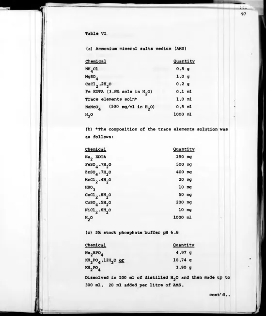
II. 1 Enrichment and isolation of microorganisms 96 growing on n-alkanes
i Media and materials 96 ii Enrichment procedures 99 iii Choice of organisms for further study 101
II .2 Morphological studies of n-alkane-grown ^q2 organisms
i External structure 102 ii Internal structure 103 iii Analysis by light microscopy 105
II .3 Routine growth of cells in batch culture ^Qg and preparation of cell extracts
II .4 Enzyme assays H I i n-alkane hydroxylase H I ii Alcohol and aldehyde dehydrogenases H 7 iii Isocitrate lyase H O iv Acetyl CoA carboxylase 120
v Fatty-acyl-CoA synthetase 121
II .5 Polyacrylamide gel electrophoresis 122 i Preparation of samples 122 ii Polyacrylamide gels and electrophoresis 123 iii Staining of polyacrylamide gels 125
iv Photography of gels 125
II .6 Preliminary genetic studies 126 i Curing experiments with acridine 126
orange and ethidium bromide
ii Mutagenesis 127
CHAPTER III Results and Discussion
111.1 Isolation of microorganisms 134
III .1.1 Enrichment and isolation of
organisms from the environment 134
III .1.2 C16/8S and TC15-1 135
III .1.3 Growth measurements of
n-alkane-grown organisms 138a
III.1.4 Discussion 139
111.2 Mode of growth on n-alkanes 141
III.2.1 Growth in relation to the
substrate 141
III .2 .2 Morphology 152
III .2 .3 Discussion 161
111.3 Enzymes associated with growth on n-alkane
hydroxylase 165
III .3 .1 n-Alkane hydroxylase 165
III .3 .2 Cytochrome P-450 in C16/8S and
TC15-1 166a
111.3.3 Alcohol and aldehyde
dehydrogenases 167
III .3 .4 The induction of n-alkane degrading
enzymes in TC15-1 171a
III .3 .5 Isocitrate lyase and other enzyme
assays 172
III .3 .6 Polyacrylamide gel electrophoresis 172
III .3 .7 Discussion 174
111.4 Genetic analyses 180
III .4 .1 Curing experiments 180
III .4 .2 Mutants of C16/8S 183
111.4.3 The large plasmid of TC15-1 188
111.4.4 Discussion 188
CHAPTER IV General Discussion 196
iv
With very many thanks to my supervisor Dr. H. Dalton
for his enthusiastic encouragment and calming influence
during more stressful moments and to Gail Dawson without
whose help and support life would have been considerably
less bearable.
Thanks also to the S .R .C. and I.C.I. Petrochemical
Division for financing the present work and to
Dr. J. Taylor and Dr. R. Higgins of I.C.I. Petrochemicals
for much useful discussion.
Finally many thanks to all members, past and present,
of the Microbiology Group of the Department of Biological
Sciences for both practical help and discussion, and
to Miss Carolyn Alderson for her excellent typing.
With very many thanks to my supervisor D r . H . Dalton
for his enthusiastic encouragment and calming influence
during more stressful moments and to Gail Dawson without
whose help and support life would have been considerably
less bearable.
Thanks also to the S.R.C. and I.C.I. Petrochemical
Division for financing the present work and to
Dr. J. Taylor and Dr. R. Higgins of I.C.I. Petrochemicals
for much useful discussion.
Finally many thanks to all members, past and present,
of the Microbiology Group of the Department of Biological
Sciences for both practical help and discussion, and
to Miss Carolyn Alderson for her excellent typing.
Declaration
I hereby declare that this thesis was composed by
myself and has not been accepted in any previous
application for a degree. The work represented was all
done by myself. All sources of information have been
acknowledged by reference.
1
I .1 Organisms capable of growth using n-alkanes
I .1.1 Sources
The occurrence of microorganisms capable of growth
using n-alkanes as sole carbon and energy source is widespread.
In a survey of microorganisms from soil and underlying shale,
n-alkane degrading organisms were found at every level
(Jones and Edington, 1968) . Perry and Scheld (1968) also
found a wide variety of n-alkane utilizing organisms when
screening microorganisms isolated from soil using non
hydrocarbon substrates.
Marine and freshwater environments have proved to be
rich sources of n-alkane utilizing microorganisms, particularly
when polluted with petroleum products. Austin et al. (1977),
from surveys of n-alkane degrading microorganisms in aquatic
environments concluded that a wide variety of taxa are
responsible for the degradation of petroleum. However, there
appeared to be a predominance of Pseudomonas. Mycobacterium
or Nocardia species, depending upon the aquatic niche
investigated. Ecological studies were confused further by
the observation of Calomiris eit al^. (1976) that enrichment
procedures significantly influence the range of organisms
isolated. Recent interest in degradation of petroleum in
aquatic environments centres upon the possibility of using
subject has been reviewed recently by Atlas (1977) .
Thermophilic microorganisms capable of growth at the
expense of n-alkanes have been isolated by Merkel et a l .
(1978 Some of the organisms isolated had an optimum
growth temperature of 60°C and were unable to utilize
substrates other than n-alkanes and 1-alkenes (Merkel,
Underwood and Perry, 1976 The capacity to grow only at
the expense of a limited range of saturated or unsaturated
hydrocarbons is rare except in the case of methylotrophs
(Anthony, 1975), although some isolated cases have
been reported (e.g. Bertrand et slL., 1976) .
Because of the wide range of bacteria that have been
shown to utilize n-alkanes, the characteristic is of only
limited taxanomic value. This was illustrated by Grange
(1974) in a study of 50 strains of Mycobacterium. He
concluded that there was considerable variation within species
as well as between species when the range of shorter chain
n-alkanes that could be utilized was tested . Teh and Lee
(1973) had shown similar variations in four isolates of
Cladosporium resinae. In yeasts, however, n-alkane
degradation was found to be a sufficiently distinct property
to aid in the identification of species (Bos and De Bruyn,
1973) .
Prom such work it is evident that the ability to degrade
micro-3
organism nor to any specific environment. This observation
reflects the considerable amounts of n-paraffinic hydrocarbon
that are produced and consumed in the biosphere. n-alkanes
are known to occur in the tissues of plants and animals and
in the cellular lipids of microorganisms, it is only under
exceptional circumstances that oil and coal deposits build
up having escaped complete microbial decomposition.
Davis (1967) in an extensive review of selected aspects
of petroleum microbiology pointed out that in most soils the
high molecular weight n-alkane content is relatively stable;
usually in the range of 0.5 - 5 ng/g of soil. From a 50 kg
sample of New Mexico soil 78 mg of heavy n-alkane were
recovered. Under laboratory conditions a sample of the same
soil was enriched with some of the extracted hydrocarbon,
increasing the amount of n-alkane present from approximately
1.5 ng/g to about 30 ng/g of soil. When incubated at 15%
humidity for two months no change in the hydrocarbon content
was observed. However, when nutrient mineral salts were added
a decrease in the soil paraffins occurred. It was concluded
that the microbial degradation of n-alkanes in the soil was
limited by deficiencies of nutrient minerals, particularly
nitrogen and phosphorus.
Since Fuhs (1961) reviewed the organisms known to that
date to utilize alkanes, the frequency of reports of
as interest in the subject widened. A similar list would
now be of such proportions as to be impractical, nonetheless,
reviews by Klug and Markovetz (1971) and Shennan and Levi
\ (1974) present sufficient examples to illustrate the range
of organisms involved. The present discussion however will
be limited to those organisms that predominate in recent
and current work on n-alkane utilization.
1.1.2 Bacteria
It was observed b y Klug and Markovetz that the number
of bacteria known to utilize n-alkanes far exceeded the
number of yeasts and filamentous fungi. They suggested that
this merely reflected the bias of workers towards bacteria;
more recent work indicates that this was the case .
Of the enormous number of bacteria isolated that are
capable of utilizing n-alkanes relatively few genera have
been used for detailed study of growth at the expense of
these substrates .
Pseudomonas strains have been used for a wide range of
investigations into hydrocarbon utilization. Their common
occurrence and their ability to grow to high cell densities
on a variety of substrates make Pseudomonas species
convenient organisms for such studies . Also of interest is
the existence of plasmid-borne catabolic pathways in many
5
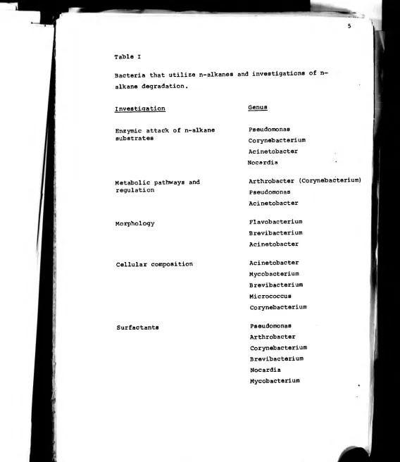
Table I
Bacteria that utilize n-alkanes and investigations of n alkane degradation.
Investigation Genus
Enzymic attack of n-alkane substrates
Pseudomonas Corynebacterium Acinetobacter Nocardia
Metabolic pathways and regulation
Arthrobacter (Corynebacterium) Pseudomonas
Acinetobacter
Morphology Flavobacterium Brevibacterium Acinetobacter
the OCT plasmid of P. putida (Chakrabarty et al., 1973)
which carries the genes for an inducible n-alkane hydroxylase
and primary alcohol dehydrogenase (Grund et al., 1975) is of
particular interest.
Strains of Acinetobacter have been extensively studied
in relation to changes in structure and lipid composition
involved in adaptation to n-alkane metabolism.
Strains of Nocardia. Mycobacterium and Corynebacterium
have also been used to investigate various aspects of
n-alkane degradation. The predominance of these genera above
others is probably due to their common occurrence amongst
n-alkane-degrading isolates. Table I gives an indication
of the range of bacteria that have been used and the nature
of the different work done.
1 .1.3 Yeasts
The amount of work done on n-alkane utilizing yeasts
has increased rapidly over the last fifteen years. Much of
the increased interest shown can be attributed to industrially
sponsored research on organisms that have some commercial
potential.
As with other n-alkane degrading microorganisms, yeasts
that utilize paraffinic hydrocarbons can be readily isolated
from garden or agricultural soils, as well as from sources
7
of yeasts that utilize n-alkane substrates was obtained by
screening cultures from the Central bureau voor
Schimmel-cultures using n-decane and n-hexadecane as growth substrates
(Scheda and Bos, 1966) . It was found that the majority of
strains of Pichia. Debaromyces and Torulopsis as well as the
better documented strains of Candida were positive when
tested.
Shennan and Levi (1974) compiled an extensive list of
organisms when reviewing the growth of yeasts on hydrocarbons.
Strains from the genus Candida however have recently received
most attention. This may hardly be surprising when 63 of
the 155 species cited by Shennan and Levi were from this
genus .
Recently much work has centred upon biotechnological
aspects of growth on hydrocarbons and has been concerned
with such problems as oxygen transfer, substrate interaction
and transfer and the behaviour of cells under various cultural
conditions. Other considerations not directly related to
these problems have received relatively little attention.
Important exceptions to this general view are the isolation
and reconstitution i_n vitro of the hydroxylation systems for
n-alkanes from Candida guillermondii and Candida tropicalis
which will be discussed later.
Table II outlines genera of yeasts cited by Shennan
[image:17.598.0.589.7.687.2]containing n-alkane-utilizing species.
Ascomvcetous Yeasts Funai Imperfecti
Debaromyces Brettanomyces
Endomycopsis Candida
Hansenula* Rhodotorula
Kluyveromyces* Selenotila
Lodderomyces Sporidiobolus
Metschinkowia Sporobolomyces
Pichia Torulopsis
Saccharomyces*
Schizosaccharomyces
Schwanniomyces
Wingea
Trichosporon
*Bos and de Bruyn (1973) Basidiomvcetous Yeasts
concluded that these genera Leucosporidium
were devoid of n-alkane
utilizing species .
9
I .1.4 Filamentous fungi
The observation by Miyoshi (1895) that Botrytis cjnerea
would attack paraffin is one of the earliest examples of
n-alkane utilization in any microorganism. However, many
more species of filamentous fungi are now known to grow at
the expense of n-alkanes. Klug and Markovetz (1971)
reviewed the genera of moulds that grow on n-alkanes and
alk-l-enes; those mentioned and the substrates tested are
shown in Table III. In a more recent study by Bemmann and
Troger (1975) nearly 300 strains of mould were tested for
their ability to grow at the expense of a mixture of
n-alkanes (C^2 “ c2l ) •
Sources of n-alkane degrading moulds are similar to
bacteria and yeasts with isolates from soil being easily
obtainable. Rynearson and Peterson (1965) isolated 20
cultures that grew with paraffin as sole carbon source by a
direct soil-baiting method, the strains represented species
of Aspergillus. Chaetomium, Pénicillium. Syncephalastrum and
Cunninghamella.
Jet fuel (Prince, 1961? Edmonds and Cooney, 1967) and
diesel fuel (Flippin et al.. 1964; Koval et al., 1966) have
also been reported to support growth of filamentous fungi.
Contamination of fuel lines with fungus has caused blockages
and given rise to the need for fuel filters in some engines.
Table III
Genera of Filamentous Fungi cited by Klug and Markovetz
(1971) as being able to utilize n-alkanes.
Genus Chain lengths of n-alkane substrates
11
important differences between other n-alkane-degrading
microorganisms have been noted. In particular, Walker and
Cooney (1973a) found that the n-alkane-oxidizing apparatus
in fungi is constitutive rather than inducible.
As with yeasts however studies have mostly been
confined to specific areas such as identification of primary
oxidation products . Biochemical knowledge of n-alkane
metabolism in general is lacking.
I .2 Toxicity of lower n-alkanes
The number of microorganisms capable of growth on
n-alkanes shorter than nonane (C H ) is much smaller than
y Z\j
that of the microorganisms that can utilize n-alkanes of
greater chain length. Lower alkanes (i.e. shorter than Cg)
are relatively soluble but tend to have toxic effects,
higher alkanes exhibit lower toxicity but are extremely
insoluble in water (Table IV) . It generally appears that
increasing solubility of n-alkanes leads to increasing
toxicity and this factor is harder to overcome than transfer
problems with highly insoluble substrates .
Opinions vary as to what causes lower n-alkanes to
be toxic. It is known that alkanes are able to adsorb to
membranes and bind to hydrophobic membrane proteins . The
total effect is likely to be a combination of membrane dis
n-alkane a m eng Qf saturated solutionMolar concentration
Hexane 6 1.1 x 10"4
Octane 8 5.8 x 10⁻⁶
Decane 10 3.1 x 10⁻⁷
Dodecane 12 1.7 x 10⁻⁸
Tetradecane 14 9.8 x 10_1°
[image:22.606.0.606.9.668.2]When studying the toxicity of lower alkanes in yeasts,
Gill and Ratledge (1972) found that the toxic effect could
be reduced by addition of longer chain n-alkanes . The
explanation of this phenomenon was given as the partition
of the short chain alkanes into the more insoluble, immiscible
higher alkane phase, effectively reducing their concentration
in the medium. It was found that under these conditions
n-alkanes that were normally toxic could be oxidized by
organisms that utilized long chain molecules. Such results
have been used to explain the ability of some microorganisms
to grow at the expense of motor oils.
Observations by Finnerty et al. (1962) suggest that the
physical character of n-alkane substrates as well as the
metabolic capability of the microorganisms may affect growth.
Using strains of Micrococcus it was shown that the range of
shorter chain substrates that could be used could be
expanded if the growth temperature was lowered. One strain
able to use n-undecane as a minimum length growth substrate
at 25°C could also utilize n-decane at 20°C. Such decreases
in temperature affect the solubility and vapour pressure of
alkane substrates . It can also be argued however that such
decreases in temperature may have a more profound effect on
the biochemical activity of the organisms and upon the
fluidity of the cell membrane.
being responsible for toxicity was obtained by Johnson
(1964) . it was shown that the number of organisms capable
of growth on n-hexane increased if the hydrocarbon
concentration was kept below saturation.
I .3 Substrate interaction
I .3 .1 Initial interaction between cells and n-alkane
substrates
Before any substrate can be degraded it must be made
available to the microorganism concerned. However, for an
organism to grow on long chain n-alkanes which are only
poorly soluble it is necessary for cellular contact with the
substrate to be maximized. Three mechanisms for uptake of
sparingly soluble n-alkanes by microorganisms have been
proposed: (i) by direct contact of cells with large oil
drops, (ii) by direct contact of cells with submicron drops
of oil in the 'accommodated' form, and (iii) by direct uptake
of hydrocarbon dissolved in the aqueous phase (Nakahara et a l ..
1977) .
Although some workers have reported that uptake of
dissolved hydrocarbon was significant when n-undecane and
n-dodecane were used as substrates (Erdtsieck and Rietema,
1969) Aiba et al. (1969) and Aiba and Huang (1970) considered
15
that the ratio of n-alkane in solution to the amount
consumed during growth was approximately 10 ® .
The large amount of work done on yeasts by several
workers left opinion divided as to which of the remaining
two alternatives was dominant. Velankar et al. (1974, 1975)
suggested that submicron droplets were essential for growth
on hydrocarbons and proposed the mechanism outlined in
figures 1 and 2. High rates of growth on n-alkanes were
explained by large drops of oil acting as reservoirs for
the diffusion of substrates into micelles. When the micelles
were immediately adjacent to a cell rapid transfer of
hydrocarbon could take place. Transport of hydrocarbons
was dependent upon the amounts within the micelle and the
number of micelles present. Support for this model comes
from the work of numerous researchers . Yoshida and Yamane
(1974) found that when growing yeasts on a colloidal
emulsion of n-paraffins the growth rate was directly
proportional to the concentration of submicron droplets .
When submicron droplets and an oil phase were present the
growth rate was still solely dependent upon the submicron
droplet concentration.
Other workers have observed that in many fermentations
the bulk of cells appear to be associated with large drops
of oil. Nakahara et al_. (1977) noted that Candida lipolvtica
Figure 1
Possible structure of micelles .
Velankar et al. (1974) .
Figure 2
Model for uptake of n-alkanes by microbial cells.
17
Nutrient Ions
+ +■
© © © ©
© © © © + + i +
Micelle
with oil. Einsele et al. (1975) showed that microemulsions
did build up around cells growing on n-alkane and pointed
out that these microemulsions were formed at the water - oil
interface of an emulsified oil fraction. One interpretation
of these results is that the association of cells with large
oil drops may be due to the cells’ requirement for the
formation of microemulsions.
The role of large oil drops and submicron droplets may
change as a fermentation proceeds . The observation by
Katinger (1973) that the interfacial area per unit of the
oil fraction increased as cells grew may be accounted for
by an increase in the number of submicron droplets which
have a larger surface area relative to volume. Goma et a l .
(1973) noted that drop size decreased during the growth of
Candida lipolytica and that ' pseudosolubility' increased.
They also observed that the phenomenon was caused by cells
and appeared to exhibit a specificity for utilized substrates.
Cellular affinity for hydrocarbons was investigated by
Miura et al. (1977) . Several species of hydrocarbon
utilizing yeasts were compared with each other and with other
non-hydrocarbon utilizing species . Adsorption was only
observed in those organisms that could degrade hydrocarbons.
The degree of adsorption varied between species and it was
suggested that the ability to grow at the expense of large
19
work confirmed the finding of Einsele et a l . (1975) who
observed the adherance of submicron drops to alkane-utilizing
yeasts but not on yeasts grown on glucose.
The predominance of research into yeast is due to
their potential as a source of single cell protein and the
possibility of producing useful transformation products from
n-alkanes on an industrial scale (Ratledge, 1970) . The
situation is complicated considerably when other
micro-J
organisms are considered since their behaviour often differs
markedly from that of yeasts. Rosenburg et al. (1980) have
recently done a simple survey of adherance of bacteria to
hydrocarbons. Widely varying organisms were used and it
was shown that the majority of types that did not grow on
hydrocarbons showed little affinity for the non-agueous
layer. There were however some exceptions; Staphylococcus
aureus and early stationary phase Serratia marcescens adhered
strongly to a variety of hydrocarbons . It was also noted
that an Acinetobacter sp. tested adhered not only to
substrates but also to a number of compounds it was incapable
of metabolizing. These results were attributed to the
hydrophobic nature of the cells . Such observations agree
with the work of Kennedy eit a l . (1975) who investigated
the fine structure of an Acinetobacter species growing on
n-alkanes using electron microscopy. They also noted
Pseudomonas aeruginosa for hydrocarbons despite its ability
to use them. This had previously been noted by Hisatsuka
et a l . (1975) and emphasizes the danger of generalizations
concerning hydrocarbon uptake in such a wide variety of
organisms.
I .3.2 Emulsification of n-alkane substrates
The dispersion of oil in the aqueous phase and the
increase in interfacial area between oil drops and water
occurs in fermentations involving both yeasts and bacteria
when grown on hydrocarbons . In all cases investigated such
emulsification was greater than could be explained by simple
agitation and was thought to be induced by the presence of
cells. Many examples of cultures producing emulsifying
agents or surfactants affecting the dispersion of .oil
substrates in the aqueous phase have been reported. Suzuki
et a l . (1969) observed the formation of trehalose lipid by
strains of Arthrobacter paraffinicus when grown on n-alkanes.
It was postulated that such glycolipids may play a role in
hydrocarbon utilization. Strains of several other species
able to grow on n-alkanes were also screened and many were
found to produce lipids containing trehalose in significant
quantities (see Table V) . Although it was mentioned that
Table V
Surfactants associated with microorganisms grown on
n-alkanes (Cooper and Zajic, 1980)
Surfactants Genus
Trehalose lipids Arthrobacter Mycobacterium Brevibacterium Corynebacterium Nocardia
Phamnolipids Pseudomonas Torulopsis
Sophorose Torulopsis
Fatty acids and Neutral lipids Corynebacterium Pseudomonas Mycococcus Acinetobacter Candida Aspergillus Pénicillium
Polysaccharide lipid complex Candida Arthrobacter
Lipopeptides Candida
done. However, the trehalose lipid when isolated from
Arthrobacter paraffinicus did have significant surfactant
activity when added to a mixture of oil and an aqueous solution.
Further evidence for the role of trehalose-containing
glycolipid in n-alkane utilization was obtained by treating
cultures of the organism growing on hydrocarbons with
penicillin. Free trehalose and a-branched p-hydroxy fatty
acids were produced. It was suggested that these compounds
were the precursors of the active glycolipid and that
penicillin inhibited the formation of the final product.
The accumulation of these precursors was accompanied by the
suppression of both growth and n-alkane consumption.
Hisatsuka et _al. (1971) isolated from Pseudomonas
aeruginosa a growth stimulant which acted specifically on
cells growing on n-alkanes . When added to cells growing on
glucose no effects were observed whereas when added to cells
growing on n-alkanes the lag phase was decreased and the
growth rate increased. The compound was identified as a
rhamnolipid and shown to be identical in structure to a
glycolipid previously isolated from a similar organism grown
on glycerol-bacteropeptone medium. It was shown to possess
strong surface activity and emulsifying power but did not
23
able to grow on n-alkanes .
Further work by Hisatsuka. Nakahara and Yamada (1972)
demonstrated the stimulating effect of a 'protein-like'
activator molecule produced when Pseudomonas aeruginosa
was grown on n-alkanes. It was also shown that this
activator interacted with the rhamnolipid previously isolated .
The phenomenon was observed even when sufficient amounts of
rhamnolipid were present and stimulation of growth occurred
only with n-alkane substrates . Work by the same group (1975)
suggested the existence of a complex relationship between
the rhamnolipid, the protein-like activator and bivalent
cations . Treatment of cells of Pseudomonas aeruginosa
with EDTA decreased their ability to oxidize hydrocarbon
substrates, oxidation of soluble substrates however was not
affected. The ability to oxidize unemulsified n-alkanes was
restored by addition of the protein-like activator. It was
postulated that removal of bivalent cations from the cells
with EDTA caused changes in conformation of the cell wall
and released the protein-like activator into the medium.
Studies byltohet al. (1971) and Itoh and Suzuki (1972)
confirmed the findings of Hisatsuka est al^. (1971) concerning
the formation of rhamnolipid by n-alkane grown Pseudomonas
aeruginosa but identified two different types that differed
in one rhamnose residue . Working with mutants of P s , aeruginosa
that growth on such substrates could be restored by addition
of either of the two rhamnolipids to the culture. The
mutants were shown to be unable to produce rhamnolipids.
Zajic et ed. (1977) investigated a biopolymer produced
by Corynebacterium hydrocarboclastus when grown on n-alkanes.
The polymer was also found to be present when the organism
was grown on some sugars, notably fructose, glucose and
mannitol. However the final concentrations obtained with
these substrates were not as great as the optimum amounts
produced during growth on some n-alkanes. The nature of
the polymer did not vary with the growth substrate used but
the surface activity and emulsifying power d i d . This
observation in association with the unusual relationship
between polymer concentration and surface tension led to the
hypothesis that the crude preparation was heterogeneous and
that variations in behaviour reflected variations in its
relative composition.
In yeasts glycolipids associated with sophorose have
been identified and interaction of cells with n-alkanes has
been shown to involve a lipopolysaccharide attached to the
cell surface.
More recently Kappeli and Finnerty (1979) attributed
the enhanced solubility of hexadecane in the growth medium
of hexadecane grown Acinetobacter s p . to the accumulation of
to have a phospholipid-rich lipo-polysaccharide-rich
composition with proteins similar to those found in the
Acinetobacter outer membrane.
Many of the substances isolated from cultures of
organisms grown on n-alkanes that have some effect upon the
dispersal of substrates throughout the medium have also been
reported to occur in cultures growing on other substrates .
Whether surfactants produced by cultures grown on n-alkanes
are a direct response to demands imposed by insoluble
substrates in many cases has yet to be demonstrated.
I .4 Transport and uptake of n-alkanes by cells
The uptake of n-alkanes or other highly insoluble
substrates by microorganisms, may be facilitated by initial
substrate interactions at an extracellular level. The
subsequent transport of n-alkane substrates into cells has
been shown in many cases to be rapid. In yeasts the
ability to transport n-alkanes into the cell is vital
because of the localization of the degradative enzymes in
the microsomal and mitochondrial membranes. Ultramicro
scopio pores were shown by Kozlova et al. (1973) to be
present in n-alkane-utilizing yeasts and Munk et a l .
(1969) had suggested that such pores might account for
the appearance of n-alkanes within cells 60 seconds after
pores and n-alkane uptake in yeasts may not provide the
total mechanism involved. Davidova et al. (1975) showed
that octadecane was able to penetrate into all membranes
of Candida utilis within three minutes at 30°C despite the
inability of the organism used to utilize n-alkanes . Such
observations may be accounted for by the solubility of
n-alkanes in the hydrophobic regions of cellular membranes.
Ratledge (1978) indicated that little evidence existed for
the hypothesis that active transport of n-alkanes across
membranes occurred. Hydrocarbon uptake appeared to be by
diffusion since it was unaffected by inhibitors of ATP
synthesis . It was postulated that diffusion was in fact
the mechanism by which hydrocarbons entered cells and that
interaction between the substrate and surfactants produced
by the cells themselves contributed to a process of
'facilitated diffusion' .
In the case of hydrocarbon uptake it would appear that
the transport of n-alkanes into cells does not pose problems
of the same magnitude as those that have been shown to
arise in the initiation of cell adhesion to the substrate.
As has been discussed, cells have overcome the problems of
initial interaction with the substrate by synthesis of
27
Davies, 1972) and modifications to the chemical and
structural nature of the cell surface which will be
discussed later.
I .5 Initial oxidation of n-alkanes
I .5.1 Possible routes for oxidation of n-alkanes
In most biological systems that are able to degrade •
n-alkanes or similar saturated carbon residues, the
substrate is initially converted to a primary alcohol.
This is achieved by direct hydroxylation of a terminal
methyl group. The reaction is catalyzed by complex hydroxy
lases which, in those cases investigated, consist of at
least three different components .
Although less common, reports of subterminal attack of
n-alkanes to yield secondary alcohols and the detection of
products derived from subterminal hydroxylation are numerous.
Lukins and Foster (1963) showed that Mycobacterium smegmatis
produced methyl ketones from corresponding short-chain
n-alkanes (C--C,.) and Fredericks (1967) identified several 2 o
ketones and corresponding secondary alcohols when
Pseudomonas aeruginosa oxidized n-decane. Klein et a l .
(1968) and Klein and Hemming (1969) demonstrated the ability
n-tetradecane and n-hexadecane to n-alkan-2-, -3-, and
-4- ols and the corresponding ketones. In these experiments
however n-alkane substrates were co-oxidized and were unable
to serve alone as sole carbon or energy source .
Forney and Markovetz (1970) demonstrated subterminal
oxidation by Pseudomonas aeruginosa of tridecane to
tridecan-2-01 and undecan-l-ol. They suggested a possible degradative
pathway shown in figure 3. Shum and Markovetz (1974a, 1974b)
confirmed undecyl acetate as an intermediate in this path
way by a detailed study of undecyl acetate esterase in
Pseudomonas cepacia. They found that the enzyme was
induced by growth on undecyl acetate, tridecan-2-one and
tridecan-2-ol but not on undecan-l-ol.
In filamentous fungi terminal hydroxylation is
predominant in n-alkane oxidations. Allen and Markovetz
(1970) in a study of species of Cunninghamella and Penicillium
demonstrated that the Penicillium species attacked n-alkane
substrates subterminally and proposed a pathway similar to
that in figure 3 .for n-alkane degradation. Allen est a l .
(1971) showed further that n-tetradecane was attacked by a
Penicillium species to give a mixture of subterminally
hydroxylated products. This finding was confirmed inde
pendently by Pelz and Rehm (1973) who also found products
29
Figure 3
Pathway of n-alkane degradation after subterminal attack.
c h
3
(
c h
2
)
9
c h
2
.
c h
2
.
c h
3
♦
c h
3
(
c h
2
)
9
c h
2
.
c h o h
.
c h
3
♦
c h
3
(
c h
2
^
c h
2
.
c o
.
c h
3
♦
CH
3
(CH
2
)
9
CH
2
.O.CO.CH
3
t
CH
3
(CH
2
)
9
CH2OH + HOCO.CHg
♦
c h
3
(
c h
2)9
c o o h
♦
J1
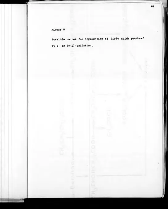
Verticillium. Ratledge (1978) in a summary of subterminal
oxidations of n-alkanes suggested that the phenomenon may
be more widespread than previously thought.
Alk-l-enes have been reported by several groups as
intermediates in the oxidation of n-alkanes. McKenna ai d
Kallio (1965) prompted by the inconclusive work of Senez and
Azoulay (1961) and Chouteau e t al. (1962) pointed out that
such a system would be thermodynamically unfavourable. An
NAD+-linked dehydrogenase would have to work under anaerobic
conditions against an unfavourable equilibrium constant.
Despite these reservations Parekh et al. (1977) purified an
NAD-dependent n-alkane dehydrogenase together with a reduced
NADP-dependent n-alkane hydroxylase from a Pseudomonas
species grown anaerobically on n-alkanes. The mechanism.
shown in figure 4(a) was proposed for the anaerobic conversion
of n-alkane to fatty acid. Ratledge (1978) indicated that
anaerobic growth on n-alkanes is rare and that the organism (1977)
used by Parekh et al.j^was a special case since it grew very
poorly aerobically. An alternative to this view was that
the alk-l-ene was perhaps produced from a hypothetical
intermediate between n-alkane and alcohol if the oxidation
of the intermediate became rate limiting. The proposed
scheme would then become that in figure4 (b) .
fatty acids (NHIP = non-haem iron protein).
Parekhet al. (1977).
(b) Modified scheme in which 1-alkenes are produced when
oxidation of an intermediate becomes rate limiting.
Q
<
0 1
û-u
ou
T
CO
I
U
c\j
internal monoalkenes by Nocardia salmonicolor.
Glucose-grown suspensions of resting cells oxidized n-hexadecane
to a mixture of internal cis-hexadecenes. The major product
when using n-hexadecane as substrate was 7-hexadecene.
When using n-octadecane, 9-octadecene was the major product.
Such unsaturated products accumulated only in trace amounts
when cells were grown on n-alkanes and it was postulated
that the insertion of the double bond represented an early
step in a novel pathway of aliphatic hydrocarbon degradation.
This dehydrogenation did not appear to be associated with
the synthesis of unsaturated fatty acids. Further
intermediates in the proposed degradative pathway were not
identified.
In many cases work with whole cells tended to suggest
that 1-alkenes were not intermediates in the oxidation of
n-alkanes although the double bond was shown to be attacked
when 1-alkenes were introduced into n-alkane degrading
systems. Markovetz et al. (1967) obtained 13-tetradeceneoic
acid when growing Pseudomonas aeruginosa on 1-tetradecene.
This indicated that the double bond was not attacked by the
organism and was unlikely to be an intermediate in n-alkane
oxidation. However, small amounts of tetradecan-2-ol were
also produced.
mechanisms for oxidation of 1-alkenes when investigating
the degradation of hydrocarbons by Candida lipolytica;
(i) Methyl group oxidation to produce w-unsaturated primary
alcohols, (ii) subterminal oxidation to give a,-unsaturated
secondary alcohols and (iii) double-bond oxidations to
produce 1-2-epoxides and 1,2-diols. The latter products
were not detected when cells were grown on n-alkanes.
Allen and Markovetz (1970) noted that when strains of
Cunninqhamella and Pénicillium were grown on 1-tetradecene
13-tetradecenoic acid and unsaturated derivatives of
subterminal oxidation were produced by the two organisms
respectively. These results show that the unsaturated
carbons were not attacked in these strains of fungi .
Overall there is little substantiated evidence for the
involvement of a 1-alkene intermediate in the oxidation
of n-alkanes .
Of the microbial systems for the degradation of
n-alkanes investigated the majority appear to involve
terminal hydroxylation of a methyl group as an initial
oxidation of the substrate. Two systems which are relatively
well understood are the cytochrome P-450 system from
Candida tropicalis and the rubredoxin system of
Pseudomonas oleovorans. both of which will be discussed
37
I .5.2 The cytochrome P-450 hydroxylating system
The term cytochrome P-450 refers to a group of haem
proteins that have several characteristics in common. An
important diagnostic feature is the formation of carbon
monoxide complexes that have a major absorption band at
about 450 nm. Such proteins occur widely in nature and
have been identified in most animal tissues and in plants-.
Their chief function is thought to be the hydroxylation of
lipophilic substrates.
Of several bacterial cytochrome P-450 systems to have
been reported, the most highly purified and characterized
is the camphor hydroxylation system of Pseudomonas putida
(see figure 5 ). This model system, together with
rat-liver microsomal cytochrome P-450, have proved to be of
great value in elucidating many aspects of substrate binding
and in particular oxygen activation, in reactions involving
relatively inert hydrophobic substrates . The subject has
been extensively reviewed by Gunsales et al. (1974) and
White and Coon (1980) .
The first bacterial cytochrome P-450 system associated
with the hydroxylation of n-alkanes was reported by Cardini
and Jurtshuk (1968, 1970) in a Corynebacterium species strain
7E1C grown at the expense of n-octane. Unlike the liver
Figure 5
Cytochrome P-450 oxygenation-reduction cycle,
native system. (S = substrate molecule.)
A postulated
electron donor was NADH rather than NADPH. Electron
transfer to the cytochrome was shown to involve a
flavo-protein dehydrogenase. The Corynebacterium also differed
from the liver microsomal and Pseudomonas putida systems
in that no iron-sulphur protein was identified and reductive
coupling to the cytochrome was unclear. Atmospheric
oxygen was shown to be incorporated into the primary alcohol 18
products using 0^ and the equivalence of NADH to oxygen
and products conformed to the stoichiometry of a mono
oxygenase .
More recently the occurrence of a cytochrome P-450 in
several strains of Acinetobacter after growth on n-alkanes
has been demonstrated (Asperger et a_l., 1981) . The presence
of the cytochrome P-450 was shown to be dependent upon the
presence of n-hexadecane in the medium. Also of interest was
the observation that only seven of the fifteen strains
investigated showed the diagnostic absorbance peak at
450 nm when reduced and in the presence of carbon monoxide.
The n-alkane hydroxylases of Candida guillermondii
(Schurket a l .. 1978; Müller et al^, 1979) and Candida
tropicalis (Lebeault et a l .. 1971) have both been shown to
involve a cytochrome P-450 moiety. In Candida tropicalis
Duppel et al. (1973) determined that the entire hydroxylation
41
P-450, an NADPH-cytochrome P-450 reductase and a heat stable
lipid fraction. The yeast reductase and lipid fractions
could be replaced by corresponding fractions obtained from
rat liver microsomes . As far as the lipid was concerned,
optimal activity was obtained by addition of a yeast
lysophosphatidylethanolamine. Under the conditions of
preparation the fractions appeared to be soluble. Lebeault
et a l . (1970) had been misled into suggesting the presence of
an NAD+ dependent n-alkane dehydrogenase by the observation
of anaerobic NAD+ reduction in the presence of n-decane.
Re-examination by Gallo et al^. (1973) concluded that the
observed NAD+ reduction could be attributed to impurities
contaminating the n-decane used in assays . The hydroxylation
system that was obtained agreed with the observations of (1973)
Duppel et al|. The enzyme was NADPH dependent and contained
cytochrome P-450 . The hydroxylation function however was
localized on microsomal membranes in close association with
alcohol and aldehyde dehydrogenases. Gallo et ¿1. (1976)
in a comparison of methods of preparation of microsomal
fractions observed the cytochromes P-450 and b_ were
D
specifically induced by growth on n-alkanes . It was also
noted that glucose grown cells contained low levels of
NADPH cytochrome c reductase.
the cytochrome P-450 n-alkane hydroxylase of Candida
tropicalis in terms of its individual components. An
NADPH cytochrome c reductase was detected and shown to be
identical with that produced by Saccharomyces cerevisiae
and glycerol grown Candida tropicalis . In n-alkane grown
Candida tropicalis however the concentration of the protein
was three to four times higher than in glycerol grown cells.
It was concluded that such an enzyme was probably involved
directly in n-alkane hydroxylation. It was impossible
initially to show conclusively that the NADPH-cytochrome c
reductase was capable of reducing cytochrome P-450 since the
cytochrome itself had not been sufficiently purified.
Studies of the purified NADPH cytochrome c reductase
showed that it was a flavoprotein with an apparent molecular
weight of 67,000 daltons. It contained one mole of FMN
and one mole of FAD per mole of protein. Under the
appropriate conditions the enzyme would reduce beef-heart
cytochrome c, dichlorophenolindolphenol and ferricyanide. NADPH
was oxidized in the presence of beef heart cytochrome c
and 2 methyl-1,4-naphthoquinone (menadione) .
The partial purification of the Candida tropicalis
cytochrome P-450 by Bertrand et al^. (1979b) allowed the
reconstruction of hydroxylation activity in vitro. The
4 J
gentle treatment with mild detergents . Use of more severe
treatments caused significant conversion of the cytochrome
P-450 to its denatured form; cytochrome P-420. Subsequent
fractionation of the hydroxylase components was achieved
by various chromatographic techniques including hydrophic
interaction chromatography and DEAE-cellulose chromatography.
It was shown that the individual components as
fractionated had very little hydroxylase activity but that
a mixture of the cytochrome P-450 and the NADPH cytochrome c
reductase reconstituted a hydroxylating activity with a
specific activity of 1.25 units. The addition of a lipid
fraction was not required. It was suggested that this lack
of requirement for lipid was due to either the non-ionic
detergents used in the purification stages which may have
had the same effect as a lipid moiety or the presence of
residual lipids in the fractionated cytochrome P-450. It
is important to note that the units of activity employed
for n-alkane hydroxylation effected by the cytochrome P-450
system were very small. It is doubtful whether activities
expressed in nanomoles per minute per milligramme of
protein may be considered to reflect those encountered in
intact cells . The labile nature of the enzyme system as
a whole and the insolubility of the substrates were major
with high activity.
More recently Riege et ad. (1981) and Honeck et al.
(1982) have demonstrated conclusively the involvement of
cytochrome P-450 in n-alkane hydroxylation on Lodderomyces
elongispora (formerly Candida guillermondii) . Both the
cytochrome P-450 and the NADPH-cytochrome P-450 reductase
were purified to electrophoretic homogeneity and used to
reconstitute hydroxylation activity using n-hexadecane as
substrate.
The purified cytochrome P-450 had absorption maxima
at 555 nm, 523 nm and 417 nm indicating the presence of a
protohaeme group. The molecular weight of the enzyme was
estimated to be 53,000 and contained one mole of haem per
mole of protein. The oxidized form of the protein exhibited
a low-spin type absorption spectrum analogous to other
low-spin type cytochrome P-450 from other organisms but the
absorption peak of the CO-complex was relatively low at
447 nm. The cytochrome P-450 was reduced by NADPH in the
presence of the cytochrome P-450 reductase and the substrate
which could be replaced by 'tween 20' .
The purified NADPH cytochrome P-450 reductase was shown
to be similar to that of Candida tropicalis and contained
one mole of both FAD and FMN per mole of protein. The
molecular weight was estimated to be 79,000.
45
nmoles of product per nmole of cytochrome P-450 per minute,
but was enhanced to 5.64 when the reaction was supplemented
with a non-ionic detergent. In this respect also, this
system bears significant similarities to other systems that
have been studied.
Cytochrome P-450 is a widespread and important
hydroxylation enzyme with a variety of roles in nature
including the initial oxidation of n-alkanes . In bacterial
systems that degrade n-alkanes however, cytochrome P-450
has been implicated only in the two cases previously
mentioned.
I .5.3 The Rubredoxin-linked w-hydrcxylase of Pseudomonas
putida
The only bacterial n-alkane hydroxylase that has been
extensively purified and characterized is the
rubredoxin-linked system isolated from Pseudomonas putida (previously
called P . oleovorans (Nieder and Shapiro, 1975) ) outlined
in figure 6 . Baptiste et ad. (1963) reported the preparation
of soluble cell-free extracts capable of oxidizing n-octane
to octan-l-ol. Kusunose et aj.. (196 4) demonstrated that
similar extracts would also ui-oxidize a series of fatty
acids. The hydroxylating activity was shown to require the
presence of dioxygen, ferrous ions and NADH. It was resolved
into two protein components both required for the formation
Figure 6
The rubredoxin-linked tu-hydroxylase of Pseudomonas putida .
(a) Cytochrome C reductase
(b) Rubredoxin
Coon eit al., 1964) . Peterson et al. (1966) further
separated the a)-hydroxylation system into three essential
protein components. These were identified as (i) a highly
purified rubredoxin-like protein containing non-haem iron
but no inorganic sulphide, (ii) an NADH-rubredoxin reductase
and (iii) the «-hydroxylase.
The ability of rubredoxin to act as an electron carrier
was demonstrated by the reduction of cytochrome c by NADPH.
in the presence of spinach NADPH-ferredoxin reductase
(Peterson ^t a l .. 1967) . when the bacterial NADH-rubredoxin
reductase replaced the NADPH-ferredoxin reductase, NADH rather
than NADPH was required for the reaction to proceed. Neither
of the reductases alone were able to reduce cytochrome c .
It was suggested that rubredoxin acted in a similar
manner in the «-hydroxylation system. Both rubredoxin and
a reductase were required with the «-hydroxylase for substrate
hydroxylation and homogeneous preparations of the
rubredoxin retained all the activity in the hydroxylation
system attributed to partially purified preparations. In
addition to this evidence it was shown that under comparable
conditions the rate of electron transfer from NADH to
cytochrome c was more than enough to account for the rate
of electron transfer to oxygen in the presence of the
u)-hydroxylase.
Further work was done on various properties of the
49
apparent molecular weight determined by gel filtration was
12,800. Two atoms of iron per molecule were shown to be
present and evidence was presented that when enzymically
reduced each iron atom accepted one electron. Apart from
a comparatively high molecular weight, the rubredoxin from
P s . putida appeared to share many properties with rubredoxins
from other sources . The rubredoxins from Clostridium
pasteurianum and Peptostreptococcus elselenii. however,
were poor substitutes for the Pseudomonas rubredoxin in the
(«-hydroxylation system. It was assumed that there was a
specificity in the transfer of electrons from the rubredoxin
to the us-hydroxylase since all the rubredoxins used were
readily reduced by NADPH in the presence of spinach
NADPH-ferredoxin reductase .
Whereas in Ps . putida a role for the rubredoxin had
been established in a pathway involving molecular oxygen,
the role of rubredoxins in the anaerobic organisms from
which they had previously been isolated had not been
established .
Improved methods employed by Lode and Coon (1971)
showed that the rubredoxin molecule was a single polypeptide
chain with a molecular weight of 19,000. It differed in
its amino-acid composition from other rubredoxins which did
not contain arginine and histidine. The molecule also
appeared to differ from the rubredoxin previously isolated
present and evidence was presented that when enzymically
reduced each iron atom accepted one electron. Apart from
a comparatively high molecular weight, the rubredoxin from
P s . putida appeared to share many properties with rubredoxins
from other sources. The rubredoxins from Clostridium
pasteurianum and Peptostreptococcus elselenii. however,
were poor substitutes for the Pseudomonas rubredoxin in the
(«-hydroxylation system. It was assumed that there was a
specificity in the transfer of electrons from the rubredoxin
to the («-hydroxylase since all the rubredoxins used were
readily reduced by NADPH in the presence of spinach
NADPH-ferredoxin reductase.
Whereas in P s . putida a role for the rubredoxin had
been established in a pathway involving molecular oxygen,
the role of rubredoxins in the anaerobic organisms from
which they had previously been isolated had not been
established .
Improved methods employed by Lode and Coon (1971)
showed that the rubredoxin molecule was a single polypeptide
chain with a molecular weight of 19,000. It differed in
its amino-acid composition from other rubredoxins which did
not contain arginine and histidine. The molecule also
appeared to differ from the rubredoxin previously isolated