A Study of Scanning Electron Microscope of Vancomycin
Resistant Enterococcus faecalis from Clinical Isolates
Ajay Kumar Oli1, Raju Sungar2, Nagaveni Shivshetty1, Rajeshwari Hosamani1, Kelmani Chandrakanth Revansiddappa1*
1Department of Biotechnology, Gulbarga University, Gulbarga, India 2ELLA Foundation, Hyderabad, India
Email: *[email protected]
Received February 4, 2012; revised March 2, 2012; accepted April 5, 2012
ABSTRACT
Vancomycin-resistant Enterococcus faecalis pose an emerging health risk, but little is known about the precise epide-miology for vancomycin resistance. The glycopeptide resistant was studied using different techniques such as broth macrodilution, agar dilution combined with agar diffusion, morphology cell changes by scanning electron microscopy. Eight VREF isolated from different clinical samples were used. Results showed low level and high level resistant to vancomycin antibiotic at concentration of 64 to 128 µg/ml, but antibacterial activity was reduced to 256 µg/ml, the SEM revaled increased in the cell size with the antibiotic compared to control and standard culture. The technique con-stitutes simple method for the detection of organism.
Keywords: VREF; SEM; Vancomycin
1. Introduction
Enterococcus spp. are natural inhabitants of the gastroin-testinal tract of humans and animals [1,2] but can be also found in soil, water, and vegetables [3]. The two most important species, Enterococcus faecium and E. faecalis, are most frequently implicated in human and animal in-fections [4]. E. faecalis is an opportunistic pathogen known to cause serious infections, such as bacteraemia, septicaemia, urinary tract infections, wound infections, meningitis, and endocarditis [2,5,6].
Prior to 1990s, enterococci also have been recognized as an important cause of bacterial endocarditis for almost a century [7]. However, during the past decade, there has been a worldwide trend in increasing occurrence of en-terococci (in the hospitals), a shift in the spectrum of enterococcal infections, and emergence of antimicrobial resistance among such isolates. Enterococci were re-ported as the second most common cause of nosocomial infections in the US. The most frequent infections caused by enterococci are urinary tract infections (UTIs) [8].
The acquisition of high level aminoglycoside resis-tance (HLAR) and vancomycin resisresis-tance has limited the therapeutic options available for clinicians. The transfer potential of vancomycin resistant genes from Enterococci to S. aureus have been reported in clinical settings, in-creases the importance of findings ways to limit the
spread of vancomycin resistant Enterococci (VRE) [9]. The problem of nosocomial enterococcal infection is compounded by emerging antibiotic resistance. The re- sistance alone does not explain the increase of Entero- cocci in nosocomial infections, microorganisms can adapt to different organic substances and other forms of envi- ronmental stress by several adaptive mechanisms. The ex- posure of bacteria to sub-MICs (Minimum Inhibition Con- centration) of Vancomycin results in a significant altera-tion of cellular morphology and disturbance of metabolic activity in resistant E. faecalis [10]. The major adaptive responses of microorganisms to externally occurring changes in the environment are modifications of the cell envelope [11] and also coupled with changes in the over-all morphology of the cells.
Scanning electron microscopy offers the unique ability to examine surface structures at relatively high resolution and proves particularly useful in the examination of the effect of antibiotics that act on the bacterial cell wall [12- 14]. The present study describes the effect of antibiotic stress on the morphology of vancomycin resistant E. fae- calis strains examined by scanning electron microscopy.
2. Materials and Method
2.1. Bacterial Strains
riod from September 2008 and January 2009 from Dis-trict Govt. hospital and diagnostic centres from Gulbarga region. The strains were isolated from blood, urine, pus and Cerebrospinal fluid sample.
Bacteria were isolated as previously described [15] and routinely grown in trypticase soy broth or agar at 37˚C. They were purified by standard methods and iden-tified to the species level by the conventional biochemi-cal identification scheme of De Marques and Suzart [16]. Confirmed isolates were stored in trypticase soy broth containing 20% glycerol at −80˚C until further charac-terisation could be performed.
2.2. Antimicrobial Susceptibility Testing
Antimicrobial susceptibility testing was performed on Mueller Hinton agar (Hi-media, India) by the standard disk diffusion method as recommended by the National Committee for Clinical Laboratory Standards [17]. The antibiotics used for the tests were vancomycin, ampicillin, oxacillin, rifamycin, ciprofloxacin, tobramycin, genta-mycin, teicoplanin and streptomycin.
2.3. Minimal Inhibitory Concentration (MIC)
All strains were screened for vancomycin (Sigma Aldrich Ltd., Bangalore) MICs by the agar dilution method while the disc diffusion method was performed for screening susceptibility to other antimicrobials, by CLSI guidelines [18]E. faecalis NCIM 5025 used as control.
2.4. Scanning Electron Microscopy
SEM is used to examine the minor changes in cell mor-phology of the populations that have adapted to antibiotic stress [19]. The selected VREF strains were grown in BHI media with increasing vancomycin concentrations as described earlier. The bacterial cells from each culture were recovered by centrifugation at 6000 rev/min and the cells were washed twice with potassium phosphate buffer (50 mM, pH 7.0). Bacterial cells were then fixed by im-mersing in 2.5% glutaraldehyde in potassium phosphate buffer (50 mM, pH 7) for overnight at 4˚C. Then the specimens were washed twice with buffer and dehy-drated by ethanol series (v/v) ranging from 30%, 40%, 50%, 60%, 70%, 80%, 90% to 100% and stored in 100% ethanol. For SEM, the specimens were dried to critical point, coated with gold and examined with an S-200C scanning electron microscope. [20] compared with stan-dard NCIM 5025 and control EF122 strain.
3. Results
3.1. Bacterial Isolates
A total of 122 Enterococcus strains were isolated from
different clinical samples on bile esculin agar. The spe-cies identities of the clinical Enterococccal isolates, in-cludes 76 (62.29%) strains were E. faecalis. The E. fae-calis was the predominant isolates from urine, pus, CSF and blood samples.
3.2. Antimicrobial Susceptibility
E. faecalis strains showed resistance to the different anti-biotics like vancomycin (77.63%), gentamycin (64.47%) and oxacillin (55.26%) antibiotics, and were found to be multi drug resistant. The isolates were found susceptible to rifamycin (61.84%), teicoplanin (55.26%) streptomy-cin (52.63%) and tobramystreptomy-cin (51.13%).
3.3. MIC’s in E. faecalis Isolates
All the vancomycin resistant E. faecalis were subjected for vancomycinMIC’s test. Twelve strains showing drug resistance to all the antibiotics tested were selected for the MIC studies. Among them, 8 strains showed MIC in the range of ≥64 μg/ml while other 4 strains exhibited MIC of ≥128 μg/ml. The bactericidal activity was ob-served at concentration of 256/256 μg/ml and low bacte-ricidal growth at 128/256 μg/ml. The concentration of antibiotic showed bacterial growth to about ten-fold at 24 hrs, with a concentration of 128/256 μg/ml. An increase in 100 fold at 24 hr was observed with a vancomycin concentration of 6/32 μg/ml.
3.4. Scanning Electron Microscope Study
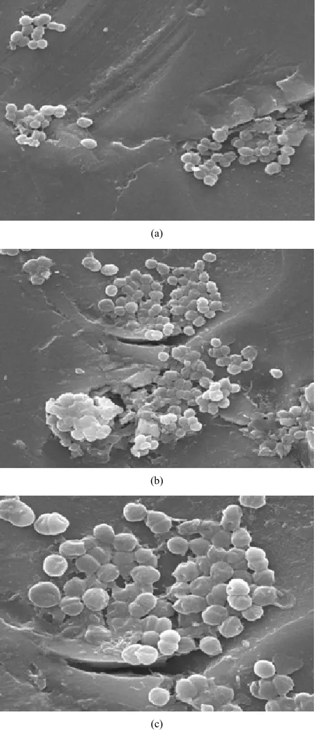
The results of cell morphology of VREF strains exam-ined by scanning electron microscopy (SEM) revealed that in the presence of vancomycin, the cells altered their morphology with respect to different concentrations of the antibiotic. In the absence of vancomycin the cell morphology of control were apparently normal (Figure 1(a)). However standard culture showed no alteration in
their cell morphology (Figure 1(b)) but enlarged,
mal-formed and rough surfaced were observed in the antibi-otic treated VREF culture with a concentration of 12 µg/ml) (Figure 1(c)).
4. Discussion
(a)
(b)
[image:3.595.58.288.85.616.2](c)
Figure 1. Scanning electron micrographs of VREF cells Mag- nification at 10,000 × 5 µm. (a) Control-strain EF 122 with- out antibiotic; (b) Standard culture NCIM 5025; (c) Strain EF 122 treated with antibiotic concentration of 12 µg/ml.
tance have increased. Determination of glycopeptides activity has a significant role in guiding antibiotic usage. The results of this study confirms that E. faecalis were more resistant to the vancomycin (77.63%), gentamycin (64.47%) and oxacillin (55.26%) and were sensitive to
rifamycin (61.84%), teicoplanin (55.26%) and strepto- mycin (52.63%) The multidrug-resistant Enterococci are being increasingly reported from all over world. Many studies have demonstrated that E. faecium is compara-tively mores resistant than E. faecalis. [14]. The E.
fae-calis isolates investigated demonstrated resistance to
vancomycin (MICs, 32 to 64, uglml). Similarly, Uttely et al. [23] reported prevalence of enterococci isolates resis-tant to both vancomycin and teicoplanin, and the vanco-mycin MICs were >64 µg/ml. The E. faecalis strain re-ported by shales et al. [24] had vanocmyin and teico- planin MICs of 256 and 16 µg/ml, respectively. In our studies MIC’s for vancomycin among 12 E. faecalis strains 8 strains as showed ≥64 μg/ml and 4 strains had MIC of ≥128 μg/ml. The bactericidal activity was ob-served at concentration of 256/256 μg/ml and low bacte-ricidal growth at 128/256 μg/ml. The concentration of antibiotic showed bacterial growth of about 10-fold at 24 hrs, of 128/256.
Morphological changes of organisms under stressful conditions are the most visible parameters of bacterial adaptation. The changes in morphology as an adaptive response to adverse environmental conditions have al-ready been reported with several bacterial species [25- 27]. In our study the cell morphology of vancomycin treated cells of E. faecalis under SEM provided strong evidence that the presence concentration of Vancomycin is stressful for the bacterial populations, characterized by the large size. The increase in cell size reduces the rela-tive contact surface and consequently reduces the attach- able surface for organic (antibiotic) compounds. There-fore, bigger cells can tolerate the stress conditions better than normal cells of the same species.
Our study reveals that bacteria have evolved an adap-tive response to the antibiotic stress and have developed drug resistance. This would be an alarming situation as Vancomycin is one of the few drugs used to treat patients with Enterococcus infection.
REFERENCES
[1] R. Creti, M. Imperi, L. Bertuccini, F. Fabretti, G. Orefici, D. R. Rosa and L. Baldassarri, “Survey for Virulence De- terminants among Enterococcus faecalis Isolated from Different Sources,” Journal of Medical Microbiology, Vol. 53, No. 1, 2004, pp. 13-20.
doi:10.1099/jmm.0.05353-0
[2] E. B. De Marques and S. Suzart, “Occurrence of Viru- lence-Associated Genes in Clinical Enterococcus faecalis Strains Isolated in Londrina, Brazil,” Journal of Medical Microbiology, Vol. 53, No. 11, 2004, pp. 1069-1073. doi:10.1099/jmm.0.45654-0
Sequence Types,” Foodborne Pathogens and Disease, Vol. 6, No. 3, 2009, pp. 321-327.
doi:10.1089/fpd.2008.0169
[4] A. Aakra, H. Vebø, L. Snipen, H. Hirt, A. Aastveit, V. Kapur, G. Dunny, B. Murray and I. F. Nes, “Transcrip-tional Response of Enterococcus faecalis V583 to Eryth- romycin,” Antimicrobial Agents and Chemotherapy, Vol. 49, No. 6, 2005. pp. 2246-2259.
doi:10.1128/AAC.49.6.2246-2259.2005
[5] A. Giacometti, O. Cirioni, A. M. Schimizzi, M. S. Del Prete, F. Barchiesi, M. M. D’errico, E. Petrelli and G. Scalise, “Epidemiology and Microbiology of Surgical Wound In- fections,” Journal of Medical Microbiology, Vol. 38, No. 2, 2000, pp. 918-922.
[6] A. Hällgren, C. Claesson, B. Saeedi, H.-J. Isaksson, H. Hanberger and L. E. Nilsson, “Molecular Detection of Ag- gregation Substance, Enterococcal Surface Protein, and Cytolysin Genes and in Vitro Adhesion to Urinary Cathe- ters of Enterococcus faecalis and E. faecium of Clinical Origin,”International Journal of Medical Microbiology, Vol. 299, No. 5, 2009, pp. 323-332.
doi:10.1128/AAC.49.6.2246-2259.2005
[7] D. R. Schaberg, D. H. Culver and R. P. Gaynes, “Major Trends in the Microbial Etiology of Nosocomial Infec- tion,” The American Journal of Medicine, Vol. 91, No. 3, 1991, pp. 72S-75S. doi:10.1016/0002-9343(91)90346-Y [8] D. E. Low, N. Keller and A. Barth, R. N. Jones, “Clinical
Prevalence, Antimicrobial Susceptibility, and Geographic Resistance Patterns of Enterococci: Results from the SEN- TRY Antimicrobial Surveillance Program, 1997-1999,” Clinical Infectious Diseases, Vol. 32, Suppl. 2, 2001, pp. S133-S145.
[9] S. Mohanty, S. Jose, R. Singhal, S. Sood, B. Dhawan, B. K. Das, et al., “Species Prevalence and Antimicrobial Susceptibility of Enterococci Isolated in a Tertiary Care Hospital of North India,” Southeast Asian Journal of Tro- pical Medicine and Public Health, Vol. 36, No. 4, 2005, pp. 962-965.
[10] L. V. Thomas and J. W. T. Wimpenny, “Investigation of the Effect of Combined Variations in Temperature, pH and NaCl Concentrations on Nisin Inhibition of Listeria monocytogenes and Staphylococcus aureus,” Applied and Environmental Microbiology, Vol. 62, No. 3, 1996, pp. 2006-2012.
[11] H. J. Heipieper, F. J. Weber, J. Sikkema, H. Keweloh and J. A. M. de Bont, “Mechanism Behind Resistance of Whole Cells to Toxic Organic Solvents,” Trends in Biotechnol- ogy, Vol. 12, No. 10, 1993, pp. 409-415.
doi:10.1016/0167-7799(94)90029-9
[12] D. Greenwood and F. O’Grady, “Antibiotic-Induced Sur- face Changes in Microorganisms Demonstrated by Scan- ning Electron Microscopy,” Science, Vol. 163, No. 3871, 1969, pp. 1076-1078. doi:10.1126/science.163.3871.1076 [13] D. Greenwood and F. O’Grady, “Scanning Electron Mi-
croscopy of Staphylococcus aureus Exposed to Some Com- mon Anti-Staphylococcal Agents,” Journal of General Microbiology, Vol. 70, No. 2, 1972, pp. 263-270.
[14] T. S. J. Elliott and D. Greenwood, “The Response of Pseu- domonas aeruginosa to Azlocillin, Ticarcillin and Cefsu-
lodin,” Journal of General Microbiology, Vol. 16, No. 3, 1983, pp. 351-362. doi:10.1099/00222615-16-3-351 [15] C. R. Jackson, P. J. Fedorka-Cray, J. B. Barrett and S. R.
Ladely, “Effects of Tylosin Use on Erythromycin Resis-tance in Enterococci Isolated from Swine,” Applied and Environmental Microbiology, Vol. 70, No. 7, 2004, pp. 4205-4210. doi:10.1128/AEM.70.7.4205-4210.2004 [16] E. B. De Marques, S. Suzart, “Occurrence of Virulence-
Associated Genes in Clinical Enterococcus faecalis Strains Isolated in Londrina, Brazil,”Journal of Medical Micro- biology, Vol. 53, No. 11, 2004, pp. 1069-1073.
doi:10.1099/jmm.0.45654-0
[17] National Committee for Clinical Laboratory Standards, “Performance Standards for Antimicrobial Disk Suscepti- bility Testing,” Twelfth Informational Supplement (M100- S12), ACCLS, Wayne, 2002.
[18] Clinical and Laboratory Standards Institute, “M07-A.B. Methods for Dilution Antimicrobial Susceptility Tests for Bacteria that Grow Aerobically; Approved Standard: 8th Edition,” CLSI, Wayne, 2009.
[19] F. Lanzarini, “Effect of Teicoplanin and Vancomycin on Staphylococcus Ultrastructure,” Microbiologica, Vol. 13, 1990, pp. 231-237.
[20] S. Raju, G. Rao, S. A. Patil and C. R. Kelmani, “Increase in Cell Size and Acid Tolerance Reponse in a Stepwise- Adapted Methicillin Resistant Staphylococcus aureus Mu- tant,” World Journal of Microbiology and Biotechnology, Vol. 23, No. 9, 2007, pp. 1227-1232.
doi:10.1007/s11274-007-9352-4
[21] I. Dupre, S. Zanetti, A. M. Schito, G. Fadda and L. A. Sechi, “Incidence of Virulence Determinants in Clinical Entero- coccus faecium and Enterococcus faecalis Isolates Col- lected in Sardinia (Italy),” Journal of Medical Microbiol- ogy, Vol. 52, No. 6, 2003, pp. 491-498.
doi:10.1099/jmm.0.05038-0
[22] A. H. C. Uttley, C. H. Collins, J. Naidou and R. C. George, “Vancomycin-Resistant Enterococci,” The Lancet, Vol. 331, No. 8575, 1988, pp. 57-58.
doi:10.1016/S0140-6736(88)91037-9
[23] D. M. Shales, A. Bouvet, C. Devine, J. H. Shales, S. Al- Obeid and R. Williamson, “Inducible, Transferable Re- sistance to Vancomycin in Enterococcus faecalis A256,” Antimicrobial Agents and Chemotherapy, Vol. 33, No. 2, 1989, pp. 198-203.
[24] M. O. Clements and S. J. Foster, “Stress Resistance in Staphylococcus aureus,” Trends in Microbiology, Vol. 7, No. 11, 1999, pp. 458-462.
doi:10.1016/S0966-842X(99)01607-8
[25] G. W. O’Hara and A. R. Glenn, “The Adaptive Acid Tol- erance Response in Root Nodule Bacteria and Escherichia coli,” Archives of Microbiology, Vol. 161, No. 4, 1994, pp. 286-292. doi:10.1007/BF00303582
[26] M. Ritz, J. L. Tholozan, M. Federighi and M. F. Pilet, “Morphological and Physiological Characterization of Listeria monocytogenes Subjected to High Hydrostatic Pressure,” Applied and Environmental Microbiology, Vol. 67, No. 5, 2001, pp. 2240-2247.
[27] G. Neumann, Y. Veeranagouda, T. B. Karegoudar, O. Sahin, I. Mausezahl, N. Kabelitz, U. Kappelmeyer and H. J. Heipieper, “Cells of Pseudomonas putida and Entero-