Int J Clin Exp Med 2018;11(3):2313-2318
www.ijcem.com /ISSN:1940-5901/IJCEM0062584
Original Article
An optimized protocol for whole mount
in situ hybridization of mouse brain
Wei Wu, Bingmin Luo, Zhongju Xiao
Department of Physiology, School of Basic Medical Science, Southern Medical University, Key Laboratory of Psy-chiatric Disorders of Guangdong Province, Guangzhou 510515, PR China
Received July 29, 2017; Accepted August 27, 2017; Epub March 15, 2018; Published March 30, 2018
Abstract: In this study, we describe an optimized protocol of whole mount in situ hybridization (WMISH) which is a preferred method for transcript distributions detection in whole embryos, tissues and organs, for the detection of c-fos mRNA in fresh frozen brain tissue. Therefore, critical steps of WMISH were optimized including proteinase K digestion time, composition of hybridization buffer as well as durations of incubation and washing steps. The ex-pression of c-fos mRNA and protein was detected in mouse brains with the modified WMISH protocol. As a result, the expression patterns of c-fos mRNA and protein in mouse brains were successfully detected with great specificity and low background signal.
Keywords: Whole mount in situ hybridization, c-fos mRNA, mouse brain
Introduction
Since in situ hybridization (ISH) technique was applied as a means of the detection of specific RNA or DNA sequences in the cytological prep-arations, a lot of researches and technological breakthroughs have promoted the application of ISH to a broader range [1]. The ISH technique not only processes the ability of precise tem- poral and spatial localization to target RNA by using complementary RNA (cRNA) probes, which also named as antisense, in the hybrid-ization with mRNA in target tissue, but also can identify different classes of RNA that visualized inside cells. In addition, ISH can be applied for the spatial and temporal distribution analysis of transcripts in the tissues, quantitation of gene expression levels or copy number and tracking the physical location of mRNAs or chromosomal segments inside the cell nucleus [2-4]. All of these confirm that ISH is a useful method for biological study.
Whole mount in situ hybridization (WMISH) was first applied in vertebrate embryos in 1990s [5]. After that it was successfully applied for the detection of expression patterns of temporally
and spatially restricted gene in the researches focusing on the embryonic development of vari-ous species, such as imaginal discs, salivary glands, testes, Xenopus, zebrafish, cow, mouse, etc. [6], as WMISH can detect the expression patterns of genes in tissues or embryos and analysis the position and intensity of mRNA expression with high sensitivity and specificity [7].
Meanwhile, the combination of ISH and immu-nohistochemistry (IHC) which has the advan-tage in the detection of specific brain proteins, can preserve anatomical construction and ch- aracterize distinct neuronal populations, verify the expression pattern of genes and detect the molecule expression in many experimental models. Additionally, this combination method can also visualize multiple targets located in one or different subcellular compartments [8, 9].
Materials and methods
General
All animal procedures were approved by the Animal Care and Use Committee of Southern Medical University and applicable guidelines for the care and use of laboratory animals were followed. Female C57BL/6J mice aged 5-7 weeks were purchased from the Experimental Animal Center of Southern Medical University, Guangzhou, China. RNase-free or DEPC-treated water was adopted for all the buffers used for the ISH procedures and after ISH, the auto-claved double-distilled water sterilized by filter -ing is suggested.
Tissue preparation
The process is as follows: 1) sacrifice mice by anesthesia with an overdose intraperitoneal injection of pentobarbital sodium (15 mg/kg) and the mice were transcardially perfused with normal saline and fixative (4% paraformalde -hyde in 0.1M PB, pH7.4) in the mice brains; 2) take out the brains and post-fix in 4% parafor -maldehyde for 24 h at 4°C; 3) after fixation, wash samples three times in PBS; 4) brains were cut to 2-3 mm thickness in a coronal plane in cold DEPC-treated PBS; 5) wash with 50% methyl alcohol in PBST (PBS with 0.1%
Tween 20) for 5 min; 6) wash with 100% methyl alcohol in PBST for 5 min; 6) fix in fresh 100% methyl alcohol and store at -20°C.
WMISH for mouse brain
The process is as follows: 1) rehydrate the brains by successive washes in a series of methanol/PBST solutions (75% methanol in PBST, 50% methanol in PBST and then 25% methanol in PBST) for 5-10 minutes in each concentration; 2) wash with PBST three times, each time for 5 min; 3) replace the last PBST wash solution with 10 μg/mL proteinase K (EO0491, Fermentas) in PBST for 20-30 min. Incubation time needs to be optimized by each researcher; 4) rinse briefly in PBST; 5) rinse the samples with 0.1% Tween 20 in 4% PFA-DEPC-treated PBS solution briefly; 6) re-fix the sam -ples 20 min with 0.1% Tween 20 in 4% PFA-DEPC-treated PBS solution at RT; 7) wash with PBST twice, each time for 10 min; 8) incubate with hybridization mix at 60°C for 1 h; 9) replace the hybridization mix with 300-500 μL digoxi -genin labeled RNA probe (MK1055, Boster); 10) incubate at 60°C overnight.
Post-hybridization washes
[image:2.612.91.522.71.302.2]An optimized protocol for WMISH of mouse brain
(50% formamide, 2X SSC buffer) at 60°C and wash with the washing buffer at 60°C twice, each time for 30 min; 2) wash with 2X SSC buf-fer at 60°C twice, each time for 20 min; 3) wash with 0.2X SSC buffer at 60°C twice, each time for 20 min; 4) wash with PBST twice at room temperature, each time for 50 min; 5) coronal sections (20 μm thickness) were cut by freezing microtome after overnight immersed in 30% sucrose for cryoprotection.
Detection and visualization
The process is as follows: 1) sections were washed with 0.01M PBST twice, 5 min each rinse; 2) incubate sections with blocking buffer, at least 1 h; 3) add biotin peroxidase conjugat-ed mouse anti-digoxigenin Fab fragments (100 μL per section), and incubate for 1 days at 4°C; 5) wash sections in 1X PBST at room tempera-ture 3 times, each time for 10 min; 5) add 100-200 μL of the appropriate SABC-cy3 secondary antibody to the sections, and incubate 2 h at room temperature or 4°C overnight; 6) wash sections in 1x PBST at room temperature 3 times, each time for 10 min.
Immunohistochemistry (IHC) after ISH detec-tion
The process is as follows: 1) to block nonspe-cific binding, add 5% normal goat serum on each slide and incubate for 2 h at room tem-perature; 2) incubate sections with the fos anti-body (rabbit anti-fos polyclonal IgG, SC-52, Santa Cruz, 500X in normal goat serum)
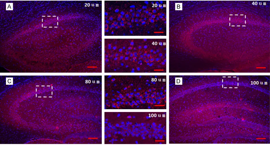
over-As shown in Figure 1, different thickness of froze sections (20 μm, 40 μm, 80 μm, 100 μm) were performed fluorescence ISH for c-fos mRNA in hippocampus, and the result showed that 20 μm section is better for detecting hybridization signals as the cell nuclei was clear and the fluorescence intensity of c-fos mRNA was proper with lowest background signal.
The representative fluorescence in situ hybrid -ization results
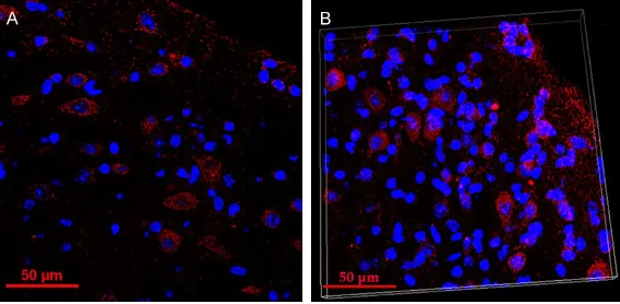
Representative confocal image from 20 μm sections showing c-fos mRNA labeling in audi-tory cortex and three-D rendering of a fluores -cence ISH-processed whole-mouse brain imag- ed by light-sheet fluorescence microscopy were shown in Figure 2.
The influence of different times digested with
proteinase K to the results of double labeling
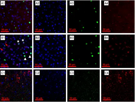
[image:3.612.91.374.72.211.2]As shown in Figure 3, the whole-mouse brain tissues were treated with proteinase K for 15, 30, 45 and 60 min, respectively, only the se- ction which was treated for 30 min showed c-fos protein + neurons, c-fos mRNA + neurons and representative neurons co-expressing both transcripts of interest simultaneously. In the section that treated for 15 min, the fluores -cence intensity is not strong enough, especially for the c-fos mRNA labeling, while in the section that treated for 15 min, the cell nuclei were damaged meanwhile, the fluorescence intensi -ty of c-fos protein was degraded to pieces and fluorescence intensity of c-fos mRNA was too Figure 2. Representative fluorescence in situ hybridization results. (A) Repre
-sentative confocal image from 20 μm sections showing c-fos mRNA labeling in auditory cortex, Scale bar = 50 μm. (B) Three-D rendering of a fluores -cence ISH-processed whole-mouse brain imaged by light-sheet fluores-cence microscopy. Width: 210.27 μm, height: 210.27 μm, depth: 18.00 μm.
night at 4°C; 3) wash sections in 1X PBS at room tempera-ture 3 times, each time for 10 min; 4) incubate with the goat anti- rabbit antibody (500X) for 2 h at room temperature; 5) after three rounds of wash-ing in 0.01M PBS, the sections were mounted and stored at 4°C for further analysis.
Results
Effects of fluorescence ISH
strong. Additionally, the 10 μg/Ml is the most proper treatment concentration.
Discussion
WMISH has been widely applied in the spatial and temporal order of gene expression and the establishment of the profile of gene expression pattern [10-13]. Considering the structural fea-tures of the brain tissue limits the application of conventional WMISH protocol to some ex- tent, we optimized WMISF for c-fos mRNA and protein detection in whole mouse brains, in- cluding the fixation and post-fixation methods, proteinase K digestion time, the section thick-ness, dying method, composition of hybridiza-tion buffer as well as durahybridiza-tions of incubahybridiza-tion and washing steps. As the conventional
proto-col has been confirmed by thousands of schol -ars, all the data in this study are intercompared without the control group.
[image:4.612.91.523.71.398.2]An optimized protocol for WMISH of mouse brain
meabilization and high-temperature washing in order to preserve the tissue morphology. Briefly, in the fixation step, the brain tissue was fix in 4% (wt/vol) paraformal-dehyde for 2 h, which could preserve the tissue’s morphology by the sufficient penetration of fixative. Subse-quently, the brain tissue was treated with in- creasing concentrations of methanol for better and long-term storage of samples.
When we need to start latter experiment, the tissue was rehydrated following by the permea-bilization step by using proteinase K which is more aggressive compared with other agen-cies used in most fluorescence ISH protocols, such as acetylation solution [11-12]. while so- me researches even omit the permeabilization step [13]. Most protocols for ISH and fluores -cence ISH in brain tissues use proteinase K with the concentration ranging from 20 to 100 μg/mL and treatment time ranging from a few minutes to 1 h. The permeabilization is a vital step in WHISH which allows the probe to pene-trate into tissue so as to generate a strong sig-nal. Therefore, the incubation time of protein-ase K should be precisely measured. To be specific, on the one hand, the short incubation time will decrease the ratio of probe that pen-etrated into the tissue (Figure 3A); on the other hand, proteinase K over-treatment will damage the brain tissues leading to the gradual disinte-gration of tissues in remaining steps (Figure 3C). To order to minimize the affecting factors of proteinase K digestion effect and increase the reliability of results, a proper concentration (10 μg/mL) and a suitable incubation time (30 min) were set in our protocol, followed by a 20 min post-fixation step (Figure 3B).
After the probe was combined with the target gene, the gradient elution was carried out, and kept in sucrose. Then, froze sections of brain tissue were prepared and hybridization signal were studied. With the comparison of the dif-ferent thickness of froze sections (20 μm, 40 μm, 80 μm, 100 μm), we found that 20 μm sec -tion is better for detecting hybridiza-tion signals (Figure 1), and can be performed ISH and IHC double staining (Figures 2, 3). Similar to immu-nohistochemistry, there are two methods for dyeing process in fluorescence ISH: stick sec -tion and free-floating sec-tion. The former is that the slices are pasted on the slide glass to be stained by immunohistochemistry [17]; the latter is that slices are floated in a solution con
-tainer (such as 24 well plates) to be dyed [18]. In our studies, we found that the free-floating section of brain tissue in fluorescence ISH has high sensitivity and good repeatability after hybridization.
After the hybridization, the needed brain sec-tions were selected for the next step, and the other brain sections were stored at -20°C. Therefore, to a certain extent, the use of experi-mental reagents is reduced, and experiexperi-mental funds and experiment time was saved. For example, the 3 mm thick of brain tissues in flu -orescence ISH only uses 300 μL of digoxigenin labeled oligonucleotide probe mixture, and its dosage is greatly reduced compared with con-ventional method [19-21]. Furthermore, the experimental steps before hybridization were shortened to avoid the pollution.
In this study, these experimental steps such as the digestion time of proteinase K, temperature control, post-hybridization washing, kept in su- crose and slice thickness adjustment were opti-mized. The procedure was simplified, the exper -imental cost was reduced, and the color ren- dering specificity was improved. A more practi -cal method of WMISH technique of adult mouse brain was successfully established. Detection and analysis of signals throughout the brain can be used for maintaining morphology and histological examination.
Acknowledgements
This work was supported by grants from a 973 program (2014CB943002) and the National Natural Science Foundation of China (U130- 1225, 31529003, 31671083) to ZX.
Disclosure of conflict of interest
None.
Address correspondence to: Zhongju Xiao, Depart- ment of Physiology, School of Basic Medical Sci- ence, Southern Medical University, Key Laboratory of Psychiatric Disorders of Guangdong Province, No.1023-1063 Shatai South Road, Baiyun District, Guangzhou 510515, PR China. Tel: +86-18620848059; E-mail: [email protected]
References
cytologi-cal preparations. Proc Natl Acad Sci U S A 1969; 63: 378-383.
[2] Tautz D and Pfeifle C. A non-radioactive in situ hybridization method for the localization of specific RNAs in Drosophila embryos reveals translational control of the segmentation gene hunchback. Chromosoma 1989; 98: 81-85. [3] Spaliviero M, Stratton KL, Donahue TF, Gow-
rishankar B, Ma C, Durack JC, Solomon SB, Houldsworth J and Jonathan A. Fluorescence in situ hybridization (FISH) and array-compara-tive genomic hybridization (a-CGH) from per- cutaneous needle biopsy compared to renal mass histology. Journal of Urology 2013; 189: 1857-1863.
[4] Kaufer BB. Detection of integrated herpesvirus genomes by fluorescence in situ, hybridization (FISH). Humana Press 2013; 141-152.
[5] Hemmati-Brivanlou A, Frank D, Bolce ME, Brown BD, Sive HL and Harland RM. Localiza-tion of specific mRNAs in Xenopus embryos by whole-mount in situ hybridization. Develop-ment 1990; 110: 325-330.
[6] Zimmerman SG, Peters NC, Altaras AE, Berg CA. Optimized RNA ISH, RNA FISH and protein-RNA double labeling (IF/FISH) in Drosophila ovaries. Nature Protocols 2013; 8: 2158. [7] Joeng KS, Regan J, Long F. Radioactive in situ
hybridization to detect gene expression in skel-etal tissue sections. Humana Press 2014; 217-232.
[8] Xiu J, Zhang Q, Zhou T, Zhou TT, Chen Y and Hu H. Visualizing an emotional valence map in the limbic forebrain by TAI-FISH. Nat Neurosci 2014; 17: 1552-1559.
[9] Yue M, Charles Richard JL, Yamada N, Ogawa A, Ogawa Y. Quick fluorescent in situ hybridiza -tion protocol for Xist RNA combined with im- munofluorescence of histone modification in X-chromosome inactivation. J Vis Exp 2014; 19: e52053.
[10] Bastien D and Lacroix S. In situ hybridization within the CNS tissue: combining in situ hyb- ridization with immunofluorescence. Visualiza -tion Techniques 2012; 53-70.
[11] Sui QQ, Zhu J, Li X, Knight GE, He C, Burnstock G, Yuan H, Xiang Z. A modified protocol for the detection of three different mRNAs with a new-generation in situ hybridization chain reaction on frozen sections. Journal of Molecular Histol-ogy 2016; 47: 511-529.
[12] Tholouli E, Hoyland JA, Byers RJ. Quantitative multiplexed quantum dot basedin situ hybrid-ization in formalin-fixed paraffin-Embedded Tissue. Neuromethods 2015; 99: 427-449. [13] Toledano H, D’Alterio C, Loza-Coll M and Jones
DL. Dual fluorescence detection of protein and RNA in Drosophila tissues. Nat Protoc 2012; 7: 1808-1817.
[14] Dakou E, Vanbekbergen N, Corradi S, Kemp CR, Willems E, Leyns L. Whole-mount in situ hybridization (WISH) optimized for gene ex-pression analysis in mouse embryos and em-bryoid bodies. Methods in Molecular Biology 2014; 1211: 27-40.
[15] Stempel AJ, Morgans CW, Stout JT, Appukuttan B. Simultaneous visualization and cell-specific confirmation of RNA and protein in the mouse retina. Molecular Vision 2014; 20: 1366-1373. [16] Shiura H, Okamoto A, Sasaki H, Abe K. Whole-mount MeFISH: a novel technique for simulta-neous visualization of specific DNA methyla -tion and protein/RNA expression. Plos One 2014; 9: e95750.
[17] Jeong JK, Chen Z, Tremere LA and Pinaud R. Double fluorescence in situ hybridization in fresh brain sections. J Vis Exp 2010; 1357-1365.
[18] Grabinski TM, Kneynsberg A, Manfredsson FP, Kanaan NM. A method for combining RNA-scope in situ hybridization with immunohisto-chemistry in thick free-floating brain sections and primary neuronal cultures. Plos One 2015; 10: e0120120.
[19] Nieto MA, Patel K and Wilkinson DG. In situ hy-bridization analysis of chick embryos in whole mount and tissue sections. Methods Cell Biol 1996; 51: 219-235.
[20] Pineau I, Barrette B, Vallieres N and Lacroix S. A novel method for multiple labeling combining in situ hybridization with immunofluorescence. J Histochem Cytochem 2006; 54: 1303-1313. [21] Lecuyer E, Parthasarathy N and Krause HM.