Role of Oculoproprioception in Coding
the Locus of Attention
Bartholomaeus Odoj
1,2and Daniela Balslev
3Abstract
■ The most common neural representations for spatial atten-tion encode locaatten-tions retinotopically, relative to center of gaze. To keep track of visual objects across saccades or to orient toward sounds, retinotopic representations must be com-bined with information about the rotation of one’s own eyes in the orbits. Although gaze input is critical for a correct allo-cation of attention, the source of this input has so far re-mained unidentified. Two main signals are available: corollary discharge (copy of oculomotor command) and oculopro-prioception (feedback from extraocular muscles). Here we asked whether the oculoproprioceptive signal relayed from the somatosensory cortex contributes to coding the locus of attention. We used continuous theta burst stimulation (cTBS) over a human oculoproprioceptive area in the postcentral
gyrus (S1EYE). S1EYE-cTBS reduces proprioceptive processing, causing∼1° underestimation of gaze angle. Participants dis-criminated visual targets whose location was cued in a non-visual modality. Throughout the non-visual space, S1EYE-cTBS shifted the locus of attention away from the cue by∼1°, in the same direction and by the same magnitude as the oculo-proprioceptive bias. This systematic shift cannot be attributed to visual mislocalization. Accuracy of open-loop pointing to the same visual targets, a function thought to rely mainly on the corollary discharge, was unchanged. We argue that oculo-proprioception is selective for attention maps. By identifying a potential substrate for the coupling between eye and attention, this study contributes to the theoretical models for spatial attention. ■
INTRODUCTION
Attention allows organisms to focus on relevant stimuli. In monkeys, neurons that respond to attended stimuli have a retinotopic receptive field, which reference loca-tion relative to the direcloca-tion of gaze (Gottlieb, Kusunoki, & Goldberg, 1998; Andersen & Mountcastle, 1983). A re-tinotopic code for attended stimuli has also been observed in humans. Indeed, although visual perception is normally facilitated at the location indicated by an attention cue, just after an eye movement, attention can be transiently allo-cated away from the cue, at its previous retinotopic coor-dinates (Talsma, White, Mathôt, Munoz, & Theeuwes, 2013; Golomb, Nguyen-Phuc, Mazer, McCarthy, & Chun, 2010; Mathôt & Theeuwes, 2010; Golomb, Chun, & Mazer, 2008).
Retinotopic representations alone cannot support cross-modal interactions in spatial attention between vi-sual and nonvivi-sual modalities or maintain a stable focus of attention across eye movements. One solution to this problem could be to create coregistered representations in multiple reference frames (Pouget, Deneve, & Duhamel, 2002; Andersen, Snyder, Bradley, & Xing, 1997). Another solution could be to update the retinotopic representation for each eye movement (Duhamel, Colby, & Goldberg,
1992). Importantly, both solutions require information about the rotation of the eyes in the orbits. Despite the im-portance of the gaze information in the brain’s representa-tions for spatial attention, the sources of this gaze input to the attention maps remain unknown. The main sources of eye position are the feedback from the extraocular muscles or oculoproprioception (Sherrington, 1918) and an inter-nal model that predicts future eye rotation based on the copy of the oculomotor command or corollary discharge (von Helmholtz, 1925).
Does oculoproprioception play a role in coding the lo-cus of attention? On the one hand, some studies fail to find a decrease in accuracy or precision of visual localization in conditions when oculoproprioception is reduced or abnor-mal (Balslev, Himmelbach, Karnath, Borchers, & Odoj, 2012; Lewis, Gaymard, & Tamargo, 1998; Guthrie, Porter, & Sparks, 1983). Such findings support the suggestion that oculoproprioception does not normally contribute to the estimate of eye rotation but rather calibrates the oculomotor command in the long term ( Wurtz, 2008; Steinbach, 1986).
On the other hand, some behavioral studies (Talsma et al., 2013; Golomb et al., 2008, 2010; Mathôt & Theeuwes, 2010) show delays in updating the retinotopic coordinates where attention is deployed after a saccade. These delays are compatible with the delay in the ascending fibers (Xu, Wang, Peck, & Goldberg, 2011; Wang, Zhang, Cohen, &
1
Goldberg, 2007) and therefore leave open the possibility that oculoproprioception may contribute. Furthermore, we have observed alterations in visual sensitivity in condi-tions that distort the oculoproprioceptive signal. The oculo-proprioceptive signal was distorted in the ocular periphery, using passive eye rotation (Balslev, Newman, & Knox, 2012), or centrally, in the somatosensory cortex, after repetitive TMS (Odoj & Balslev, 2013; Balslev, Gowen, & Miall, 2011) or after a focal lesion (Balslev, Odoj, & Karnath, 2013). These previous studies suggest a link between the oculoproprioceptive signal in the somatosensory cortex and spatial attention; however, the nature of this link is still unclear. One possibility is that theattention map incorporates oculoproprioception, so that a distortion of this signal causes a systematic shift in the locus of atten-tion relative to a cue. Another possibility is that the visual map incoporates oculoproprioception, so that a distortion of this signal causes a systematic error in locating visual targets relative to the body. Visual stimuli presented nearer the hand (Reed, Grubb, & Steele, 2006), the head midline (Durand, Camors, Trotter, & Celebrini, 2012; Durand, Trotter, & Celebrini, 2010), or the trunk midline (Grubb, Reed, Bate, Garza, & Roberts, 2008) have a privileged access to neural processing resources compared with visual stimuli presented elsewhere. A mislocalization of the visual stimuli relative to these landmarks could, in our previous studies, have changed their neural processing priority.
Here, we investigated whether oculoproprioception is incorporated in attention maps.
Oculoproprioception was manipulated using continu-ous theta burst stimulation (cTBS), a form of inhibitory TMS, over an oculoproprioceptive area in the human postcentral gyrus (S1EYE). Inhibitory repetitive TMS over S1EYEinterferes with the ability to correct for passive eye movement during a visual localization task (Balslev & Miall, 2008) and results in an underestimation of the ro-tation of the eye in the orbit by ∼1° (Odoj & Balslev, 2013). We used S1EYE-cTBS over the left postcentral gyrus to alter the oculoproprioceptive signal. Participants dis-criminated visual targets whose location was cued by the position of their unseen left index finger or pointed to these targets using the same finger. Both tasks depend critically on eye position information to match the loca-tion of the visual target with the localoca-tion of the finger that acted either as an attention cue or as an effector.
We report a systematic error in the locus of attention and no pointing error after S1EYE-cTBS. We argue therefore that oculoproprioception is the eye position signal that is selective for the attention map.
METHODS
Overview of the Experiments
Experiment 1 examined the allocation of attention using a cross-modal task. Participants discriminated a visual tar-get. The location of this target was cued by the location
of the unseen left index finger. The locus of attention was defined as the location in the visual space where the cue had the largest effect on the RT for target discrimination. If oculoproprioception is incorporated into the atten-tion maps, one would predict a systematic error in the locus of attention relative to the location of the cue after S1EYE-cTBS.
Experiment 2 examined visuospatial localization using an open-loop pointing task. Participants used their un-seen left index finger to reach to visual targets in the ab-sence of visual feedback. If oculoproprioception is incorporated into visual maps, one would predict a sys-tematic error in open-loop pointing after S1EYE-cTBS.
Experiment 3 examined participants’ ability to locate their unseen left index finger using an open-loop point-ing task. This experiment controlled for an effect of S1EYE-cTBS on the ability to locate the nonvisual cue in Experiment 1.
All experiments were conducted with the participants fixating rightward (Experiments 1A–3A) and leftward (Ex-periments 1B–3B). The reason for repeating the experi-ment for different directions of gaze was the following. The effect of S1EYE-cTBS on visual localization is gaze de-pendent, that is, a shift in perceived eye rotation toward left in rightward gaze and toward right in leftward gaze (Odoj & Balslev, 2013), reflecting the underestimation of the rotation of the eyes in the orbits. We predicted therefore that the change in the direction of gaze would reverse the effect. In this way, one can separate a specific, gaze-dependent effect, from a general influence of ante-rior parietal cortex on attention (Experiment 1) or reach-ing (Experiments 2–3).
Participants
We tested 10 participants (five women) in each of three different experiments (Experiments 1–3). All were right-handed with normal or corrected-to-normal vision. For Experiments 1A–3A, participants had an age range from 22 to 32 years (median: 28.5 years). For Experiments 1B–3B, participants had an age range from 27 to 32 years (median: 29 years). Five participants took part in all ex-periments (A and B). All participants gave their informed consent. The study was approved by the local ethics com-mittee at the University of Tübingen.
Experiment 1. Attention Map
This experiment investigated whether oculopropriocep-tion is included in the eye posioculopropriocep-tion estimate necessary for orienting attention in the visual space in response to a nonvisual cue.
only a small effect, if any on perceiving finger location. This assumption was explicitly tested in Experiment 2. The experimenter placed the participant’s index finger just below the horizontal line where targets were presented. To assess the benefit of the cue, we calculated the differ-ence in RT for visual discrimination in the presdiffer-ence versus the absence of the cue. The locus of attention was the location with the largest benefit of the cue.“Cueing error” was defined as the distance between the locus of attention and the actual location of the cue. If eye proprioception con-tributes to coding the locus of attention, S1EYE-cTBS should increase cueing error by causing a shift in the locus of attention away from the cue and toward the center of the orbit, in the same direction as the shift in perceived eye position.
Setup
Participants sat with their head fixed in a chin rest and cheek pads. A cathode ray tube (CRT) display was placed at 45 cm in front of them (Figure 1A). The CRT was cen-tered at +19° (Experiment 1A) or at −19° (Experiment 1B, Figure 1) from center of the right orbit. Participants performed the task in right eye vision, with the left eye patched. The experiment was conducted in monocular vision for the following reason. In the macaque, the pri-mary oculoproprioceptive area 3a receives propriocep-tive information from the contralateral eye only ( Wang et al., 2007). In humans, although proprioceptive in-formation from both eyes is relayed to each brain hemi-sphere (Balslev, Albert, & Miall, 2011), the input from the contralateral eye is functionally more important (Balslev, Himmelbach, et al., 2012). Therefore, we assumed that after cTBS of one hemisphere (left), the effects would be strongest and easier to measure for the contralateral eye (right). As a somatosensory cue, we used the posi-tion of the participants’ left index finger, ipsilateral to the hemisphere where TMS was applied. A transparent (Plexiglas) sheet was mounted 5 cm in front of the CRT
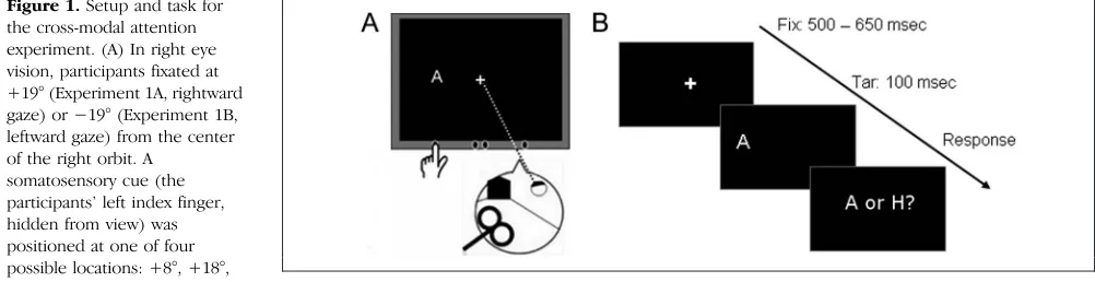
screen. Participants had their left index finger on a sup-port (a wooden ledge) attached to the Plexiglas immedi-ately under the location where the targets would appear. The finger was placed at one of four possible cue posi-tions, 8°, 18°, 20°, or 30° to the right (Experiment 1A, rightward gaze) or left (Experiment 1B, leftward gaze) from orbit midline of the right eye and covered with black cloth by the experimenter. The experiment took place in total darkness. The participants had no visual in-formation about finger location (somatosensory cue) to prompt the use of gaze information to locate the visual targets relative to the cue.
At the beginning of the trial, participants fixated on a central cross (white, 1° × 1°) presented on black back-ground (Figure 1B). Fixation was verified with a head-mounted eye tracker (EyeLink II, SR Research Ltd., Ottawa, Canada). After 500–650 msec (randomized), the fixation cross disappeared. At 100 msec later, a target letter (“A” or“H,”1° visual angle) appeared for 100 msec. The target appeared at one of seven possible locations, at−3°,−2°, −1°, 0°, 1°, 2°, and 3° from the somatosensory cue. Partici-pants were told that the target letter is most likely to appear at the location indicated by the cue. The target letter was however presented with equal probability (eight times) at each of the seven possible target locations. Additionally, three trials showed target letters at random locations out-side this range so that the participants could not predict the location of the nonvisual cue from the spatial distribu-tion of the visual targets. The participants were instructed to name the target letter as fast and accurately as possible. Voice RT was recorded.
[image:3.612.59.560.557.686.2]Trials with the same cue location were grouped in blocks. Each block consisted of 59 trials (8 trials for each of the 7 target positions + 3 random positions). Trial or-der was pseudorandomized. At the end of each block, participants were instructed to close their eyes. Then the experimenter moved the participants’index finger at the next cue location and started a new block. The par-ticipants completed four cued blocks (cue at +8°, +18°,
Figure 1.Setup and task for the cross-modal attention experiment. (A) In right eye vision, participants fixated at +19° (Experiment 1A, rightward gaze) or−19° (Experiment 1B, leftward gaze) from the center of the right orbit. A
somatosensory cue (the participants’left index finger, hidden from view) was positioned at one of four possible locations: +8°, +18°, +20°, or +30° (Experiment 1A)
or−8°,−18°,−20°, or−30° (Experiment 1B) from orbit midline (here, Experiment 1B: fixation at−19°, cue at−30° from orbit midline). (B) A target letter,“A”or“H,”was presented for 100 msec at one of seven possible locations, at−3°,−2°,−1°, 0, +1°, +2°, and +3° horizontally from the cue. Participants named the letter as fast and correct as possible. Voice RT and accuracy were recorded. The solid line shows eye position at fixation. The dotted line shows perceived eye position after S1EYE-cTBS according to Odoj and Balslev (2013). We predicted a shift of the locus of attention
+20° or +30° from orbit midline in Experiment 1A and −8°, −18°,−20° or−30° in Experiment 1B). The order of the blocks was pseudorandomized.
To assess the baseline distribution of attention as well as visual accuracy, participants performed the same visual discrimination task in the absence of a cue. Participants’ left index finger rested in front of their body midline. Tar-get letters were presented on the screen at all locations tested in the cued blocks. These locations were probed in random order, four times each. The uncued block con-sisted of 92 trials. This block was performed either before or after the cued blocks, randomized across participants.
Data Analysis
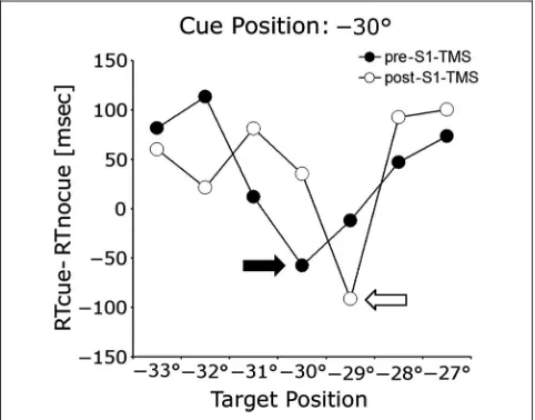
We calculated the difference in RT for visual discrimina-tion in the presence versus the absence of the cue. The locus of attention was the location with the largest ben-efit of the cue (the largest decrease in RT in the presence vs. the absence of the cue). The cueing error was calcu-lated as the distance between the locus of attention and the location of the cue (Figure 2).
Cueing error for each cue location was compared be-fore and after cTBS using paired-samples t tests. Mean cueing error was analyzed using a repeated-measures ANOVA with factors (1) TMS run (pre vs. post), (2) Stimu-lation area (S1EYEvs. P3), and (3) Gaze direction (leftward vs. rightward). If oculoproprioception is incorporated into attention maps, one would predict a significant three-way interaction, driven by an increased cueing error after S1EYE-cTBS. The cueing error was expected to have
oppo-site sign for leftward and rightward gaze, mirroring the underestimation of the angle of gaze after S1EYE-cTBS (Ex-periment 1 in Odoj & Balslev, 2013).
To assess whether the results are robust, we repeated the analysis using a different method for calculating the lo-cus of attention. We now defined the lolo-cus of attention as the center of mass of all locations that showed a cueing benefit. First, we identified all locations showing a benefit of the cue, indicated by a faster RT with the cue than with-out the cue. Then, we calculated the mean of these loca-tions after weighting each location with the magnitude of the cueing effect there. To separate out an eventual prac-tice effect (i.e., an improvement of RT that was common to all target locations within a block), we preprocessed the data by subtracting the mean RT for each block of trials.
Experiment 2. Visual Map
Experiment 2 examined the ability to locate visual targets relative to the left index finger after S1EYE-cTBS using a pointing task. This experiment also controlled for an er-ror in hand proprioception after cTBS in the postcentral gyrus, to rule out a systematic error in the perception of the nonvisual cue in Experiment 1.
Participants pointed with their left index finger, the same finger that was used as a nonvisual cue in Experi-ment 1. Visual targets were presented at the same loca-tions as the cues in Experiment 1. If locating visual targets for reaching takes oculoproprioception into account or if cTBS of the left postcentral gyrus alters perceived pos-ture of the ipsilateral hand, one would expect pointing errors after S1EYE-cTBS.
Setup
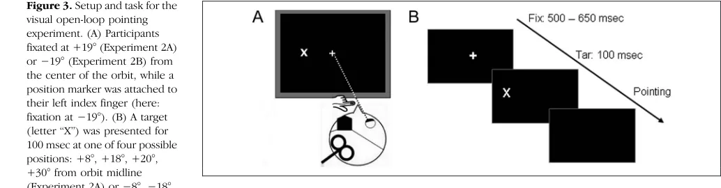
[image:4.612.52.293.472.661.2]The setup was identical with that used in Experiment 1. Additionally, a position sensor (Polhemus Fastrak, Colchester, VT) was fixed on the tip of the participants left index finger. At the beginning of the trial, the participants fixated on a central cross (1° × 1°, white on black background). Fix-ation was verified with the head-mounted eye tracker. Participants’left index finger rested in front of their body midline (Figure 3A). After 500–650 msec (randomized), the fixation cross disappeared. At 100 msec later, a target letter (“X”) appeared for 100 msec. The target could ap-pear at four possible locations: at 8°, 18°, 20°, or 30° from orbit midline in Experiment 2A or at−8°,−18°,−20°, or −30° from orbit midline in Experiment 2B. The partici-pants were instructed to close their eyes, then point with their left index finger as accurately as possible at the re-membered location of the target. The reaching move-ment stopped on the wooden ledge when the finger touched the plexiglass. Participants were allowed to ad-just the position of their finger until they felt the finger was pointing to the target. After participants confirmed that this was the case, finger position was recorded, and the experimenter moved the finger back to the
Figure 2.Cueing error in one example participant with a somatosensory cue at−30° from orbit midline, before and after S1EYE-cTBS. Before
cTBS over S1EYE(●), the largest decrease in RT in the presence versus in
the absence of the cue occurred for the target that appeared at the same location as the cue (black arrow, cueing error = 0°). After S1EYE-cTBS
resting position at body-midline. Trials for each target position were grouped in blocks. Each block consisted of six trials. Block order was pseudorandomized.
Data Analysis
Pointing error was calculated as the signed distance be-tween target and finger location at the end of the move-ment. Pointing error for each target location was compared before and after cTBS using paired-samples t tests. Mean pointing error across all target locations was analyzed using a repeated-measures ANOVA with fac-tors (1) TMS run (pre vs. post), (2) Stimulation area (S1EYE vs. P3), and (3) Gaze direction (leftward vs. right-ward). If oculoproprioception is incorporated into visual maps, one would predict a significant three-way interac-tion, driven by an increased pointing error after S1EYE-cTBS. The pointing error was expected to have opposite sign for leftward and rightward gaze, mirroring the underestimation of the angle of gaze after S1EYE-cTBS (Experiment 1 in Odoj & Balslev, 2013).
Experiment 3. Perceived Finger Posture
Experiment 1 required to match the position of the un-seen finger at rest with a visual target, whereas Experi-ment 2 asked participants to point the same finger at a visual target without visual feedback. One could explain a systematic cueing error in Experiment 1 despite accu-rate open-loop pointing in Experiment 2 by an error in the perceived posture of the left index finger at rest. To investigate whether S1EYE-cTBS selectively disturbs the felt position of the left index finger at rest (rather than during movement), we conducted a third experi-ment. In this experiment, participants were asked to point with the left index finger to the remembered posi-tion of this finger. The participants had their eyes closed and thus had no visual feedback. We measured pointing error as the difference between the location of finger at the end of movement and the location where the left in-dex finger was passively placed.
The setup was identical with Experiment 2. At the be-ginning of each trial, the experimenter took the partici-pant’s left index finger from the starting position in front of the body-midline and placed it at one of the four possible locations, identical to the target locations from Experiment 2 and the cue locations from Experiment 1. After 1 sec, the index finger was moved back to start po-sition. Then the participants were instructed to point to the remembered location of their finger as accurately as possible. They confirmed verbally when they reached this location. The experimenter recorded the coordinates of the fingertip and moved the participants’ finger back to the start position. Trials for each of the four target posi-tion were grouped in blocks. The target was presented six times within one block. The order of the blocks was randomized. If S1EYE-cTBS affect perceived position of the left index finger at rest, but not during movement, then one would predict a pointing error in this experi-ment after S1EYE-cTBS.
TMS
A standard 70-mm-diameter figure-of-eight coil centered over the stimulation site was fixed in place by a coil holder. The participant’s head was restrained by a chin rest. We followed an identical procedure for locating S1EYEas in previous studies conducted by Balslev and colleagues (Odoj & Balslev, 2013; Balslev, Gowen, et al., 2011; Balslev & Miall, 2008). S1EYEwas mapped in each partici-pant in relation to the“motor hotspot”of the left hemi-sphere, which is the scalp projection of the primary motor cortex for the hand (M1). The motor hotspot was defined as the point of maximum evoked motor response in the first dorsal interosseous muscle of the right hand. The S1EYEsite of stimulation was located at 3 cm posterior to the motor hotspot, measured on a line oriented at 45° from the sagittal plane and perpendicular on the central sulcus.
[image:5.612.43.560.66.201.2]Post hoc neuronavigation has showed that that this coil location targets an area in the postcentral gyrus, at MNI coordinates (mean ± SD: −45 ± 7, −32 ± 7, 58 ± 9;
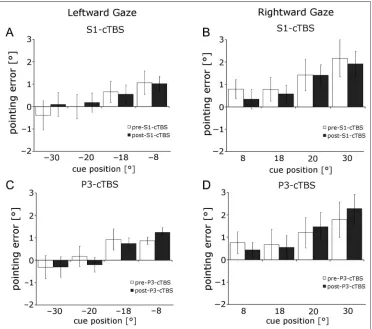
Figure 3.Setup and task for the visual open-loop pointing experiment. (A) Participants fixated at +19° (Experiment 2A) or−19° (Experiment 2B) from the center of the orbit, while a position marker was attached to their left index finger (here: fixation at−19°). (B) A target (letter“X”) was presented for 100 msec at one of four possible positions: +8°, +18°, +20°, +30° from orbit midline (Experiment 2A) or−8°,−18°,
Balslev, Siebner, Paulson, & Kassuba, 2012). The mean coordinate is associated with a probability of 46% for area 3b, 37% for area 1, and 27% for area 2 according to the probabilistic stereotaxic cytoarchitectonic atlas of the Anatomy Toolbox v2.1 (Eickhoff et al., 2005). The under-estimation of the angle of gaze after S1EYE-cTBS for both left and right directions of gaze (Experiment 1 in Odoj & Balslev, 2013) suggests that S1EYE is organized like the primary oculoproprioceptive area 3a in the macaque, where neurons encode gaze angle for all directions of gaze ( Wang et al., 2007). It is unlikely that TMS applied over the scalp can reach area 3a, located in the depth of the central sulcus (Geyer, Schleicher, & Zilles, 1999). The center–periphery principle of organization, however, may be common to all neural populations that receive an oculoproprioceptive projection. These neural populations are not limited to the depth of the central sulcus but extend into the postcentral and precentral gyri (Balslev, Albert, et al., 2011).
During stimulation, the coil was positioned tangential to the scalp with the long axis of the figure-of-eight coil ori-ented at 45° to the parasagittal plane. The current flow of the initial rising phase of the biphasic pulse in the TMS coil induced a current flowing from posterior to anterior in the brain. On the basis of the decreased amplitude of the somatosensory-evoked potentials after cTBS over a region situated at 2 cm posterior to M1 (Ishikawa et al., 2007), we assumed that cTBS over S1EYEresults in a decreased ex-citability of this area. The control site of stimulation in the parietal lobe, P3, was located by using the International
10–20 system for EEG placement. Hilgetag and colleagues found that repetitive TMS over P3 improves visuospatial at-tention in the ipsilateral hemifield (Hilgetag, Theoret, & Pascual-Leone, 2001). Therefore, we chose this area as control region to check if a possible effect, found in S1EYE-cTBS, could be explained by a spread of the induced current from S1EYEtoward P3. cTBS consisted of 600 bi-phasic stimuli produced by a Magstim Rapid2stimulator. They were delivered with a frequency of three pulses at 50 Hz repeated at 200 msec (5 Hz) for 40 sec. The stimu-lation intensity was set at 80% of active motor threshold of the right first dorsal interosseous (Huang, Edwards, Rounis, Bhatia, & Rothwell, 2005). For each experiment, participants underwent two sessions, with cTBS at either S1EYEor the control site (P3). The order of the sessions was randomized across participants and scheduled on separate days. During each session, the participant was tested before (pre-cTBS) and after (post-cTBS) on an identical task. Data collection was completed within 13 min after the cessation of the stimulation, a time interval for which the inhibitory aftereffect of cTBS in the somato-sensory cortex has been demonstrated (Ishikawa et al., 2007).
Eye Tracking
[image:6.612.178.552.61.371.2]The position of the right eye was recorded with a head mounted tracker that sampled pupil location at 250 Hz. The tracker was calibrated after each cTBS run (pre- or post-cTBS) using a 3 × 3 grid.
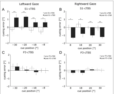
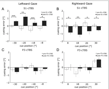
Figure 4.Systematic shift in the locus of attention after S1EYE-cTBS. The locus of
attention was defined as the location with the largest benefit of the cue. In leftward gaze, there was a rightward cueing error after S1EYE-cTBS (A) at all
cue locations. This error was specific to S1EYE-cTBS and did
not occur after the stimulation of the control site P3 (C). Likewise, in rightward gaze, there was a leftward cueing error at all cue locations after S1EYE-cTBS (B) but not after
P3-cTBS (D). All values with
Eye position time series were parsed into fixations, blinks, and saccades using the SR EyeLink detection al-gorithm, which was set to detect saccades with an am-plitude of at least 0.5°, using an acceleration threshold of 9500°/sec2 and a velocity threshold of 30°/sec and then analyzed offline. Trials with a mean deviation of more than 1.5° from fixation within 50 msec before tar-get presentation were discarded. In Experiment 1, a to-tal of 10.8 ± 7.7% (mean ± SD) trials were discarded. In Experiment 2, a total of 5.44 ± 5.28% trials were discarded.
RESULTS
Systematic Shift in the Locus of Attention Relative to the Cue after S1EYE-cTBS
Experiment 1 investigated whether oculoproprioception contributes to locating a nonvisual cue relative to a visual target in a cross-modal attention task. We predicted that, if this is the case, S1EYE-cTBS would shift the locus of at-tention to reflect the bias in the eye proprioceptive sig-nal. In accord with the prediction, after cTBS over S1EYE we found a cueing error of 0.6°–1.3° toward the midline (to the right in leftward gaze and to the left in rightward gaze; Figures 4A, B and 6A, B). The cueing error in the post S1EYE-cTBS run was significantly different from the pre-cTBS run at all tested cue locations (paired-samples t tests: allps < .03). cTBS over P3 did not significantly
change cueing error (Figure 4C and D, paired-samples t test: all ps > .32). The repeated-measures ANOVA of mean cueing error with factors cTBS run (pre vs. post) × Stimulation area (S1EYE vs. P3) × Gaze direction (left vs. right) showed a significant three-way interaction (F(1, 9) = 128.87,p< .001). Both post S1EYE-cTBS values were significantly different from pre S1EYE-cTBS data (post hoc pairwise multiple comparison using Tukey’s test, both ps < .01). In comparison, there was no significant change in cueing error after control cTBS over P3 (bothps > .05). A significant interaction was found for Gaze direction × cTBS run (F(1, 9) = 72.6,p< .001). This interaction was driven by the post-S1EYE-cTBS effect in different direc-tions for left and right gaze angles (post hoc pairwise multiple comparison using Tukey’s test:p< .01 for left post-cTBS vs. right post-cTBS, all otherps > .05). None of the other main effects or interactions was significant (allps > .231).
[image:7.612.189.561.435.738.2]This result was robust across two different methods for identifying the locus of attention. We found the same ef-fect of S1EYE-cTBS when the locus of attention was calcu-lated as the center of mass of the locations that showed a cueing benefit. After cTBS over S1EYE cueing error was 0.4–1.2° toward center (to the right in leftward gaze and to the left in rightward gaze; Figure 5A and B). The cueing error in the post S1EYE-cTBS run was signifi-cant different from the pre-cTBS run at all tested cue lo-cations (paired-samplest tests: all ps < .04). cTBS over P3 did not change cueing error (Figure 5C and D,
Figure 5.Systematic shift in the locus of attention after S1EYE-cTBS. The locus of
attention was defined as the center of mass of all locations that showed a benefit of the cue. In leftward gaze, there was a rightward cueing error after S1EYE-cTBS (A) at all cue
locations. This error was specific to S1EYE-cTBS and did
not occur after the stimulation of the control site P3 (C). Likewise, in rightward gaze, there was a leftward cueing error at all cue locations after S1EYE-cTBS (B) but not after
P3-cTBS (D). All values with
SEM. *p< .05, **p< .01, ***p< .001 in paired-samples
paired-samples ttest: all ps > .21). Repeated-measures ANOVA with factors cTBS run (pre vs. post) × Stimula-tion area (S1EYEvs. P3) × Gaze direction (left vs. right) showed a significant three-way interaction (F(1, 9) = 45.9,p< .001). For leftward gaze, the mean cueing error was−0.34 ± 0.3° (pre-S1EYE-cTBS) and−0.1 ± 0.4° (pre-P3-cTBS). For rightward gaze, these values were 0.2 ± 0.52° for pre S1EYE-cTBS and−0.07 ± 0.46° for pre P3-cTBS. After S1EYE-cTBS, the cueing error was 0.71 ± 0.51° in leftward gaze and −0.78 ± 0.42° in rightward gaze. Both post S1EYE-cTBS values were significantly dif-ferent from pre S1EYE-cTBS data (post hoc pairwise mul-tiple comparison using Tukey’s test, bothps < .02).
Here as well in the previous analysis, a significant inter-action was found for Gaze direction × cTBS run (F(1, 9) = 19.1, p< .001), driven by the post-S1EYE-cTBS effect in different directions for left and right gaze angles (post hoc pairwise multiple comparison using Tukey’s test: p< .01 for left post-cTBS vs. right post-cTBS, all other ps > .05). None of the other main effects and interactions was statistically significant (allps > .115). After P3-cTBS, no significant shift in cueing error could be observed (mean cueing error−0.33 ± 0.56° for screen left, 0.13 ± 0.52° for screen right, post hoc pairwise multiple compar-ison using Tukey’s test: bothps > .05).
Participants’accuracy was close to ceiling. They were correct in 97.85 ± 1.73% of the trials. We found no
differ-ence in accuracy across conditions. Repeated-measures ANOVA with factors cTBS run (pre vs. post) × Stimulation area (S1EYEvs. P3) × Gaze direction (left vs. right) showed no significant three-way interaction (F(1, 9) = .276, p= .612). None of the main effects or two-way interactions was significant (allps > .272).
No Change in Open-loop Pointing to Visual Targets After S1EYE-cTBS
Experiment 2 investigated whether S1EYE-cTBS impacts the perceived location of visual objects or perceived pos-ture of left hand. If this was the case, one would expect an increase in pointing error after S1EYE-cTBS.
We did not find a statistically significant increase in pointing error for any target position, gaze direction, or stimulation site (Figure 6, paired-samplesttests pre- vs. poststimulation, allps > .102). The mean pointing error over all target locations did not change significantly after cTBS (Figure 7C, D). The repeated-measures ANOVA of the pointing error with factors TMS run (pre vs. post) × Stimulation area (S1EYEvs. P3) × Gaze direction (left vs. right) showed no significant three-way interaction (F(1, 9) = 1.33,p> .27), main effects or two-way interactions (allps > .112).
[image:8.612.179.551.410.739.2]The precision of pointing was not changed either by cTBS either. The repeated-measures ANOVA with factors
Figure 6.No shift in visual pointing for any cueing position after S1EYE- or P3-cTBS. In
leftward gaze, there was a rightward pointing error after S1EYE-cTBS (A). This error was
specific to S1EYE-cTBS and did
not occur after the stimulation of the control site P3 (C). In rightward gaze, there was a leftward pointing error only after S1EYE-cTBS (B) but not
TMS run (pre vs. post) × Stimulation area (S1EYE vs. P3) × Gaze direction (left vs. right) for theSDof pointing error showed no significant three-way interaction (F(1, 9) = 0.98, p> .32) and no significant main effects or two-way interactions (allps > .181). For left gaze direction, the mean ±SDof the pointing error across participants was 0.82 ± 0.44° before S1EYE-cTBS and 0.75 ± 0.50° after S1EYE-cTBS. The values for P3-cTBS were 1.15 ± 48° (pre-cTBS) and 1.01 ± 0.35° (post c-TBS). For right gaze direc-tion, participants had SDs of 1.83 ± 0.97° before S1EYE -cTBS and 1.36 ± 0.86° after S1EYE-cTBS. The P3-cTBS values were 1.53 ± 0.84° (pre-cTBS) and 1.49 ± 0.93° (post-cTBS).
Direct comparison between the cueing and pointing error in Experiments 1 and 2 using a repeated-measures ANOVA with factors (1) Task (cross-modal attention vs. pointing), (2) TMS run (pre vs. post), (3) Stimulation area (S1EYEvs. P3), and (4) Gaze direction (leftward vs. right-ward) showed a statistically significant four-way interaction (F(1, 9) = 13.327,p< .008). No significant main effects were found (allps > .105). All significant interactions (Task × Gaze direction: F(1, 9) = 9.99, p= .012; TMS run × Gaze direction:F(1, 9) = 13.14,p= .006; task × TMS run × Gaze direction: F(1, 9) = 29.67, p < .001; TMS run × Stimulation area × Gaze direction:F(1, 9) =
27.18, p = .001) were driven by opposite effect of S1EYE-cTBS for different directions of gaze in the cross-modal attention task (post hoc pairwise multiple compar-ison using Tukey’s test:p< .01 for left post-cTBS vs. right post-cTBS and for left post-cTBS vs. right post-cTBS, all otherps > .05). A post hoc Tukey’s test, comparing pre- vs. post-cTBS, was only significant for pre- vs. post-S1EYE-cTBS in the cross-modal attention task for either gaze direction.
S1EYE-cTBS in the Left Hemisphere Does Not
Disturb Perceived Position of the Ipsilateral, Left Index Finger at Rest
Experiment 3 controlled for an effect of S1EYE-cTBS on felt position of the left index finger at rest. If perceived finger position at rest, but not during pointing, is dis-turbed by cTBS over S1EYE, one would expect an increase in pointing error after S1EYE-cTBS here.
[image:9.612.167.561.61.429.2]We found no significant change in pointing error be-tween pre- and post-cTBS over S1EYEor P3 for any of the eight tested locations (paired-samplesttests: allps > .106). Repeated-measures ANOVA of mean pointing errors with factors Gaze direction (left vs. right) × Stimulation site (S1EYEvs. P3) × Run (pre vs. post) showed neither main effects nor interactions (allps > .89).
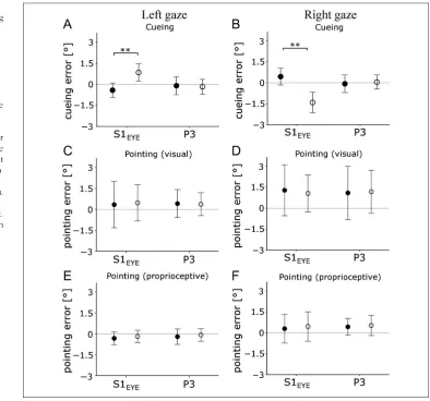
Figure 7.The effect of S1EYE-cTBS is specific for coding
the locus of attention. Upper row shows the mean cueing error in the cross-modal attention task for leftward (A) and rightward (B) gaze. The middle row shows the mean error for open-loop pointing to visual targets (C, D), and the lower row shows the pointing error to somatosensory targets (E, F). In the left column, data for left gaze direction are shown; the right column shows data for right gaze direction. Filled circles (●) show pre-cTBS data; empty circles (○) show post-cTBS data. Only changes in cueing error after S1EYE-cTBS were significant.
The mean pointing error for each participant before and after cTBS over S1EYEor P3 is shown in Figure 7E, F.
DISCUSSION
We found that altering the activity of an oculopropriocep-tive area in the human postcentral gyrus is sufficient to divert attention away from behaviorally important cues. Throughout the visual space, cueing error, or the dis-tance between the cue and the locus of attention, matched well in direction and magnitude the bias in per-ceived eye rotation measured in a previous study (Odoj & Balslev, 2013). This error cannot be explained by a mis-localization of the visual target. Direct comparison be-tween the attention and reaching tasks (Experiments 1 and 2) showed a significant interaction, driven by the larger error for cross-modal attention than for visually guided reaching. We argue therefore that oculoproprio-ception is selective for the attention map, as opposed to the visual map for reaching.
Because oculoproprioception was manipulated at cor-tical level, one could object that the effect of cTBS on at-tention was merely the result of the disruption of the cortical modules dedicated to this function. This explana-tion is unlikely. The direcexplana-tion of the cueing error was gaze dependent (leftward in right gaze and rightward in left gaze), and its magnitude at all tested locations matched well the error in perceived eye position (Odoj & Balslev, 2013).
A Role of Oculoproprioception in Coding the Locus of Attention Is Compatible with Neurophysiological Data
Two neural mechanisms have been proposed to imple-ment a locus of attention that anchors retinotopic repre-sentations to the physical location of the visual stimuli. First, populations of gain-field neurons with a retinotopic receptive field scale their activity with the angle of gaze to encode visual location relative to the body and/or the world (Pouget et al., 2002; Andersen & Mountcastle, 1983). A second mechanism is remapping of the re-tinotopic receptive field to account for eye movements (Mirpour & Bisley, 2012; Duhamel et al., 1992). A role of oculoproprioception in coding the locus of attention is compatible with both these mechanisms. A signature of the fastest of the two components of the oculoproprio-ceptive feedback can be recorded from somatosensory area 3a already at 5 msec before the end of the saccade ( Wang et al., 2007). This is in line with the time needed for a remapped representation for the locus of atten-tion to emerge in area LIP at 8–32 msec after the saccade (Mirpour & Bisley, 2012). Likewise, gain-field neurons show ∼150 msec postsaccadic delay in updating (Xu, Karachi, & Goldberg, 2012), which is compatible with
the ∼60 msec latency of the slower, tonic (Xu et al., 2011) component of the oculoproprioceptive signal.
Implications of the Current Findings for Understanding the Coupling between the Eye and Attention
Although the idea that the eye and attention systems are coupled is not new (Rizzolatti, Riggio, Dascola, & Umiltá, 1987), the mechanism of this coupling remains a current topic of debate (Smith & Schenk, 2012; Wright & Ward, 2008). We argue here that the attention maps incorpo-rate oculosensory signals to align retinotopic representa-tions to physical locarepresenta-tions. A role of the oculosensory signals in spatial attention is also suggested by observa-tions of a reduced ability to orient attention to cues in patients suffering from a disease of the extraocular mus-cles or their peripheral innervation (Gabay, Henik, & Gradstein, 2010; Smith, Rorden, & Jackson, 2004; Craighero, Carta, & Fadiga, 2001). Under the assumption that in these patients the brain areas that relay the corollary dis-charge (Sommer & Wurtz, 2008) are normal, such ob-servations can provide insight into the role of the oculoproprioceptive inflow in spatial attention. Likewise, the allocation of attention is disrupted at extreme rotations of the eyes (Smith, Schenk, & Rorden, 2012; Smith, Ball, Ellison, & Schenk, 2010; Craighero, Nascimben, & Fadiga, 2004), a condition known to cause an abnormal oculopro-prioceptive inflow (Paap & Ebenholtz, 1976; Ebenholtz, 1974).
Smith and Schenk have proposed that the oculomotor signals bias the competition among sensory inputs to fa-vor stimuli that are also the end point of a planned sac-cade (Smith & Schenk, 2012). In line with the idea that attention follows the gaze, can an alternative explanation of the current results be that oculoproprioception merely biases perception to favor visual stimuli nearer the per-ceived direction of gaze? The design of the spatial atten-tion task rules out this explanaatten-tion. This is because an unspecific bias toward perceived direction of gaze would have been identical during both conditions, with and without a cue. S1EYE-cTBS changed the difference in RT between these two conditions. Thus, cueing error after S1EYE-cTBS cannot be explained by a general bias in spa-tial attention toward the midline.
Can the Selective Role of Oculoproprioception in Spatial Attention Be Explained by the Timing of the Underlying Neural Processing?
oculoproprioception can be unreliable for up to 60 msec after an eye movement (Xu et al., 2011; Wang et al., 2007). In both tasks, participants maintained fixation for 600–750 msec before visual target onset. So, it is safe to assume that, at target onset, reliable oculopropriocep-tion had been available. The lack of a detectable effect of the oculoproprioceptive distortion on visually guided reaching is in line with previous findings. Some studies fail to find errors in locating visual targets relative to the body when oculoproprioception is reduced or abnor-mal (Balslev, Himmelbach, et al., 2012; Lewis et al., 1998) or calculate a smaller weight for oculoproprioception than for the corollary discharge in the multimodal esti-mate of eye position (Bridgeman & Stark, 1991; Gauthier, Nommay, & Vercher, 1990).
We speculate that the reason for the larger weight of oculoproprioceptive signal from the somatosensory cortex for spatial attention rather than for visually guided reaching is the timing of the underlying neural processes. If neural processing necessary to build attention representations ex-ceeds the time interval in which proprioception is unreli-able and if the neural processing for the visual map does not, then only the former would incorporate oculoproprio-ception. In support of this idea, brain areas that process shape or color (i.e., mainly dedicated to perceptual dis-crimination) have a longer latency of the visually evoked activity compared with brain areas that process luminance or motion (i.e., mainly for action; Laycock, Crewther, & Crewther, 2007; Schmolesky et al., 1998; Schroeder, Mehta, & Givre, 1998). The longer latency of neural pro-cessing for perception versus action may leave more time for computing priority versus reaching maps. The forward model based on the corollary discharge provides an early estimate of eye position that is probably reasonably accu-rate, given the predictable environment of the orbits. Therefore, fast neural processes (i.e., computing visual maps for reaching) are likely to rely on corollary discharge or, alternatively, on oculoproprioceptive input available up-stream the somatosensory cortex. Subcortical structures that could provide oculoproprioceptive signals for visually guided reaching are the superior colliculus, which in rats is connected to the trigeminal nucleus (Ndiaye, Pinganaud, VanderWerf, Buisseret-Delmas, & Buisseret, 2000) or the central thalamus that in the monkey contains neurons sensitive to eye position which discharge after a saccade (Tanaka, 2007). In contrast, neural processes that take lon-ger (i.e., computing the priority map for perception) may accommodate the delay in the ascending proprioceptive pathways and benefit from a more robust estimate of eye position by incorporating the oculoproprioceptive input from the somatosensory cortex.
Acknowledgments
This work was supported by the Danish Medical Research Councils (grant number 09-072209 to D. B.). We thank Prof.
H.-O. Karnath for hosting these experiments in the Section of Neuropsychology at the University of Tübingen.
Reprint requests should be sent to Daniela Balslev, School of Psychology and Neuroscience, University of St. Andrews, St Mary’s Quad, South Street, St. Andrews, KY16 9JP, United Kingdom, or via e-mail: [email protected].
REFERENCES
Andersen, R. A., & Mountcastle, B. (1983). The influence of the angle of gaze upon the excitability of the light-sensitive neurons of the posterior parietal cortex.Journal of Neuroscience, 3,532–548.
Andersen, R. A., Snyder, L. H., Bradley, D. C., & Xing, J. (1997). Multimodal representation of space in the posterior parietal cortex and its use in planning movements.Annual Review of Neuroscience, 20,303–330.
Balslev, D., Albert, N. B., & Miall, R. C. (2011). Eye muscle proprioception is represented bilaterally in the sensorimotor cortex.Human Brain Mapping, 32,624–631.
Balslev, D., Gowen, E., & Miall, R. C. (2011). Decreased visual attention further from the perceived direction of gaze for equidistant retinal targets.Journal of Cognitive
Neuroscience, 23,661–669.
Balslev, D., Himmelbach, M., Karnath, H.-O., Borchers, S., & Odoj, B. (2012). Eye proprioception used for visual localization only in conflict with the oculomotor plan. Journal of Neuroscience, 32,8569–8573.
Balslev, D., Siebner, H. R., Paulson, O. B., & Kassuba, T. (2012). The cortical eye proprioceptive signal modulates neural activity in higher-order visual cortex as predicted by the variation in visual sensitivity.Neuroimage, 61, 950–956.
Balslev, D., & Miall, R. C. (2008). Eye position representation in human anterior parietal cortex.Journal of Neuroscience, 28,8968–8972.
Balslev, D., Newman, W., & Knox, P. C. (2012). Extraocular muscle afferent signals modulate visual attention.Investigative Ophthalmology & Visual Science, 53,7004–7009.
Balslev, D., Odoj, B., & Karnath, H.-O. (2013). Role of somatosensory cortex in spatial attention.Journal of Neuroscience, 33,18311–18318.
Bridgeman, B., & Stark, L. (1991). Ocular proprioception and efference copy in registering visual direction.Vision Research, 31, 1903–1913.
Craighero, L., Carta, A., & Fadiga, L. (2001). Peripheral oculomotor palsy affects orienting of visuospatial attention. NeuroReport, 12,3283–3286.
Craighero, L., Nascimben, M., & Fadiga, L. (2004). Eye position affects orienting of visuospatial attention.Current Biology, 14,331–333.
Duhamel, J. R., Colby, C. L., & Goldberg, M. E. (1992). The updating of the representation of visual space in parietal cortex by intended eye movements.Science, 255,90–92. Durand, J.-B., Camors, D., Trotter, Y., & Celebrini, S. (2012).
Privileged visual processing of the straight-ahead direction in humans.Journal of Vision, 12,34.
Durand, J.-B., Trotter, Y., & Celebrini, S. (2010). Privileged processing of the straight-ahead direction in primate area V1.Neuron, 66,126–137.
Ebenholtz, S. M. (1974). The possible role of eye-muscle potentiation in several forms of prism adaptation. Perception, 3,477–485.
Gabay, S., Henik, A., & Gradstein, L. (2010). Ocular motor ability and covert attention in patients with Duane Retraction Syndrome.Neuropsychologia, 48,3102–3109.
Gauthier, G. M., Nommay, D., & Vercher, J. L. (1990). The role of ocular muscle proprioception in visual localization of targets.Science, 249,58–61.
Geyer, S., Schleicher, A., & Zilles, K. (1999). Areas 3a, 3b, and 1 of human primary somatosensory cortex.Neuroimage, 10,63–83.
Golomb, J. D., Chun, M. M., & Mazer, J. A. (2008). The native coordinate system of spatial attention is retinotopic.Journal of Neuroscience, 28,10654–10662.
Golomb, J. D., Nguyen-Phuc, A. Y., Mazer, J. A., McCarthy, G., & Chun, M. M. (2010). Attentional facilitation throughout human visual cortex lingers in retinotopic coordinates after eye movements.Journal of Neuroscience, 30,10493–10506. Gottlieb, J. P., Kusunoki, M., & Goldberg, M. E. (1998). The
representation of visual salience in monkey parietal cortex. Nature, 391,481–484.
Grubb, J. D., Reed, C. L., Bate, S., Garza, J., & Roberts, R. L. (2008). Walking reveals trunk orientation bias for visual attention.Perception & Psychophysics, 70,688–696. Guthrie, B. L., Porter, J. D., & Sparks, D. L. (1983). Corollary
discharge provides accurate eye position information to the oculomotor system.Science, 221,1193–1195.
Hilgetag, C. C., & Pascual-Leone, A. (2001). Enhanced visual spatial attention ipsilateral to rTMS-induced“virtual lesions” of human parietal cortexNature Neuroscience, 4,953–957. Huang, Y. Z., Edwards, M. J., Rounis, E., Bhatia, K. P., &
Rothwell, J. C. (2005). Theta burst stimulation of the human motor cortex.Neuron, 45,201–206.
Ishikawa, S., Matsunaga, K., Nakanishi, R., Kawahira, K., Murayama, N., Tsugi, S., et al. (2007). Effect of theta burst stimulation over the human sensorimotor cortex on motor and somatosensory evoked potentials.Clinical Neurophysiology, 118,1033–1043.
Laycock, R., Crewther, S. G., & Crewther, D. P. (2007). A role for the“magnocellular advantage”in visual impairments in neurodevelopmental and psychiatric disorders.Neuroscience and Biobehavioral Reviews, 31,363–376.
Lewis, R. F., Gaymard, B. M., & Tamargo, R. J. (1998). Efference copy provides the eye position information required for visually guided reaching.Journal of Neurophysiology, 80,1605–1608.
Mathôt, S., & Theeuwes, J. (2010). Gradual remapping results in early retinotopic and late spatiotopic inhibition of return. Psychological Science, 21,1793–1798.
Mirpour, K., & Bisley, J. W. (2012). Anticipatory remapping of attentional priority across the entire visual field.Journal of Neuroscience, 32,16449–16457.
Ndiaye, A., Pinganaud, G., VanderWerf, F., Buisseret-Delmas, C., & Buisseret, P. (2000). Connections between the trigeminal mesencephalic nucleus and the superior colliculus in the rat. Neuroscience Letters, 294,17–20.
Odoj, B., & Balslev, D. (2013). Visual sensitivity shifts with perceived eye position.Journal of Cognitive Neuroscience, 25,1180–1189.
Paap, K. R., & Ebenholtz, M. (1976). Perceptual consequences of potentiation in the extraocular muscles: An alternative explanation for adaptation to wedge prisms.Journal of Experimental Psychology: Human Perception and Performance, 2,457–468.
Pouget, A., Deneve, S., & Duhamel, J. R. (2002). A computational perspective on the neural basis of multisensory spatial representations.Nature Reviews Neuroscience, 3,741–747.
Reed, C. L., Grubb, J. D., & Steele, C. (2006). Hands up: Attentional prioritization of space near the hand.Journal of Experimental Psychology: Human Perception and Performance, 32,166–177.
Rizzolatti, G., Riggio, L., Dascola, I., & Umiltá, C. (1987). Reorienting attention across the horizontal and vertical meridians: Evidence in favor of a premotor theory of attention.Neuropsychologia, 25,31–40.
Schmolesky, M. T., Wang, Y., Hanes, D. P., Thompson, K. G., Leutgeb, S., Schall, J. D., et al. (1998). Signal timing across the macaque visual system.Journal of Neurophysiology, 79,3272–3278.
Schroeder, C. E., Mehta, A. D., & Givre, S. J. (1998). A spatiotemporal profile of visual system activation revealed by current source density analysis in the awake macaque. Cerebral Cortex, 8,575–592.
Sherrington, C. S. (1918). Observations on the sensual role of the proprioceptive nerve-supply of the extrinsic ocular muscles.Brain, 41,332–343.
Smith, D. T., Ball, K., Ellison, A., & Schenk, T. (2010). Deficits of reflexive attention induced by abduction of the eye. Neuropsychologia, 48,1269–1276.
Smith, D. T., Rorden, C., & Jackson, S. R. (2004). Exogenous orienting of attention depends upon the ability to execute eye movements.Current Biology, 14,792–795.
Smith, D. T., & Schenk, T. (2012). The premotor theory of attention: Time to move on?Neuropsychologia, 50,1104–1114.
Smith, D. T., Schenk, T., & Rorden, C. (2012). Saccade preparation is required for exogenous attention but not endogenous attention or IOR.Journal of Experimental Psychology. Human Perception and Performance, 38,1438–1447.
Sommer, M. A., & Wurtz, R. H. (2008). Brain circuits for the internal monitoring of movements.Annual Review of Neuroscience, 31,317–338.
Steinbach, M. J. (1986). Inflow as a long-term calibrator of eye position in humans.Acta Psychologica, 63,297–306. Talsma, D., White, B. J., Mathôt, S., Munoz, D. P., & Theeuwes,
J. (2013). A retinotopic attentional trace after saccadic eye movements: Evidence from event-related potentials.Journal of Cognitive Neuroscience, 25,1563–1577.
Tanaka, M. (2007). Spatiotemporal properties of eye position signals in the primate central thalamus.Cerebral Cortex, 17,1504–1515.
von Helmholtz, H. (1925). In J. P. C. Southhall (Ed.),Helmholtz’s treatise on physiological optics(Vol. 3, pp. 242–270). New York: Optical Society of America, Electronic edition (2001): (pp. 242–270). University of Pennsylvania.
Wang, X., Zhang, M., Cohen, I. S., & Goldberg, M. E. (2007). The proprioceptive representation of eye position in monkey primary somatosensory cortex.Nature Neuroscience, 10,640–646.
Wright, R. D., & Ward, L. M. (2008).Orienting of attention. New York: Oxford University Press.
Wurtz, R. H. (2008). Neuronal mechanisms of visual stability. Vision Research, 48,2070–2089.
Xu, B. Y., Karachi, C., & Goldberg, M. E. (2012). The
postsaccadic unreliability of gain fields renders it unlikely that the motor system can use them to calculate target position in space.Neuron, 76,1201–1209.