Review Article
Potential diagnostic value of circulating miR-30a-3p for
non-small cell lung cancers: meta-analysis
from microarray datasets
Lihua Yang1, Rongquan He1, Linxia Zou2, Xinggu Lin3, Binliang Gan4, Ling Jiang4, Yiwu Dang4, Gang Chen4,
Huawei Zhu1, Tingqing Gan1
Departments of 1Medical Oncology, 4Pathology, The First Affiliated Hospital of Guangxi Medical University, Nanning, P. R. China; 2Department of Children Rehabilitation Medicine, Guangxi Matemal and Child Health
Hospital, Nanning, P. R. China; 3Center for Genomic and Personalized Medicine, Guangxi Medical University, Nanning, P. R. China
Received June 15, 2016; Accepted October 27, 2016; Epub December 15, 2016; Published December 30, 2016
Abstract:Background and objective: Aberrant microRNAs (miRNAs) have been reported to play vital roles in the tumorigenesis and progression of non-small cell lung cancers (NSCLCs). Several circulating miRNAs have also been demonstrated as potential biomarkers in early diagnosis of NSCLCs. However, the clinical diagnostic value of circu-lating microRNA-30a-3p (miR-30a-3p) of NSCLCs has not been clarified. Thus, the current meta-analysis was carried out to assess the possibility of circulating miR-30a-3p as a biomarker with microarray datasets for early detection of NSCLCs. Material and Methods: The NCBI Gene Expression Omnibus (GEO) database and EBI ArrayExpress data-base were searched to collect NSCLC–related miRNA microarray datasets for detection of circulating miRNA levels until 30th September, 2015. Limma package and ExiMiR package in R were used to evaluate the quality control of the data output. Standardized mean difference (SMD) together with 95% confidence intervals (CIs) of miR-30a-3p level from included datasets was pooled with STATA 12.0. Heterogeneity was evaluated by Cochran’s Q test and the I2 statistic. A p value<0.005 or I2>50% was regarded as significant heterogeneity. Additionally, sensitivity analysis was performed to estimate the stability of the pooled results. Results: Six miRNA datasets (GSE61741, GSE46729, GSE40738, GSE24709, GSE17681 and GSE27486) from blood samples were selected, including 250 NSCLC patients and 242 healthy controls. A marginal alteration of circulating miR-30a-3p level was observed be-tween NSCLC cases and control groups; however, the p value did not reach the statistically significant standard (SMD = 0.169; 95% CI, -0.012 to 0.350; P = 0.067). No significant heterogeneity was generated by random-effects model (P = 0.186, I2 = 33.4%). Furthermore, sensitivity analysis showed stable results of the current meta-analysis. Conclusions: MiR-30a-3p expression levels in whole blood and peripheral blood cells show no significant differences between NSCLC patients and healthy controls; thus it might be ineffective to detect circulating miR-30a-3p expres-sion for an early diagnosis of lung cancer. However, larger cohorts are required to verify this finding.
Keywords: Biomarker, non-small cell lung cancer, meta-analysis, microarray datasets, miR-30a-3p
Introduction
Non-small cell lung cancer (NSCLC) is one of the most common leading causes of cancer related death worldwide [1, 2]. The determining factors of the high mortality of NSCLC include a late clinical manifestation, tumor heterogene-ities from histological and molecular subtypes,
and the insufficient understanding of the tumor
biology [3]. The early diagnosis has been one of the key elements in the clinic management of NSCLCs, which can be realized with the
discov-ery of sensitive and specific biomarkers [4].
MicroRNAs (miRNAs), a family of short
endoge-nous non-coding RNAs, have been confirmed to play significant roles in cell growth, cell differen -tiation, and tumorigenesis and progress of many classes of cancers [5-7]. Several reviews have summarized that circulating miRNAs have been demonstrated as stable and potential bio-markers in early diagnosis of NSCLCs [8-10], for instance, miR-21, miR-25, miR-126, miR-141, miR-155, etc. However, it is still premature to apply these candidate miRNAs in clinical
pro-cess due to lack of potent clinical confirmation.
candi-date biomarkers for the precise early diagnosis for NSCLC patients.
The relationship between miR-30a-3p and NSCLC has been investigated by only one research group [11]. Cazzoli R et al. [11] stud-ied the predictive power of miRNAs with 2 patient sets: 1 training set with 10 adenocarci-nomas of lung, 10 granulomas of lung and 10 former smokers as healthy controls, and anoth-er validation set with 50 cases of lung adeno-carcinomas, 30 cases of lung granulomas and 25 former smokers. Among the aberrant miR-NAs, miR-30a-3p was found to be one of the best microRNAs by CfsSubsetEval analysis, which was discovered by Cazzoli R et al. [11] with microRNA Ready-to-Use PCR, Human panel I+II, V2.M and quantitative RT-PCR, respectively. MiR-30a-3p was slightly downreg-ulated in lung granulomas, while it was moder-ately upregulated in lung adenocarcinomas. However, the small sample size in this individu-al study [11] has restricted the reliability of any conclusion that miR-30a-3p expression level in blood samples can be considered as a diagnos-tic marker for NSCLCs. Hence, the diagnosdiagnos-tic effect of circulating miR-30a-3p on NSCLC remains unclear.
Several microarray datasets from blood sam-ples are available to detect the differential lev-els of miRNAs between NSCLCs and non-can-cer controls. The two frequently used databas-es are the National Center of Biotechnology Information (NCBI) Gene Expression Omnibus (GEO; http://www.ncbi.nlm.nih.gov/geo/) and the European Bioinformatics Institute (EBI) Array Express (http://www.ebi.ac.uk/arrayex-press/). Thus, the current meta-analysis was carried out to assess the possibility of circulat-ing miR-30a-3p as a biomarker with microarray datasets for early detection of NSCLCs.
Materials and methods
Data acquisition
Two databases of GEO and Array Express were searched for NSCLC-related miRNA microarray datasets from blood samples until 30th Sep-
[image:2.612.92.523.98.188.2]tember 2015. The following keywords were applied in the study screening of the meta-anal-ysis: lung OR bronchi OR bronchioles OR pulmo-nary OR alveoli OR respiratory, cancer OR carci-noma OR tumor OR neoplas* OR malignan* OR adenocarcima, ser* OR plasma OR blood OR circulating, microRNA OR miRNA OR non-coding RNA.
Table 1. Characteristics of hsa-miR-30a-3p expression profiling datasets included in the current
meta-analysis
Series Country Tissue Platform Lung cancer types Sample of Lung Cancer Patients Healthy ControlsSample of Citation (ref.) Year
GSE61741 Germany Peripheral blood GPL9040 NSCLC 73 109 Keller A. 2014 GSE46729 USA Serum GPL8786 NSCLC 24 24 Godrey A, et al. 2014 GSE40738 USA Whole blood GPL16016 NSCLC 86 59 Patnaik SK, et al. 2012 GSE27486 USA Whole blood GPL11432 NSCLC* 22 12 Patnaik SK, et al. 2011 GSE24709 Germany Peripheral blood GPL9040 NSCLC 28 19 Keller A, et al. 2011 GSE17681 Germany Peripheral blood GPL9040 NSCLC 17 19 Keller A, et al. 2009
NSCLC: non-small cell lung carcinoma. *Only adenocarcinomas were involved.
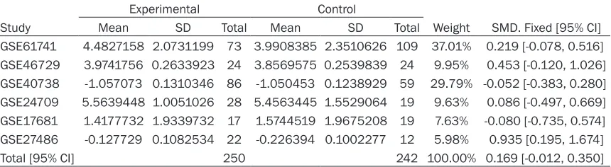
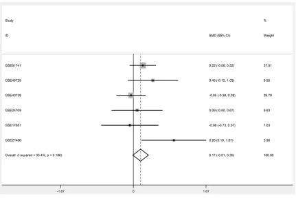
Table 2. Forest plot of studies evaluating standard mean difference (SMD) of hsa-miR-30a-3p
expres-sion between NSCLC and control group (a fixed-effects model)
Experimental Control
Study Mean SD Total Mean SD Total Weight SMD. Fixed [95% CI] GSE61741 4.4827158 2.0731199 73 3.9908385 2.3510626 109 37.01% 0.219 [-0.078, 0.516] GSE46729 3.9741756 0.2633923 24 3.8569575 0.2539839 24 9.95% 0.453 [-0.120, 1.026] GSE40738 -1.057073 0.1310346 86 -1.050453 0.1238929 59 29.79% -0.052 [-0.383, 0.280] GSE24709 5.5639448 1.0051026 28 5.4563445 1.5529064 19 9.63% 0.086 [-0.497, 0.669] GSE17681 1.4177732 1.9339732 17 1.5744519 1.9675208 19 7.63% -0.080 [-0.735, 0.574] GSE27486 -0.127729 0.1082534 22 -0.226394 0.1002277 12 5.98% 0.935 [0.195, 1.674]
Total [95% CI] 250 242 100.00% 0.169 [-0.012, 0.350]
[image:2.612.92.522.247.365.2]Inclusion criteria
Eligible datasets were included if they met the criteria as follows: (i) both NSCLC patients and non-cancerous healthy controls were included in each dataset, and each group contained at least 3 samples; (ii) the original expression
pro-filing data of miRNAs from both of the case and
control groups were available or could be calcu-lated; (iii) the subjects involved in the current meta-analysis were only humans.
Quality control and data extraction
Two investigators (Xing-gu Lin and Gang Chen) extracted the data independently from all
quali-fied datasets according to the aforementioned
requirements. Disagreements were resolved via cautious deliberation with the third and fourth authors (Rong-quan He and Li-hua Yang). Quality control was conducted with Limma package and ExiMiR package in R, including background correction and normalization pro-cessing [12, 13]. Expression values of miR-30a-3p and sample size were extracted in both case and control groups. If multiple probes
were mapped to a single miRNA, average value of these levels was regarded as the expression value of a certain miRNA. Furthermore, means and standard deviations (SD) of these values were calculated.
Statistical analysis
The meta package in R was employed to per-form the current meta-analysis [12, 14]. Conti- nuous outcomes were presented as standard
mean difference (SMD) with 95% confidence
interval (CI), and effect sizes were pooled with
random- or fixed-effects model according to dif -ferent conditions. Heterogeneity across studies was assessed with the chi-square test of Q and the I2 statistic [12, 15, 16]. A p value<0.05 or
I2>50% was considered as heterogeneous, if
so, the random-effects model (DerSimonian-Laird method) would be selected to calculate
the pooled SMD. Or else, the fixed-effects
model (Mantel-Haenszel method) was pre-ferred for the pooling process [17].
[image:3.612.92.523.69.357.2]To further elucidate whether the pooled result was achieved due to one large study or a single
study with an extremely divergent result, sensi-tivity analysis was applied to omit one study at a time. In addition, the potential publication bias was assessed with Begg’s and Egger’s tests. When P<0.05, the results would be regarded as publication bias.
Results
Features of the included datasets
The features of the enrolled datasets were shown in Table 1. In total, 6 eligible datasets, including GSE61741 (Germany), GSE46729
group (SMD = 0.169; 95% CI, -0.012 to 0.350; P = 0.067), the p value was close to 0.05. The results suggested that high circulating miR-30a-3p expression might have potential to dis-tinguish NSCLC patients from non-cancer con-trols. However, this hypothesis needs to be
veri-fied with larger cohorts.
Sensitivity analysis and publication bias as -sessment
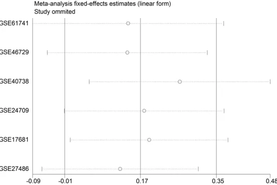
According to sensitivity analysis, the results indicated that no studies had the most
[image:4.612.96.372.74.258.2]nega-tive or posinega-tive influence on the summary SMD
[image:4.612.93.372.325.512.2]Figure 2. Sensitivity analysis of the value of miR-30a-3p on the early diag-nosis of NSCLC. No studies had the most negative or positive influence on the summary SMD.
Figure 3. Begg’s funnel plot for the assessment of potential publication bias. No potential publication bias was found.
(USA), GSE40738 (USA), GSE- 27486 (USA), GSE24709 (Ger- many), and GSE17681 (Germ- any), were involved in the meta-analysis. The data for GSE61741 (Germany), GSE24- 709 (Germany), and GSE17- 681 (Germany) were all de- rived from peripheral blood cells, and the data for GSE- 46729 (USA) was from serum, while the data for GSE40738 (USA) and GSE27486 (USA) were from whole blood. Alto- gether, 250 NSCLC patients and 242 healthy controls were enrolled in these 6 datasets.
Potential diagnostic value of circulating microRNA-30a-3p as a biomarker for lung
cancer
The means and SD of both experimental and control gro- ups were calculated for all 6 included microarray datasets (Table 2). The SMD ranged from -0.080 to 0.935 (Table 2). According to the
heteroge-neity test, no significant het -erogeneity was found among these individual datasets (P = 0.186, I2 = 33.4%, Table 2;
Figure 1). Thus, the
fixed-effects model was selected to calculate the pooled SMD and 95% CI.
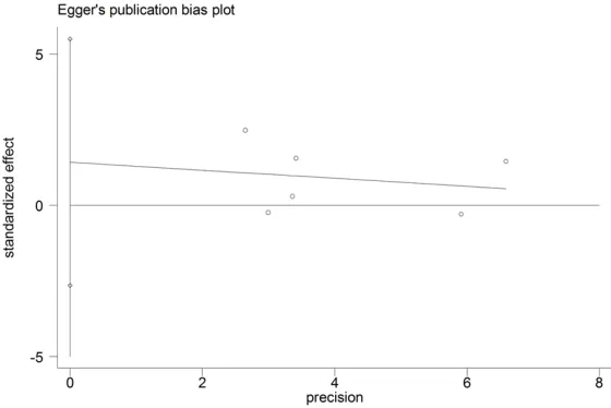
(Figure 2). The results of the meta-analysis were stable. Thus, no study omission was per-formed for the sensitivity analysis. Similarly, the Begg’s funnel plot showed symmetry distri-bution and no obvious publication bias was found in this meta-analysis (Figure 3, P = 0.707). Additionally, Egger’s test revealed no potential publication bias (Figure 4, P = 0.388). Discussion
Lung cancer, including NSCLC and SCLC, is a risk factor in cancer death and late diagnosis is one of the most essential causes for the high mortality rate [1, 2, 18]. Circulating miRNAs represent stable and reproducible markers for various solid tumors, including NSCLCs [19-21]. Hence, circulating miRNAs in serum, plas-ma or whole peripheral blood have been hypothesized as non-invasive diagnostic mark-ers for NSCLC [19, 22, 23]. However, no extremely effective miRNA has been discov-ered as biomarker in clinical settings for the early diagnosis of NSCLC [8, 19, 22].
In addition to some miRNA candidates, the
clin-ical significance of miR-30a-3p in NSCLCs was first investigated by the group of Cazzoli R et al.
[11]. In a training set with 30 frozen plasma samples, including 10 NSCLCs (adenocarcino-mas), 20 non-cancerous controls (10 granulo-mas and 10 healthy smokers), Cazzoli R et al.
[11] performed microRNA Ready-to-Use PCR and found that miR-30a-3p was slightly down-regulated in lung granulomas and on the
con-to the small number of patients involved, the validity of the conclusion was questioned. Therefore, the current meta-analysis was
car-ried out to explore the clinical significance of
circulating miR-30a-3p for the early detection of NSCLCs.
In the present meta-analysis, a total of 6miRNA
expression profiling datasets including 250
NSCLC patients and 242 healthy controls were enrolled. The pooled SMD revealed that miR-30a-3p expression in patients’ whole blood, peripheral blood cells and serum showed no
significant difference between NSCLC patients
and healthy controls. However, the pooled SMD was 0.169 and p value was a borderline as 0.067, which suggested a potential trend that high level of circulating miR-30a-3p occurred more likely in NSCLCs, as compared to non-cancer people. This showed the concordant direction with the report of Cazzoli R et al. [11].
Of course, the diagnostic significance of circu
-lating miR-30a-3p level needs further
verifi-cation.
To the best of our knowledge, this is the first
meta-analysis so far to assess the suitability of miR-30a-3p as a blood-based biomarker for early revealing of NSCLCs based on the micro-array datasets. Two meta-analyses were con-ducted to summarize the known different
microRNA expression profiles between lung
[image:5.612.92.372.74.261.2]cancer tissues and non-tumor lung by vote-counting strategy with miRNA microarray assays [24, 25]. Both of these 2 meta-analyses
Figure 4. Egger’s funnel plot for the assessment of potential publication bias. No potential publication bias was observed.
trary, miR-30a-3p was upregu-lated moderately in lung ade-nocarcinomas. Furthermore, consistent expression pattern
of miR-30a-3p was confirmed
in another validation set with another 50 NSCLCs and 55 non-cancerous controls with quantitative RT-PCR. Student t-test comparing the differ-ence of miR-30a-3p between controls and NSCLCs showed
the significance of 8 miRNAs
demonstrated that 30a-5p, but not miR-30a-3p, was among the most frequent lowly expressed miRNAs in NSCLC tissues rather than in non-cancerous lung tissues. However, the miR-30a-3p level in NSCLC tissues and its
clinical significance remain yet uncertain.
Either, the correlation between miR-30a-3p
level in tissues and in body fluid has not been clarified. Further experiments are required to figure out the clinical role of miR-30a-3p in
NSCLC tissue samples.
Nevertheless, several studies have been per-formed to investigate the characteristic and molecular mechanism of miR-30a-3p in differ-ent malignancies. Most of the studies revealed a lower expression of miR-30a-3p in the tumor tissues and regarded miR-30a-3p as a sup-pressor miRNA. For example, in breast cancer tissues, downregulation of miR-30a-3p was
identified [26, 27] and miR-30a-3p was also
downregulated in tumors from breast cancer patients with early recurrence [26]. In clear cell renal cell carcinoma tissues, miR-30a-3p down-regulation promoted increased expression of
HIF2α [28]. The expression of miR-30a-3p was also significantly downregulated in tumors in
HCC patients compared to adjacent normal tis-sues. Downregulation of miR-30a-3p was asso-ciated with notably higher occurrence of portal vein tumor thrombus in HCC. Furthermore, miR-30a-3p overexpression in vitro showed a sup-pressive effect on cell proliferation, an induced effect on apoptosis and an increased effect on arrest of cells in the S phase in HCC, via target-ing vimentin, MMP3 and E-cadherin in HCC [29]. Among different types of ovarian carcino-mas, miR-30a-3p expression was found to be the lowest in mucinous carcinoma, but highest in clear cell carcinoma. Also, expression level of miR-30a-3p was up-regulated in well-ated tumor as compared with poorly differenti-ated ones of ovarian carcinomas [30]. Addi- tionally, the expression of miR-30a-3pwas decreased in bladder cancer, colorectal cancer, as well as esophageal squamous cell carcino-ma tissues [31-33]. Being inconsistent with aforementioned studies, upregulation of miR-30a-3p was reported in squamous cell carci-noma of the tongue [34]. Thus, miR-30a-3p
may act as a tumor-specific miRNA, playing vari -ous roles in different cancers.
In addition to the level of miR-30a-3p in tumor tissues, another study demonstrated that
circu-lating level of miR-30a-3p in plasma significant -ly decreased after hysterectomy of endometri-oid endometrial carcinoma [35]. Due to the non-invasiveness and stability, detection of cir-culating miRNAs gains the potential value in clinical application for tumor early diagnosis. Further research is needed to study the clinical role of circulating miR-30a-3p level in different classes of malignancies.
Several limitations should be noted in the cur-rent meta-analysis. Firstly, the small sample size limited the robustness of the meta-analy-sis. Only 6 datasets were included. The power of the funnel plots in estimating publication bias might be misguided owing to the limited
number of qualified studies for meta-analysis.
Further studies involving large sample size
should be designed to confirm miR-30a-3p
expression levels in NSCLCs. Secondly, beca- use the miR-30a-3p expression data were only extracted from microarray assays without extra
confirmation by more accurate methods (for
instance, real time RT-qPCR), the level of circu-lating miR-30a-3p needs to be further validat-ed. Thirdly, additional source of a relevant bias might include the regions involved in the meta-analysis, since only data sets from USA and Germany were included. Therefore, the current result has to be interpreted cautiously.
In conclusion, even though high level of circu-lating miR-30a-3p expression showed a poten-tial relationship with the risk of NSCLC, the
evi-dence of significant difference of circulating
miR-30a-3p between NSCLC patients and
healthy controls is insufficient. Further studies
of high quality in the future are desired to evalu-ate the predictive power of circulating miR-30a-3p in larger cohorts of samples with validated detection methods.
Acknowledgements
The study was supported partly by the Fund of Guangxi Zhuang Autonomous Region University Student Innovative Plan (No. 201510598016),
the Scientific Research Project of the Basic
Disclosure of conflict of interest
None.
Address correspondence to: Tingqing Gan, Depart- ment of Medical Oncology, The First Affiliated Hos-pital of Guangxi Medical University, 6 Shuangyong Road, Nanning 530021, P. R. China. Tel: 0086-771-5353121; Fax: 0086-771-0086-771-5353121; E-mail: 7782- [email protected]
References
[1] Brothers JF, Hijazi K, Mascaux C, El-Zein RA, Spitz MR and Spira A. Bridging the clinical gaps: genetic, epigenetic and transcriptomic biomarkers for the early detection of lung can-cer in the post-National Lung Screening Trial era. BMC Med 2013; 11: 168.
[2] Damjanov N, Nurmohamed MT and Szekanecz Z. Biologics, cardiovascular effects and cancer. BMC Med 2014; 12: 48.
[3] Wang Z. Selection of chemotherapy for non-small cell lung cancer is facilitated by new therapeutic strategies. Int J Clin Exp Med 2014; 7: 3833-3842.
[4] Luo H, Qiao L, Liang N and Zhang J. Risk fac-tors for recurrence in patients with resected N1 non-small cell lung cancer-a systematic re-view and meta-analysis. J BUON 2015; 20: 791-799.
[5] Chen QW, Zhu XY, Li YY and Meng ZQ. Epigenetic regulation and cancer (review). Oncol Rep 2014; 31: 523-532.
[6] Sen R, Ghosal S, Das S, Balti S and Chakrabarti J. Competing endogenous RNA: the key to posttranscriptional regulation. Scientific World Journal 2014; 2014: 896206.
[7] Jackson BL, Grabowska A and Ratan HL. MicroRNA in prostate cancer: functional impor-tance and potential as circulating biomarkers. BMC Cancer 2014; 14: 930.
[8] Qin X, Xu H, Gong W and Deng W. The Tumor Cytosol miRNAs, Fluid miRNAs, and Exosome miRNAs in Lung Cancer. Front Oncol 2014; 4: 357.
[9] Jeong HC. Clinical Aspect of MicroRNA in Lung Cancer. Tuberc Respir Dis (Seoul) 2014; 77: 60-64.
[10] Del Vescovo V, Grasso M, Barbareschi M and Denti MA. MicroRNAs as lung cancer biomark-ers. World J Clin Oncol 2014; 5: 604-620. [11] Cazzoli R, Buttitta F, Di Nicola M, Malatesta S,
Marchetti A, Rom WN and Pass HI. microRNAs derived from circulating exosomes as noninva-sive biomarkers for screening and diagnosing lung cancer. J Thorac Oncol 2013; 8: 1156-1162.
[12] Meng X, Xiao C, Zhao Y, Jia L, Tang Y and Li D. Meta-analysis of microarrays: diagnostic value of microRNA-21 as a biomarker for lung can-cer. Int J Biol Markers 2015; 30: e282-285. [13] Sewer A, Gubian S, Kogel U, Veljkovic E, Han W,
Hengstermann A, Peitsch MC and Hoeng J. Assessment of a novel multi-array normaliza-tion method based on spike-in control probes suitable for microRNA datasets with global de-creases in expression. BMC Res Notes 2014; 7: 302.
[14] Cheung SF and Chan DK. Meta-analyzing de-pendent correlations: an SPSS macro and an R script. Behav Res Methods 2014; 46: 331-345.
[15] Lau J, Ioannidis JP and Schmid CH. Quantitative synthesis in systematic reviews. Ann Intern Med 1997; 127: 820-826.
[16] Higgins JP, Thompson SG, Deeks JJ and Altman DG. Measuring inconsistency in meta-analy-ses. BMJ 2003; 327: 557-560.
[17] Zamora J, Abraira V, Muriel A, Khan K and Coomarasamy A. Meta-DiSc: a software for meta-analysis of test accuracy data. BMC Med Res Methodol 2006; 6: 31.
[18] Han RX, Liu X, Pan P, Jia YJ and Yu JC. Effectiv- eness and safety of chemotherapy combined with dendritic cells co-cultured with cytokine-induced killer cells in the treatment of ad-vanced non-small-cell lung cancer: a system-atic review and meta-analysis. PLoS One 2014; 9: e108958.
[19] Huang Y, Hu Q, Deng Z, Hang Y, Wang J and Wang K. MicroRNAs in body fluids as biomark -ers for non-small cell lung cancer: a systematic review. Technol Cancer Res Treat 2014; 13: 277-287.
[20] Wang RJ, Zheng YH, Wang P and Zhang JZ. Serum miR-125a-5p, miR-145 and miR-146a as diagnostic biomarkers in non-small cell lung cancer. Int J Clin Exp Pathol 2015; 8: 765-771. [21] Yan HJ, Ma JY, Wang L and Gu W. Expression
and significance of circulating microRNA-31 in lung cancer patients. Med Sci Monit 2015; 21: 722-726.
[22] Fujita Y, Kuwano K, Ochiya T and Takeshita F. The impact of extracellular vesicle-encapsulat-ed circulating microRNAs in lung cancer re-search. Biomed Res Int 2014; 2014: 486413. [23] Ramshankar V and Krishnamurthy A. Lung
cancer detection by screening-presenting cir-culating miRNAs as a promising next genera-tion biomarker breakthrough. Asian Pac J Cancer Prev 2013; 14: 2167-2172.
[25] Guan P, Yin Z, Li X, Wu W and Zhou B. Meta-analysis of human lung cancer microRNA ex-pression profiling studies comparing cancer tissues with normal tissues. J Exp Clin Cancer Res 2012; 31: 54.
[26] Perez-Rivas LG, Jerez JM, Carmona R, de Luque V, Vicioso L, Claros MG, Viguera E, Pajares B, Sanchez A, Ribelles N, Alba E and Lozano J. A microRNA signature associated with early recurrence in breast cancer. PLoS One 2014; 9: e91884.
[27] Yan LX, Huang XF, Shao Q, Huang MY, Deng L, Wu QL, Zeng YX and Shao JY. MicroRNA miR-21 overexpression in human breast cancer is as-sociated with advanced clinical stage, lymph node metastasis and patient poor prognosis. RNA 2008; 14: 2348-2360.
[28] Mathew LK, Lee SS, Skuli N, Rao S, Keith B, Nathanson KL, Lal P and Simon MC. Restricted expression of miR-30c-2-3p and miR-30a-3p in clear cell renal cell carcinomas enhances HIF2alpha activity. Cancer Discov 2014; 4: 53-60.
[29] Wang W, Lin H, Zhou L, Zhu Q, Gao S, Xie H, Liu Z, Xu Z, Wei J, Huang X and Zheng S. MicroRNA-30a-3p inhibits tumor proliferation, invasive-ness and metastasis and is downregulated in hepatocellular carcinoma. Eur J Surg Oncol 2014; 40: 1586-1594.
[30] Lee H, Park CS, Deftereos G, Morihara J, Stern JE, Hawes SE, Swisher E, Kiviat NB and Feng Q. MicroRNA expression in ovarian carcinoma and its correlation with clinicopathological fea-tures. World J Surg Oncol 2012; 10: 174.
[31] Ma Y, Zhang P, Yang J, Liu Z, Yang Z and Qin H. Candidate microRNA biomarkers in human colorectal cancer: systematic review profiling studies and experimental validation. Int J Cancer 2012; 130: 2077-2087.
[32] Kano M, Seki N, Kikkawa N, Fujimura L, Hoshino I, Akutsu Y, Chiyomaru T, Enokida H, Nakagawa M and Matsubara H. 145, 133a and 133b: Tumor-suppressive miR-NAs target FSCN1 in esophageal squamous cell carcinoma. Int J Cancer 2010; 127: 2804-2814.
[33] Ichimi T, Enokida H, Okuno Y, Kunimoto R, Chiyomaru T, Kawamoto K, Kawahara K, Toki K, Kawakami K, Nishiyama K, Tsujimoto G, Nakagawa M and Seki N. Identification of nov -el microRNA targets based on microRNA signa-tures in bladder cancer. Int J Cancer 2009; 125: 345-352.
[34] Wong TS, Liu XB, Wong BY, Ng RW, Yuen AP and Wei WI. Mature miR-184 as potential on-cogenic microRNA of squamous cell carcinoma of tongue. Clin Cancer Res 2008; 14: 2588-2592.