7-Nitroquinazolin-4(3
H
)-one
Jian-Ping Yong,a,bGuan-Ping Yu,a,bJiu-Ming Li,a,b Xue-Ling Houaand Haji Akber Aisaa*
a
Xinjiang Technical Institute of Physics and Chemistry, Chinese Academy of Science, Urumqi 830011, People’s Republic of China, andbGraduate School of Chinese Academy of Science, Beijing 100039, People’s Republic of China
Correspondence e-mail: [email protected]
Received 14 November 2007; accepted 23 November 2007
Key indicators: single-crystal X-ray study;T= 153 K; mean(C–C) = 0.002 A˚; Rfactor = 0.034;wRfactor = 0.096; data-to-parameter ratio = 10.6.
In the crystal structure of the title compound, C8H5N3O3,
intermolecular N—H O hydrogen bonds link molecules into centrosymmetric dimers. These dimers are, in turn, linked though weak intermolecular C—H O and C—H N hydrogen bonds and – stacking interactions, with centroid–centroid distances of 3.678 (3) A˚ , into a three-dimensional network.
Related literature
For related literature on biological activity, see: Masanoriet al.
(2003); Wolfe et al.(1990). For related structures, see: Chad-wick & Easton (1983); Etter (1983).
Experimental
Crystal data
C8H5N3O3
Mr= 191.15
Monoclinic,P21=n
a= 5.1063 (10) A˚
b= 11.206 (2) A˚ c= 13.528 (3) A˚ = 99.19 (3)
V= 764.1 (3) A˚3
MoKradiation = 0.13 mm1
0.240.180.16 mm
Data collection
Rigaku R-AXIS RAPID IP area-detector diffractometer Absorption correction: multi-scan
(ABSCOR; Higashi, 1995) Tmin= 0.969,Tmax= 0.979
5749 measured reflections 1340 independent reflections 1215 reflections withI> 2(I) Rint= 0.021
Refinement
R[F2> 2(F2)] = 0.034
wR(F2) = 0.096
S= 1.11 1340 reflections
127 parameters
H-atom parameters constrained
max= 0.15 e A˚3
min=0.31 e A˚
3
Table 1
Hydrogen-bond geometry (A˚ ,).
D—H A D—H H A D A D—H A
N2—H2A O3i
0.88 1.98 2.8514 (14) 169
C1—H1B O2ii
0.95 2.54 3.2703 (17) 134
C1—H1B O1iii
0.95 2.55 3.0978 (17) 117
C5—H5A O2iv
0.95 2.49 3.2846 (16) 142
C7—H7A N1ii
0.95 2.55 3.4402 (18) 155
Symmetry codes: (i) xþ2;y;zþ1; (ii) xþ1;yþ1;zþ1; (iii)
xþ3 2;yþ
1 2;zþ
1 2; (iv)x
1 2;y
1 2;zþ
1 2.
Data collection:RAPID-AUTO (Rigaku, 2004); cell refinement: RAPID-AUTO; data reduction:RAPID-AUTO; program(s) used to solve structure: SHELXTL (Sheldrick, 2001); program(s) used to refine structure: SHELXTL; molecular graphics: SHELXTL; soft-ware used to prepare material for publication:SHELXTL.
This work is financially supported the Foundation of Xinjiang Key Laboratory of Plant Resources and Natural Products Chemistry (No. 2006–6).
Supplementary data and figures for this paper are available from the IUCr electronic archives (Reference: LH2576).
References
Chadwick, D. J. & Easton, I. W. (1983).Acta Cryst.C39, 454–456. Etter, M. C. (1983).J. Chem. Soc. Perkin Trans. 2, pp. 115–121. Higashi, T. (1995).ABSCOR. Rigaku Corporation, Tokyo, Japan.
Masanori, T., Yoshiaki, I., Hideyuki, T., Takahiro, N., Hirotada, T., Tominaga, F. & Hideya, H. (2003).Bioorg. Med. Chem.11, 383–391.
Rigaku (2004). RAPID-AUTO. Version 3.0. Rigaku Corporation, Tokyo, Japan.
Sheldrick, G. M. (2001).SHELXTL. Version 5.0. Bruker AXS Inc., Madison, Wisconsin, USA.
Wolfe, J. F., Rathman, T. L., Sleevi, M. C., Campbell, J. A. & Greenwood, T. D. (1990).J. Med. Chem.33, 161–166.
Structure Reports
Online
supporting information
Acta Cryst. (2008). E64, o427 [doi:10.1107/S1600536807062666]
7-Nitroquinazolin-4(3
H
)-one
Jian-Ping Yong, Guan-Ping Yu, Jiu-Ming Li, Xue-Ling Hou and Haji Akber Aisa
S1. Comment
7-Nitro-4(3H)-Quinazolinone (I), is an important intermediate for drugs synthesis and its derivatives show many
biological activities including anti-fungal, anti-convulsant (Masanori et al., 2003), bacterial, cancer,
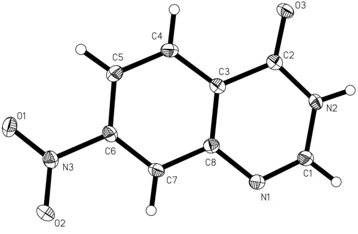
anti-inflammatory, and anti-tumor (Wolfe et al., 1990). We report here the crystal structure of (I) (Fig. 1).
In (I) (Fig. 1), all bond lengths and angles are normal and in a good agreement with those reported previously
(Chadwick & Easton, 1983; Etter, 1983). Atoms N3 and O3 lie in the 1,2-dihydroquinazoline ring (C1—C8/N1/N2)
plane, and the deviations from the least-squares plane through the ring atoms are all smaller than 0.026 (2) Å. The
relatively short distances of 3.678 (3)Å between the centroids of 1,2-dihydropyrimidine (C1/C2/C3/C8/N1/N2) and
benzene (C3—C8) rings related by (1 + x, y, x) indicates the presence of weak π-π interactions. In the crystal structure,
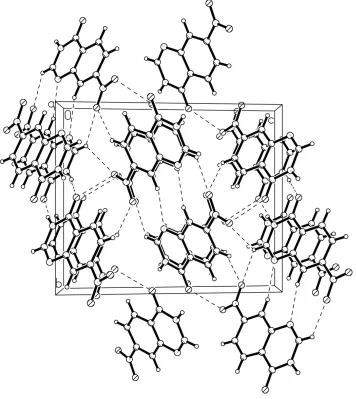
intermolecular N—H···O hydrogen bonds link molecules into centrosymmetric dimers. These dimers, are in turn, linked
though weak intermolecular C—H···O and C—H···N hydrogen bonds and π···π stacking interactions into a
three-dimensional network.
S2. Experimental
The title compound was synthesized by the reaction of 4-nitro-2-amino-benezic acid 18.2 g (0.1 mol) and formamidine
acetate 10.1 g (0.2 mol) in 100 mL andryous EtOH, refulxing for 6 h. The solid filtrated and washed with 20 ml H2O, cool
30 ml EtOH and 30 ml e ther, respectively, dried under vacuum to obtain the title compound 15.8 g, yield: 82.8%.
Crystals suitable for X-ray diffraction analysis were obtained by slow evaporation the solution of 7-Nitro-4(3H
)-Quinazolinone in EtOH/acetone/THF (1:1:1 V/V/V) at room temperature over a period of one week.
S3. Refinement
The H atoms were placed in calculated positions, with C—H = 0.95 Å, N—H = 0.88Å and included in the final cycles of
Figure 1
Figure 2
The packing of the title compound with hydrogen bonds shown as dashed lines.
7-NitroQuinazolin-4(3H)-one
Crystal data
C8H5N3O3
Mr = 191.15
Monoclinic, P21/n
Hall symbol: -P 2yn
a = 5.1063 (10) Å
b = 11.206 (2) Å
c = 13.528 (3) Å
β = 99.19 (3)°
V = 764.1 (3) Å3
Z = 4
F(000) = 392
Dx = 1.662 Mg m−3
Mo Kα radiation, λ = 0.71073 Å
Cell parameters from 6207 reflections
θ = 6.0–55.0°
µ = 0.13 mm−1
T = 153 K
Rigaku R-AXIS RAPID IP area-detector diffractometer
Radiation source: Rotating Anode Graphite monochromator
ω Oscillation scans
Absorption correction: multi-scan (ABSCOR; Higashi, 1995)
Tmin = 0.969, Tmax = 0.979
5749 measured reflections 1340 independent reflections 1215 reflections with I > 2σ(I)
Rint = 0.021
θmax = 25.0°, θmin = 3.1°
h = −6→6
k = −13→13
l = −16→15
Refinement
Refinement on F2
Least-squares matrix: full
R[F2 > 2σ(F2)] = 0.034
wR(F2) = 0.096
S = 1.11
1340 reflections 127 parameters 0 restraints
Primary atom site location: structure-invariant direct methods
Secondary atom site location: difference Fourier map
Hydrogen site location: inferred from neighbouring sites
H-atom parameters constrained
w = 1/[σ2(Fo2) + (0.064P)2 + 0.0923P]
where P = (Fo2 + 2Fc2)/3
(Δ/σ)max < 0.001
Δρmax = 0.15 e Å−3
Δρmin = −0.31 e Å−3
Special details
Geometry. All e.s.d.'s (except the e.s.d. in the dihedral angle between two l.s. planes) are estimated using the full covariance matrix. The cell e.s.d.'s are taken into account individually in the estimation of e.s.d.'s in distances, angles and torsion angles; correlations between e.s.d.'s in cell parameters are only used when they are defined by crystal symmetry. An approximate (isotropic) treatment of cell e.s.d.'s is used for estimating e.s.d.'s involving l.s. planes.
Refinement. Refinement of F2 against ALL reflections. The weighted R-factor wR and goodness of fit S are based on F2,
conventional R-factors R are based on F, with F set to zero for negative F2. The threshold expression of F2 > σ(F2) is used
only for calculating R-factors(gt) etc. and is not relevant to the choice of reflections for refinement. R-factors based on F2
are statistically about twice as large as those based on F, and R- factors based on ALL data will be even larger.
Fractional atomic coordinates and isotropic or equivalent isotropic displacement parameters (Å2)
x y z Uiso*/Ueq
O1 −0.37255 (17) 0.37262 (8) 0.24708 (6) 0.0228 (3)
O2 −0.1637 (2) 0.51746 (9) 0.33145 (8) 0.0324 (3)
O3 0.69259 (17) −0.00398 (7) 0.41669 (6) 0.0203 (3)
N1 0.6334 (2) 0.33230 (10) 0.53066 (8) 0.0214 (3)
N2 0.8443 (2) 0.14538 (9) 0.52489 (7) 0.0183 (3)
H2A 0.9807 0.1015 0.5512 0.022*
C1 0.8209 (2) 0.25752 (11) 0.56195 (9) 0.0206 (3)
H1B 0.9536 0.2827 0.6152 0.025*
C2 0.6655 (2) 0.09785 (11) 0.44863 (8) 0.0163 (3)
C3 0.4471 (2) 0.17821 (10) 0.40975 (8) 0.0160 (3)
C4 0.2491 (2) 0.14293 (11) 0.33110 (9) 0.0188 (3)
H4A 0.2557 0.0658 0.3025 0.023*
C5 0.0451 (2) 0.21960 (11) 0.29509 (9) 0.0194 (3)
H5A −0.0904 0.1964 0.2421 0.023*
H7A 0.2274 0.4498 0.4421 0.021*
C8 0.4409 (2) 0.29261 (11) 0.45270 (8) 0.0166 (3)
N3 −0.17982 (19) 0.41378 (10) 0.30265 (7) 0.0194 (3)
Atomic displacement parameters (Å2)
U11 U22 U33 U12 U13 U23
O1 0.0179 (5) 0.0261 (6) 0.0224 (5) −0.0023 (4) −0.0028 (3) 0.0042 (3)
O2 0.0344 (6) 0.0166 (5) 0.0416 (6) 0.0077 (4) −0.0086 (4) −0.0043 (4)
O3 0.0219 (5) 0.0143 (5) 0.0248 (5) 0.0033 (4) 0.0043 (3) −0.0018 (3)
N1 0.0228 (6) 0.0167 (6) 0.0227 (5) 0.0021 (4) −0.0023 (4) −0.0026 (4)
N2 0.0167 (5) 0.0155 (6) 0.0219 (5) 0.0034 (4) 0.0003 (4) 0.0017 (4)
C1 0.0225 (7) 0.0170 (7) 0.0208 (6) 0.0016 (5) −0.0005 (5) −0.0008 (4)
C2 0.0170 (6) 0.0146 (7) 0.0183 (6) −0.0009 (5) 0.0062 (4) 0.0020 (4)
C3 0.0171 (6) 0.0146 (7) 0.0174 (6) −0.0001 (5) 0.0056 (4) 0.0017 (4)
C4 0.0208 (7) 0.0145 (7) 0.0217 (6) −0.0011 (5) 0.0049 (5) −0.0034 (5)
C5 0.0192 (6) 0.0196 (7) 0.0185 (6) −0.0032 (5) 0.0010 (4) −0.0011 (5)
C6 0.0165 (6) 0.0156 (6) 0.0195 (6) 0.0014 (5) 0.0036 (4) 0.0033 (5)
C7 0.0201 (7) 0.0127 (6) 0.0199 (6) 0.0010 (5) 0.0033 (5) −0.0006 (4)
C8 0.0182 (6) 0.0150 (6) 0.0168 (6) −0.0016 (5) 0.0034 (4) 0.0009 (5)
N3 0.0192 (6) 0.0191 (6) 0.0197 (5) 0.0013 (4) 0.0020 (4) 0.0037 (4)
Geometric parameters (Å, º)
O1—N3 1.2289 (14) C3—C4 1.4023 (17)
O2—N3 1.2240 (15) C3—C8 1.4099 (17)
O3—C2 1.2358 (15) C4—C5 1.3779 (18)
N1—C1 1.2916 (17) C4—H4A 0.9500
N1—C8 1.3946 (16) C5—C6 1.3982 (18)
N2—C1 1.3652 (16) C5—H5A 0.9500
N2—C2 1.3713 (16) C6—C7 1.3750 (17)
N2—H2A 0.8800 C6—N3 1.4799 (16)
C1—H1B 0.9500 C7—C8 1.4076 (18)
C2—C3 1.4644 (17) C7—H7A 0.9500
C1—N1—C8 115.91 (11) C4—C5—C6 118.03 (11)
C1—N2—C2 123.16 (10) C4—C5—H5A 121.0
C1—N2—H2A 118.4 C6—C5—H5A 121.0
C2—N2—H2A 118.4 C7—C6—C5 123.78 (11)
N1—C1—N2 125.49 (11) C7—C6—N3 118.00 (11)
N1—C1—H1B 117.3 C5—C6—N3 118.21 (11)
N2—C1—H1B 117.3 C6—C7—C8 118.02 (11)
O3—C2—N2 121.57 (11) C6—C7—H7A 121.0
O3—C2—C3 124.30 (11) C8—C7—H7A 121.0
N2—C2—C3 114.12 (11) N1—C8—C7 117.89 (11)
C4—C3—C8 120.54 (11) N1—C8—C3 122.83 (11)
C4—C3—C2 120.97 (11) C7—C8—C3 119.28 (11)
C5—C4—H4A 119.8 O1—N3—C6 118.10 (10)
C3—C4—H4A 119.8
C8—N1—C1—N2 0.28 (19) N3—C6—C7—C8 177.39 (10)
C2—N2—C1—N1 −0.5 (2) C1—N1—C8—C7 −179.22 (11)
C1—N2—C2—O3 179.92 (11) C1—N1—C8—C3 0.03 (18)
C1—N2—C2—C3 0.33 (16) C6—C7—C8—N1 −179.57 (10)
O3—C2—C3—C4 −0.08 (19) C6—C7—C8—C3 1.15 (17)
N2—C2—C3—C4 179.50 (10) C4—C3—C8—N1 −179.68 (11)
O3—C2—C3—C8 −179.62 (10) C2—C3—C8—N1 −0.14 (17)
N2—C2—C3—C8 −0.04 (16) C4—C3—C8—C7 −0.44 (18)
C8—C3—C4—C5 −0.31 (18) C2—C3—C8—C7 179.10 (10)
C2—C3—C4—C5 −179.83 (11) C7—C6—N3—O2 10.75 (16)
C3—C4—C5—C6 0.30 (18) C5—C6—N3—O2 −170.57 (11)
C4—C5—C6—C7 0.47 (19) C7—C6—N3—O1 −167.81 (10)
C4—C5—C6—N3 −178.12 (10) C5—C6—N3—O1 10.87 (16)
C5—C6—C7—C8 −1.21 (18)
Hydrogen-bond geometry (Å, º)
D—H···A D—H H···A D···A D—H···A
N2—H2A···O3i 0.88 1.98 2.8514 (14) 169
C1—H1B···O2ii 0.95 2.54 3.2703 (17) 134
C1—H1B···O1iii 0.95 2.55 3.0978 (17) 117
C5—H5A···O2iv 0.95 2.49 3.2846 (16) 142
C7—H7A···N1ii 0.95 2.55 3.4402 (18) 155