Nijmegen
The following full text is a publisher's version.
For additional information about this publication click this link.
http://hdl.handle.net/2066/136060
Please be advised that this information was generated on 2017-12-05 and may be subject to
change.
Neurobiology of Disease
Resting-State Functional Connectivity Changes in Aging
apoE4 and apoE-KO Mice
Valerio Zerbi,
1,2Maximilian Wiesmann,
1,3Tim L. Emmerzaal,
1Diane Jansen,
1Maarten Van Beek,
1X
Martina P.C. Mutsaers,
1Christian F. Beckmann,
4,5Arend Heerschap,
2and Amanda J. Kiliaan
1Departments of1Anatomy, Donders Institute for Brain Cognition and Behaviour,2Radiology,3Geriatric Medicine, and4Donders Centre for Cognitive Neuroimaging, Radboud university medical center, 6525 EZ Nijmegen, The Netherlands, and5MIRA Institute for Biomedical Technology and Technical Medicine, University of Twente, 7500 AE Enschede, The Netherlands
It is well established that the cholesterol-transporter apolipoprotein
(APOE) genotype is associated with the risk of developing
neuro-degenerative diseases. Recently, brain functional connectivity (FC) in apoE-
4 carriers has been investigated by means of resting-state
fMRI, showing a marked differentiation in several functional networks at different ages compared with carriers of other apoE isoforms.
The causes of such hampered FC are not understood. We hypothesize that vascular function and synaptic repair processes, which are both
impaired in carriers of
4, are the major contributors to the loss of FC during aging. To test this hypothesis, we integrated several different
MRI techniques with immunohistochemistry and investigated FC changes in relation with perfusion, diffusion, and synaptic density in
apoE4 and apoE-knock-out (KO) mice at 12 (adult) and 18 months of age.
Compared with wild-type mice, we detected FC deficits in both adult and old apoE4 and apoE-KO mice. In apoE4 mice, these changes
occurred concomitant with increased mean diffusivity in the hippocampus, whereas perfusion deficits appear only later in life, together
with reduced postsynaptic density levels. Instead, in apoE-KO mice FC deficits were mirrored by strongly reduced brain perfusion since
adulthood. In conclusion, we provide new evidence for a relation between apoE and brain connectivity, possibly mediated by vascular risk
factors and by the efficiency of APOE as synaptic modulator in the brain. Our results show that multimodal MR neuroimaging is an
excellent tool to assess brain function and to investigate early neuropathology and aging effects in translational research.
Key words:
apoE; apoE4 mice; cerebral blood flow; diffusion tensor imaging; functional connectivity; resting-state fMRI
Introduction
The only gene currently associated to sporadic Alzheimer’s
dis-ease (AD) is the
4 allele of the apolipoprotein E (APOE) gene
(
Mahley and Rall, 2000
). Several mechanisms by which APOE-
4
promotes AD have been proposed; after brain injury, apoE is
produced by astrocytes to transport cholesterol to the damaged
neuronal and synaptic membranes; however, the repair and
re-modelling of damaged synapses appears to be less effective by
apoE-
4 than other isoforms (
Mahley et al., 2006
;
Verghese et al.,
2011
). Moreover, APOE-
4 carriers are more susceptible to
vas-cular brain damages (e.g., stroke, brain hemorrhage;
Zlokovic,
2011
,
2013
;
Liu et al., 2013
). This, in the end, can result in a
permanent loss of synaptic contacts, with gradual loss of
neuro-nal connectivity (
Bu, 2009
;
Verghese et al., 2011
).
Investigating functional connectivity is nowadays possible by
resting-state functional MRI (rsfMRI). rsfMRI examines the
tem-poral correlations of blood oxygen level-dependent (BOLD)
fluc-tuations between brain regions at rest, which is thought to reflect
resting neuronal activity and is often referred to functional
con-nectivity (FC;
Biswal et al., 1995
;
Damoiseaux et al., 2006
;
De
Luca et al., 2006
). This MR technique has generated a great deal of
interest among neuroscientists and has been widely used to
in-vestigate neurological disorders (
Greicius, 2008
).
Many studies have reported a correlation between APOE-
4,
AD, and abnormalities in functional connectivity measured with
rsfMRI or task-based fMRI (
Trachtenberg et al., 2012
);
cogni-tively normal young APOE-
4 carriers showed elevated
resting-state activity in the default mode network (DMN) and high
hippocampal activation during memory tasks; both areas that are
preferentially affected in early AD (
Bookheimer et al., 2000
;
Fil-ippini et al., 2009
). This hippocampal hyperactivation is thought
to represent a compensatory response, in which increased
cogni-tive effort is required to achieve an equal level of performance to
that of non-
4 carriers (
Bondi et al., 2005
). Such hyperactivation
is followed by a decline in FC and structural interconnectivity
between cortical regions at older age (
O’Brien et al., 2010
;
Brown
et al., 2011
;
Machulda et al., 2011
). This is in-line with studies
Received Feb. 14, 2014; revised Aug. 26, 2014; accepted Sept. 5, 2014.
Author contributions: V.Z., C.F.B., A.H., and A.J.K. designed research; V.Z., M.W., T.L.E., D.J., M.V.B., and M.P.M. performed research; V.Z., M.W., T.L.E., and M.V.B. analyzed data; V.Z. and A.J.K. wrote the paper.
This work was supported by the European Community’s Seventh Framework Programme (FP7/2007-2013) un-der Grant agreement no. 211696 and NWO investment Grants 91106021 and BIG (VISTA). We thank Ilse Arnoldus-sen, Jos Dederen, Roy Haast, Karin Vos, Sabine DenisArnoldus-sen, and Laura Mellendijk for their laboratory work; and Henk Arnts and Bianca Lemmers for their excellent care giving of our mice.
The authors declare no competing financial interests.
Correspondence should be addressed to Dr Amanda J. Kiliaan, Department of Anatomy, Donders Institute for Brain Cognition and Behaviour, Radboud university medical center, Geert Grooteplein noord 21, 6525 EZ Nijmegen, The Netherlands. E-mail: [email protected].
DOI:10.1523/JNEUROSCI.0684-14.2014
showing that elderly APOE-
4 carriers have reduced FC
com-pared with APOE-
3 carriers, even in absence of amyloid-

plaques (
Sheline et al., 2010
). Alterations in the DMN have also
been reported in
4-carriers (
Fleisher et al., 2009
), and similarly
in AD patients (
Greicius et al., 2004
).
Despite an increasing amount of evidence for an association
between apoE genotype and FC changes, the mechanisms
under-lying this relationship remain elusive (
Verghese et al., 2011
).
Spe-cifically, it is not clear whether changes in FC in APOE-
4 carriers
have neural origins or are driven by other copathologies, such as
impaired neurovascular coupling.
To determine the underlying neural or vascular origin of
such changes, we investigate the relation between functional
connectivity, cerebral perfusion, brain tissue microstructure,
and postsynaptic density in target-replacement apoE4- and
apoE-deficient mice; these mice represent mild and severe AD
vascular risk factors, respectively, and may provide insights on
the role of APOE genotype on FC changes, with or without the
presence of vascular deficits.
Materials and Methods
Animals
The apoE4 founder mice were originally obtained from Taconic Trans-genic Models and a colony was established at the Radboud University Medical Center (Radboud UMC). ApoE4 mice were created by targeting
the murine APOE gene for replacement with the human APOE-4 alleles
cultured in E14TG2a embryonic stem (ES) cells as described previously (Sullivan et al., 1997). Resulting chimeras were backcrossed to C57BL/6J (B6) mice for eight generations. The line was derived by embryo transfer and is maintained by incrossing homozygous mice. For the present study, male and female apoE4 breeder mice were used to generate homozygous apoE4 offspring (third generation).
The apoE-deficient (B6,129P2-Apoetm1Unc/J) founders were
origi-nally obtained from Jackson Laboratories and a colony was established at the Radboud UMC. In ApoE-knock-out (KO) mice the APOE fragment was targeted with an apoE-specific probe (a SacI/BglII fragment) isolated from a mouse APOE cDNA clone. The strongly hybridizing phage clones obtained in this screening, a 7.8 kilobase (kb) EcoRI fragment was iso-lated and compared with the restriction map. Subsequently these tar-geted cells were cultured in E14TG2a ES and injected into C57BL/6J (B6) mice. Resultant chimeras were backcrossed for 11 generations and inter-crossed to homozygosity. The line was derived by embryo transfer and is maintained by incrossing homozygous mice (Piedrahita et al., 1992). For the present work, male and female apoE-KO breeder mice were used to generate homozygous apoE-KO offspring (third generation).
C57BL/6J wild-type mice, obtained from our colony at the Radboud UMC were used as controls. Throughout the experiment animals were housed in groups of two to seven mice per cage in a controlled environ-ment, homogenously illuminated by normal fluorescent room light at 60 lux, with room temperature at 21°C, and an artificial 12 h light/dark cycle (lights on at 7:00 A.M.). Food and water were availablead libitum.
The experiments were performed according to Dutch federal regula-tions for animal protection. The Veterinary Authority of the Radboud University Nijmegen Medical Centre, the Netherlands, approved all the protocols within this study.
MRI
Two cohorts of apoE4, apoE-KO, and wild-type male mice of 12 months of age (number of animals for each genotype:n⫽8, 10, and 9, respec-tively) and of 18 months of age (n⫽9, 9, and 10, respectively) were used for this cross-sectional study. To study genotype and aging related dif-ferences in brain function and structure, rsfMRI, cerebral blood flow (CBF), and diffusion tensor imaging (DT-MRI) were measured in each cohort.
MRI measurements were performed on an 11.7 T BioSpec Avance III small animal MR system (Bruker BioSpin) equipped with an ac-tively shielded gradient set of 600 mT/m and operated by Paravision
5.1 software. We used a circular polarized volume resonator for signal transmission and an actively decoupled mouse brain quadrature sur-face coil for signal reception (Bruker BioSpin). During the MR
exper-iments, low-dose isoflurane was used (3.5% for induction and⬃1.5%
for maintenance), slightly adjusted throughout the experiment to
maintain a fast and stable breathing frequency (⬎130 bpm). The mice
were placed in a stereotactic device with earbars and toothholder to immobilize the head. Great care was taken to fix the mouse head, as this is important to avoid movement-related artifacts, particularly when applying multishot fast acquisition MR sequences. As we did not detect movement artifacts in the EPI images, we decided not to use further methodologies to limit artifacts from movements, such as the respiratory gating, that would have increased the acquisition time. Body temperature was measured with a rectal thermometer and maintained at 37°C by a heated airflow device.
Gradient echo (GE) T2*-weighted images covering the entire mouse brain were acquired in three directions for anatomical reference. Subse-quently, rsfMRI datasets were acquired using a single-shot spin-echo sequence combined with echo-planar imaging (SE-EPI) sequence. Al-though its sensitivity to image the BOLD effect is slightly reduced, SE-EPI has less susceptibility artifacts compared with GE-EPI; in addition, it is less sensitive to geometric distortion and physiological noise, which are known to generate confounding results in resting-state FC, also in ro-dents (Kalthoff et al., 2011). Six hundred repetitions with a repetition time (TR) of 1.8 s and echo time of 16.9 ms were recorded for a total acquisition time of 18 min.
To study brain perfusion under resting conditions, a flow-sensitive alternating inversion recovery arterial spin labeling (FAIR ASL) tech-nique was used (Kim, 1995;Zerbi et al., 2014). Fifteen images with in-creasing inversion times (TIs; 40 –3000 ms) were obtained for the T1 calculations, amounting to a total scan time of 12 min. Inversion recov-ery data from the imaging slice were acquired after selective inversion interleaved with nonselective inversion.
Diffusion of water was imaged as described previously (Harsan et
al., 2010;Zerbi et al., 2013). In short, 22 axial slices covering the whole brain were acquired with a four-shot SE-EPI protocol. B0 shift com-pensation, navigator echoes, and an automatic correction algorithm to limit the occurrence of ghosts and artifacts were implemented.
Encodingbfactors of 0 s/mm2(fiveb⫽0 images) and 1000 s/mm2
were used and diffusion-sensitizing gradients were applied along 30 non-collinear directions in three-dimensional space. All other
imag-ing parameters are listed inTable 1. Before each sequence, a
whole-brain automatic shim protocol was applied; this includes the adjustment of field homogeneity, the adjustment of the basic reso-nance frequency and the adjustment of the reference pulse gain. The
FWHM achieved for a square box of 6⫻6⫻6 mm3was⬍35 Hz,
in-line with other studies (Nasrallah et al., 2014).
FC measurements
The rsfMRI datasets were first realigned using a least-squares method and rigid-body transformation with Statistical Parametric Mapping (SPM) mouse toolbox (SPM5, University College London; http://www.
fil.ion.ucl.ac.uk/spm/;Sawiak et al., 2009). Mean and maximum
dis-placement across the six degrees of freedom (along thex-,y-, andz-axes and on three rotation parameters pitch, roll, and yaw) were measured in each mouse. The mean SE-EPI images of each mouse were then used to generate a study-specific template through linear affine and nonlinear diffeomorphic transformation (ANTs. v1.9; http://picsl.upenn.edu/ ANTS/). Visual inspection of the normalized dataset was performed to screen for possible normalization biases. On the template, 15 areas were selected in left and right hemisphere and back-transformed in each sub-ject space using the inverse of the affine and diffeomorphic transforma-tions. The selected regions were based on previous work in functional connectivity in mice (Jonckers et al., 2011), and includes: dorsal hip-pocampus (DH), ventral hiphip-pocampus (VH), auditory cortex (AU), mo-tor cortex (M1), somatosensory cortex (S1),visual cortex (V1), and retrosplenial cortex (RS). All cortical ROI were selected 1–2 voxels away from the edge of the cortex, to minimize the impact of
susceptibility-weighted artifacts, which are more prominent in areas of different tissues interface (e.g., near the skull or near the ear canals).
In-plane spatial smoothing (0.4⫻0.4 mm), linear detrending, and
temporal high-pass filtering (cutoff at 0.01 Hz) were applied to com-pensate for small across-mouse misregistration and temporal low-frequency noise. Head movement components detected from the rigid-body transformation were regressed using FSL (the FMRIB soft-ware library;Jenkinson et al., 2012). In VH, S1, and AU, voxelwise FC
maps were computed using the REST MATLAB toolkit (Song et al.,
2011) and group comparisons were assessed voxelwise using SPM5
with the SPMMouse toolbox. In both 12- and 18-month-old mice,
twottests were performed to identify genotype differences in the
framework of the general linear model. Statistical significance for voxels exceeding a minimum cluster size of 4, to achieve cluster size
⬇0.05 mm3as byDubois et al. (2008), was established atp⬍0.05,
uncorrected for multiple comparisons.
FC group comparison between ROIs were calculated from the BOLD time series using total correlation and partial correlation analyses imple-mented in FSLNets (FSLNets v0.3; www.fmrib.ox.ac.uk/fsl). Pearson’s correlation values were Fisher transformed to Z-scores for group com-parisons and statistical analysis.
For an optimal characterization of the resting-state networks (RSNs), detected with our methodology in the mouse brain, a group-level esti-mation of functional connectivity was performed in the normalized da-taset of the 12-month-old wild-type mice using an independent component analysis (ICA) method with GIFT v2.0a toolbox (group ICA of fMRI toolbox, http://icatb.sourceforge.net/; Jonckers et al., 2011;
Zhou et al., 2014). The number of components for all rsfMRI data were set to be either 30, 20, or 15. Group ICA is performed using the Infomax algorithm. The group-level spatial ICA maps of independent RSNs were scaled to Z-scores with a threshold of兩Z兩⬎1.96, corresponding to un-correctedp⬍0.025 for two-tailed test. The ICA maps were then visually inspected and labeled based on the spatial pattern identified via an ana-tomical atlas (Ullmann et al., 2013).
CBF calculation
For each mouse, the FAIR images with different TIs were realigned over the first TI using a rigid-body model, implemented in SPM. Determina-tion of T1selectiveand T1nonselectivewas performed by fitting the averaged
signal intensities in each ROI with a three-parameters monoexponential T1 relaxation curve. CBF was determined in cortex, hippocampus, and thalamus using the following equation:
CBF ⫽ T1non-selective T1blood
冉
1 T1selective⫺ 1 T1non-selective冊 ,whereis the blood/tissue partition coefficient for water, assumed to be 0.9 ml/g (Herscovitch and Raichle, 1985;Leithner et al., 2010) and T1 blood was assumed to be 2.75 s at 11.7T (Lin et al., 2012).
Diffusion tensor MRI parameter estimation and
group comparisons
The calculation of four commonly used DT-MRI parameters, mean dif-fusivity (MD), fractional anisotropy (FA), radial difdif-fusivity (RD), and parallel diffusivity (1), was performed following a protocol as described previously (Zerbi et al., 2013). Briefly, the diffusion tensor was estimated
for every voxel using the PATCH algorithm (Zwiers, 2010). Thereafter,
FA, MD, RD, and1 maps were normalized to a study-specific template
through linear affine and nonlinear diffeomorphic transformation using ANTs. Regional differences between apoE4 mice and wild-type, and be-tween apoE-KO and wild-type in spatially normalized diffusion maps were assessed voxelwise using SPM5 following the same procedure as described byZerbi et al. (2013). Statistical significance for an individual
voxel was established atp⬍0.05, with a minimum cluster size of 4
interconnected voxels (to achieve cluster size⬇0.05 mm3as by (Dubois et al. (2008). As we considered the voxel based analysis (VBA) an explor-ative approach to investigate structural differences in the whole brain, we did not correct for multiple-comparison.
In addition, ROI of several white matter (WM) and gray matter (GM) areas were drawn on the template image based on an anatomical atlas (Paxinos and Franklin, 2004) and the resulting diffusion-related param-eters were measured for further statistical analyses.
Immunohistochemistry
Directly following the MR measurements at 12 and 18 months of age, anesthetized mice were killed by transcardial perfusion with 0.1MPBS. The perfused brains were collected and postfixed for 15 h at 4°C in 4% paraformaldehyde fixative and thereafter stored in 0.1MPBS with 0.01% sodium azide at 4°C for immunohistochemical staining. Eight series of 30
m coronal sections were cut through the brain using a sliding
mi-crotome (Microm HM 440 E) equipped with an object table for freeze sectioning at⫺60°C. The tissue was stained for postsynaptic density with PSD95 antibody using one complete series of brain sections. Immuno-histochemistry was performed using standard free-floating labeling pro-cedures, as described previously (Jansen et al., 2013).
PSD95
Polyclonal rabbit anti-PSD95 (1:2000; Abcam, catalog #ab18258, RRID: AB_444362) was used as a primary antibody. The sections were first pretreated with 0.9% H2O2in PBS to block endogenous peroxidise and
then incubated overnight at room temperature on a shaker table. After
incubation, the sections were rinsed three times with 0.1MPBS and
incubated with the secondary antibody, donkey anti-rabbit biotin (1:
1500; Biotin-SP-AffiniPure Donkey Anti-Rabbit IgG (H⫹L), Jackson
ImmunoResearch). After 90 min, the sections were rinsed three times again and transferred to a solution containing Vector ABC-elite (1:800; Vector Laboratories) for 90 min. Thereafter, visualization of postsynap-tic density was achieved by incubation with DAB-Ni solution. Stained sections were mounted on gelatin-coated glass slides, dried overnight in a stove at 37°C, dehydrated in alcohol series, cleared with xylol, and mounted in Entellan.
Quantification.The stained sections were analyzed using a Zeiss Ax-ioskop microscope equipped with hardware and software of Micro-brightfield. Brain regions were based on the mouse brain atlas ofPaxinos and Franklin (2004)and quantified in five regions of the hippocampus: the inner molecular layer (IML), outer molecular layer (OML), cornus ammonis 1 (CA1), CA2, and CA3. Additionally, two regions in the cortex corresponding to the visual and somatosensory cortex were analyzed. The relevant regions were digitized at 100 times magnification with im-mersion oil using Stereo Investigator. The quantification of the photo-graphs was performed using ImageJ (NIH). The contrast was manually
Table 1. List of parameters used in each MRI scan Imaging sequences
Anatomical T2*w rsfMRI CBF Diffusion tensor imaging
Imaging method GE Spin-echo EPI FAIR-ASL 4-shot spin-echo EPI
Echo time (ms) 5 16.9 11.8 20
Repetition time 630 ms 1.8 s 13.75 s 7.55 s
Image matrix 512⫻512 96⫻96 128⫻128 128⫻128
Field-of-view (mm) 40⫻40 25⫻25 30⫻30 20⫻20
Spatial resolution (m/pixel) 78⫻78⫻340 260⫻260⫻500 234⫻234⫻1000 156⫻156⫻500
No. of slices 20⫻3 9 1 22
enhanced, following the same procedure for all digitized images, and the amount of tissue stained was measured with a threshold-based approach.
Statistics
For the statistical analysis, IBM SPSS 20 soft-ware was used. Because the setup of the current study was designed to determine the effect of aging and the extent to which apoE4 and apoE-KO mice develop neuropathological traits of AD and not to study the effects of the apoE allele itself, statistical analyses were per-formed separately for the apoE4 and apoE-KO
mice (apoE-4 vs wild-type, and apoE-KO vs
wild-type). Multivariate ANOVA (MANOVA) with Bonferroni corrections was conducted with between-group factors genotype and age of the animals. If the Bonferronipost hoctest indicated a significant interaction between ge-notype and age, the data were split for the con-cerning factor and thereafter analyzed again with the MANOVA. For the rsfMRI analysis, respiration of the animals was considered as covariate, to remove its confounding effect in the statistical analysis. Statistical significance was set atpⱕ0.05. Correlation analyses be-tween perfusion, diffusion parameters, and PSD-95 were performed with the bivariate Spearman’s correlation method in the cortex (averaged value in the S1, AU, and V1 for rs-fMRI, DT-MRI, and PSD-95) and hippocam-pus. To avoid false-positive correlations, the statistical significance for the correlation anal-yses was set atpⱕ0.01. All values used are
expressed as mean⫾SEM.
Results
The breathing of the mice was constantly
monitored during the MR acquisition and
used as a measure of the level of sedation
of each individual mouse. During the
rs-fMRI and the CBF scans, the averaged
res-piration rate for all mice was 153
⫾
4 and
146
⫾
5 breaths per minutes (BPM),
respec-tively. During the DT-MRI scan, which is
the last scan of our protocol after
⬃
2 h
an-esthesia, the averaged respiration rate for all
mice was 84
⫾
3 BPM. No statistical group
differences were found in respiration rate
between either genotype or age, suggesting
that an equal dose of anesthesia was
per-ceived by the mice during the acquisition.
Individual breathing rate changes were also
uncorrelated with FC strength and CBF levels.
The displacement across the six
degrees-of-freedom did not show significant
differ-ences between groups, suggesting that all
mice were equally influenced by motion artifacts. The higher
move-ments were detected in the up– down direction (0.23
⫾
0.03 mm
averaged in all mice), whereas the displacement in other directions
was lower than the voxel size. No mice were excluded after visual
inspection of the normalized rsfMRI and DTI datasets.
rsfMRI: independent component analysis
Results of the ICA in 12-month-old wild-type mice are shown in
Figure 1
. The number of components is arbitrarily chosen based
on previous studies in mouse rsfMRI (
Jonckers et al., 2011
,
2014
;
Zhou et al., 2014
). With 30 components, most of the components
properly match specific anatomical and functional brain areas on
both hemispheres (
Fig. 1
b
); with this analysis, most of the cortical
components were found to be mainly unilateral, with the
excep-tion of the motor 1 and 2 regions (M1, M2) and the
motor-somatosensory-visual cortices (M1, S1, V1), which display a
single band across the two hemispheres (
Fig. 1
b
). By reducing the
number of components to 20 and 15, more cortical regions are
Figure 1. a, Anatomical locations of the cortical ROI, based onUllmann et al., 2013.b, FC maps defined in the 12-month-old male wild-type mice by group ICA based on 30 components. The spatial color-codedz-maps of these components are overlaid on the template SE-EPI image. Components in same anatomical locations but in contralateral regions are shown together with two different color-codes, with a higher Z-score (yellow or light blue, thresholded for兩Z兩⬎1.96) representing a higher correlation between the time course of that voxel and the mean time course for this component. Mean components comprise: M1–M2; M1–S1; M1–S1-V1; S1–V1; S2-AU; RS; piriform cortex (Pir); Cingulum (Cg); HC; CPu; Lateral septal nuclei (LS)-Th; Th-CPu, hypo-thalamus (HTc).c, By reducing the number of components to 15, many areas displayed an extended connectivity pattern across the same anatomical location, revealing a certain degree of interhemispheric connectivity.d, Other components having low amount of voxels at the edge of the brain, or displayed in only one slice, are considered artifacts.
covered by one band bilaterally; other brain regions that
dis-played a highly unilateral pattern in the 30-component analysis,
such as the HC, the caudate-putamen (CPu), and the thalamic
nuclei (Th), showed more prominent bilateral activity (
Fig. 1
c
).
Mean FC patterns in the different mice groups from seeds in
VH, S1, and AU are shown in
Figure 2
. Overall, a strong bilateral
connectivity is notable when seeding in hippocampal regions. In
the cortex, we detected a strong connectivity covering
somato-sensory, auditory, and motor cortices, with some extent also in
their corresponding contralateral regions.
The VBA of these patterns revealed a widespread FC reduction
in both apoE4 and apoE-KO mice compared with wild-type,
par-ticularly visible at younger age. No voxels indicated an increase
FC in these genotypes.
Total correlation analyses
To compare the FC patterns in different genotype and ages,
rs-fMRI data were statistically analyzed based on total correlation
(
Fig. 3
) and partial correlation (
Fig. 4
).
The multivariate ANOVA showed overall significant aging
and genotype effects when comparing apoE4 and apoE-KO with
wild-type mice (
Fig. 3
b
,
c
). In both apoE4, apoE-KO, and
wild-type mice, 18-month-old animals showed decreased FC levels
compared with 12-month-old mice. Most striking aging
differ-Figure 2. Mean FC maps defined in three ROIs in wild-type, apoE4, and apoE-KO mice. Voxel-based FC maps of the different seeds-area positioned in the VH, somatosensory cortex (SS), and AU in the left hemisphere reveal functionally connected areas. The spatial color-coded FC maps are overlaid on the SE-EPI images of the same mouse. A higherpscore (yellow) represents a higher correlation between the BOLD time course of the seed area and the other voxels in the brain. Voxels representing a significant reduction in FC (p⬍0.05, minimum voxel cluster size 0.05 mm3) between apoE4, apoE-KO compared with wild-type are shown in blue. No voxels indicated an increased FC in these genotypes at both ages.a, At 12 months of age, a strong interhemispheric connectivity between right and left hippocampus is notable. In the cortical regions, the connectivity is covering SS, AU, and motor cortices, mainly unilaterally distributed but with extent also in contralateral areas, in the hippocampus and in few thalamic nuclei. VBA defined the location of significant FC reduction in the selected ROI in apoE4 and apoE-KO mice.b, At 18 months of age, the strength of correlation and the interhemispheric connectivity were visibly reduced but still present due to aging. Differences between genotypes were less visible and present mainly in the apoE-KO mice.
ences were located in the connectivity between motor cortices
(left and right) and hippocampus, and, specifically for apoE4 and
wild-type mice, between motor cortex and retrosplenial cortex
(
Fig. 3
b
,
c
, top-right). Reduced FC between auditory cortices was
also detected in apoE-KO and wild-type animals.
The pairwise comparison also revealed significant lower FC in
apoE4 and apoE-KO mice, independent of age. These reductions
in FC commonly affected auditory, motor, and somatosensory
cortices and hippocampal areas (
Fig. 3
b
,
c
, bottom-left).
How-ever, stronger reductions of FC were seen in the apoE-KO mice
compared with wild-type, also including the motor-visual
con-nectivity and retrosplenial cortex.
In both group comparisons, the MANOVA revealed no
sig-nificant genotype
⫻
age interactions.
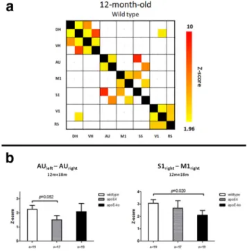
Partial correlation analyses
The multivariate ANOVA of the partial correlation values (with
Z
-value thresholded at
兩
z
兩
⬎
1.96), showed overall significant
genotype effects (
Fig. 4
); the apoE4 had lower FC compared with
wild-type in the direct connectivity between auditory cortices
although not significant,
p
⫽
0.052. In the apoE-KO mice, a more
severe reduction was found between somatosensory and motor
cortices (
Fig. 4
b
). Reduced partial correlation was found in the
motor-somatosensory cortices connectivity for apoE-KO and
wild-type mice due to aging (
p
⫽
0.034, data not shown). Also in
the partial correlation analyses, the MANOVA, revealed no
sig-nificant genotype
⫻
age interactions.
CBF
To study differences in cerebrovascular health between the mice
groups, we measured CBF with a FAIR ASL. Three ROIs on the
left and right brain hemispheres were analyzed: cortex,
hip-pocampus, and thalamus. Because no intraindividual differences
in CBF between right and left hemispheres were detected between
mice groups (data not shown), values from both sides were
averaged.
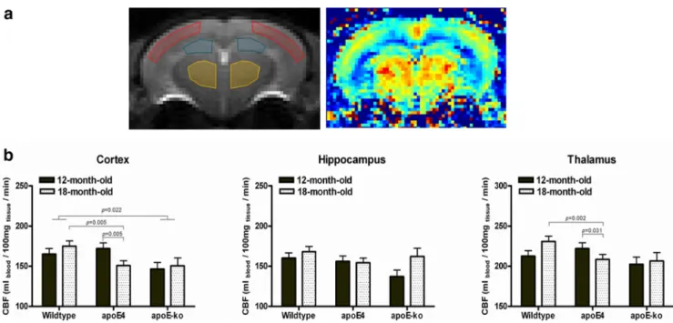
In the comparison of apoE4 with wild-type, the MANOVA
revealed a genotype
⫻
age interaction in the cortex and in
thala-mus (
p
⫽
0.027 and
p
⫽
0.026, respectively); after splitting the
data for age and for genotype, we found that 18-month-old
apoE4 mice have significantly lower CBF in these ROIs compared
with wild-type at the same age (
p
⫽
0.005 for the cortex and
p
⫽
0.002 for the thalamus), and also compared with 12-month-old
apoE4 animals (
p
⫽
0.005 and
p
⫽
0.002, respectively;
Fig. 5
).
Contrarily to what is seen in the FC results, in the 12-month-old
mice we did not detect any difference between wild-type and
apoE4.
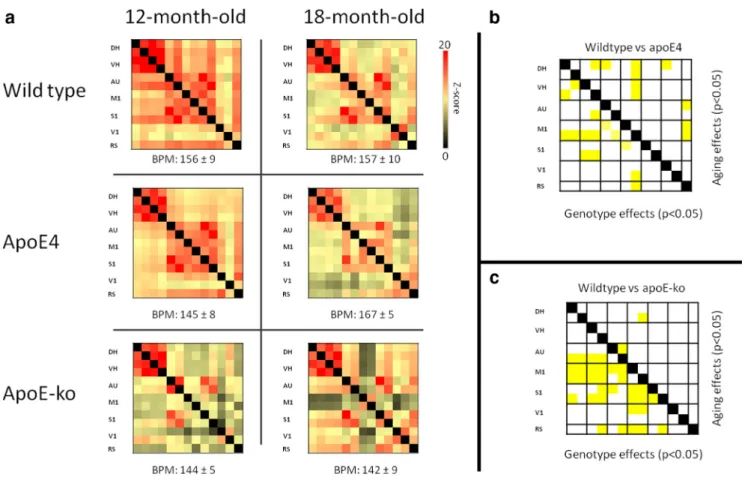
Figure 3. Resting-state FC based on total correlation analyses of 15 ROI in the mouse brain.a, Total correlation matrices of wild-type, apoE4 and apoE-KO mice at 12- and 18 months of age. Some characteristics of the mouse brain connectivity are consistent within different mouse models and age-effect; for example, it is visible a strong bilateral connectivity between DH and VH. AU, M1, S1, and RS also show a high degree of interconnectivity, as well as high FC between V1 (BPM; average⫾SE).b, Statistical analyses of FC is shown as a matrix, to reveal significant differences between genotypes (bottom-right) and between aging (top-right) for each ROI–ROI connection. Because no genotype⫻age interactions were seen, data were analyzed as genotype effects, independent of age, and aging effects, independent of genotype. ApoE4 mice displayed a reduction of FC at both ages, compared with wild-type, between several regions; particularly, a reduced FC is seen between dorsal and ventral hippocampus and M1, AU, and SS. Intracortical FC reduction are also seen, affecting primarily the AU-M1-SS system. Aging effects are also shown, revealing a reduction of FC over time between 12- and 18-month-old mice. Most striking differences are found between M1 and DH, VH and RS.c, Statistical analyses revealed strong reduction of FC in apoE-KO mice compared with wild-type mice, at both ages. These deficits seem to occur primarily in the hippocampal-cortical connectivity, but also between M1, AU, S1, and RS. Reduced FC due to aging is seen between M1 and DH, and between AU cortices.
Compared with wild-type, the apoE-KO mice showed a
sig-nificant lower CBF in the cortex (
p
⫽
0.022), independent of age.
In the hippocampus and in the thalamus a slight reduction of
CBF was also seen, although it did not reach statistical
signifi-cance (
p
⫽
0.117 and
p
⫽
0.084, respectively). No aging effects
were seen in both genotypes.
DT-MRI
In DT-MRI the water diffusivity, assessed in multiple directions,
is used to reconstruct an ellipsoid for every voxel to model the
diffusion. The MD describes the size of the ellipsoid, while its
shape is quantified by the FA. RD and
1 further describe the
shape of the ellipsoid and its directionality with respect to the
main fiber orientation. Differences of diffusion tensor derived
indices were determined separately in each age group with an
explorative VBA and, with a ROI-based approach. The VBA can
detect the occurrence of diffusion changes at higher spatial
reso-lution and is the option of choice when there is no prior
knowl-edge about the expected changes; however, these changes must
then be confirmed by a proper statistical analysis in a ROI-based
approach. For the VBA,
t
value maps (for a
p
⬍
0.05, and
mini-mum voxel cluster size set at 0.05 mm
3) were overlaid with FA
and MD template images (
Figs. 6
a
,
b
,
7
a
,
b
).
Several differences in diffusion parameters were detected
from the VBA. Among them, the most striking seem to be an
increased MD in the cortex and hippocampus, commonly found
in 18-month-old apoE4 and apoE-KO mice compared with
wild-type (
Fig. 6
a
,
b
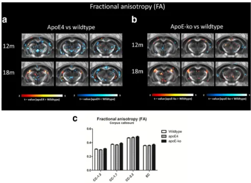
). In the apoE4 mice, a reduction of FA is also
noticeable in the molecular layer of the hippocampus, and in
other cortical regions such as in the retrosplenial and in the
piri-form cortices. In the apoE-KO mice, an increased FA in the
ex-ternal capsule is seen in 12-month-old mice. In both apoE4 and
apoE-KO mice, differences in FA and MD were seen near to the
ventricle area, possibly due to partial volume effects.
With the ROI-based approach, we measured a higher MD in
the DH in apoE4 mice (
p
⫽
0.038), reflected by increases (not
significant) in both RD (
p
⫽
0.056) and
1 (
p
⫽
0.231). An
increased MD in the apoE4 mice was also found in the corpus
callosum (CC) at
⫺
1.7 from bregma (
p
⫽
0.016), driven by a
higher
1 (
p
⫽
0.001); similar differences were seen also in the
external capsule (EC; higher MD:
p
⫽
0.035; higher
1:
p
⫽
0.015); however, no FA changes were measured in these WM
regions. The FA differences found by VBA, but not confirmed by
the ROI-based approach could therefore be: (1) indicative for a
minor change in diffusion proprieties, and (2) true only for a
limited part of the structure of interest.
Few other differences were found, such as an increase MD in
the DH in apoE-KO mice and an increased MD in the MEctx in
apoE4 mice, although were not significant (
p
⫽
0.08). No
signif-icant aging effect or genotype
⫻
age interactions were seen in the
MANOVA.
PSD-95
Levels of postsynaptic density were visualized and quantified with
polyclonal rabbit anti-PSD95 and are shown as relative values
compared with wild-type mice (
Fig. 8
). In the 12-month-old
an-imal group, we found a significant reduction of PDS-95 staining
in apoE-KO mice compared with wild-type in the IML (
p
⫽
0.005) and in the CA3 (
p
⫽
0.046;
Fig. 8
c
). In 18-month-old
animals, reduced PSD-95 levels in both apoE-KO and apoE4
mice compared with wild-type was seen in the IML and OML;
however, these differences were only significant in the apoE4
group (
p
⫽
0.043 and
p
⫽
0.039, respectively;
Fig. 8
d
). Because
the staining of the two groups was not performed at the same
time, we could not assess aging effects on the different genotypes.
The two-tailed Spearman’s correlation test revealed a strong
positive correlation between PSD-95 levels and CBF in both the
hippocampal region (
p
⫽
0.003) and in the cortex (
p
⫽
0.006).
When we split the data for genotype and age, we detected a similar
positive correlation for all groups, but smaller in magnitude. The
correlation in the cortex was nearly significant at both 12 and 18
months of age (
p
⫽
0.072 and
p
⫽
0.043), and more pronounced
in the apoE4 mice (
p
⫽
0.014) than in apoE-KO (
p
⫽
0.087) and
wild-type (
p
⫽
0.033).
No correlations were found between PSD-95 and DT-MRI
parameters, neither a significant correlation between DT-MRI
and CBF for the same regions.
Discussion
Target-replacement apoE4 and apoE-KO mice are attractive
models to investigate the role of apoE and vascular risk factors in
relationship with AD-like pathology, such as changes in brain FC.
However, the technical challenges to obtain good-quality MR
images and the lack of knowledge of murine brain network
sys-tems have been strong limiting factors for these studies. Recently,
new dedicated hardware and methods for acquisition and data
analysis enabled the analysis of rsfMRI in mice, resulting in a
growing number of publications (
Jonckers et al., 2011
,
2014
;
Guilfoyle et al., 2013
;
Nasrallah et al., 2014
).
Figure 4. Resting-state FC based on partial correlation analyses of 17 ROIs in the mouse brain.a, Averaged partial correlation matrix of 12-month-old wild-type mice. Compared with total correlation analysis, the partial correlation highlights the direct connectivity between two regions, by regressing the temporal BOLD signal from all other ROI. These matrices were highly similar between different genotypes and ages (data not shown) and revealed unique features of mouse brain connectivity; in particular, a significant direct interhemispheric connectivity is seen within the DH, VH, AU, M1, and V1. Among the cortical regions, we could not detect a significant direct interhemispheric connectivity only in the S1 region. However, a strong intrahemispheric connectivity is seen between S1, M1, and AU and between V1 and RS. Resulting connectivity were thresholded at兩Z兩⬎1.96, corresponding to uncorrectedp⬍0.025 for two-tailed test, based on the 12-month-old wild-type data.b, From the selected areas, reduced interhemi-spheric direct connectivity between auditory cortices was detected in apoE4 mice; significant reduced FC in the apoE-KO mice, independent of age, was seen between the S1 and the M1. Data represent mean⫾SE.
The strongest potential confounding
factor when performing FC analysis in
an-imals, still remains the impact of
anesthet-ics. Some studies in rats have used
analgesic muscle relaxants, such as
dex-medetomidine (
Lu et al., 2012
) or
me-detomidine (
Zhao et al., 2008
), to limit
the sedation level of the animals.
How-ever, maintaining a stable dose of these
anesthetics for prolonged experiments
(
⬇
2 h) is barely reliable. More recently, it
has been demonstrated that robust FC
measures in rats and mice could be
de-tected using a regime of low-dose
isoflu-rane (
Guilfoyle et al., 2013
;
Zhou et al.,
2014
). Compared with other anesthetic
regimes, low-dose isoflurane seems to
preserve resting-state networks in mice,
with similar results to those obtained in
awake animals (
Jonckers et al., 2014
).
Iso-flurane is also known to increase the
rest-ing blood flow, due to its moderate
systemic vasodilator effect (
Iida et al.,
1998
). In a separate experiment, we
con-firmed that both CBF and FC are
depen-dent on isoflurane level, and they both
rapidly decline with concentrations of
an-esthetic
⬎
2.2%, comparable to previous
findings (
Liu et al., 2011
). We also
mea-sured an increased CBF and a reduced FC
by stimulating the maximum vessel vasodilation using a higher
percentage of N
2O in the gas composition (
Drummond et al.,
1987
). However, the connectivity patterns, particularly between
hippocampus and cortical structures, and the CBF were restored
by switching the gas concentration to the standard experimental
condition; together, these data indicate that the methodology used
for this study is replicable and valid to measure functional
connec-tivity, without reaching the condition of maximum vasodilation and
without disrupting the neurovascular coupling. Furthermore, there
are no reports on a possible effect of isoflurane on apoE-signaling,
suggesting that this anesthetic does not interfere with the outcome of
this study, as all animals were kept under the same conditions.
Figure 6. MD comparison between wild-type, apoE4, and apoE-KO mice. VBA indicate significant differences in MD at 12 and 18 months of age. Three rostral to caudal averaged MD maps are overlaid with voxels that showed a significant difference (p⬍0.05, minimum voxel cluster size 0.05 mm3). The voxel color indicates a negative or positive change in the apoE4 (a) or in the apoE-KO (b) mice compared with the wild-type mice of the same age. Few areas of significant differences are depicted in the explorative VBA; among them, an increase in MD in the DH of the apoE4 mice, independent of age, was also confirmed by ROI-based MANOVA (c). Increase in MD in the DH was also observed in the apoE-KO mice, although not significant (p⬎0.05).
Figure 5. a, CBF was measured in apoE4, apoE-KO, and wild-type mice at 12 and 18 months of age in three different ROIs: cortex (including somatosensory, auditory, and visual cortices), hippocampus, and thalamus. The area corresponding to the retrosplenial cortex was excluded, because it overlaps with the azygos pericallosal artery (azPA) of the anterior cerebral artery (ACA). To measure the CBF we used a FAIR ASL MRI technique.b, A reduced CBF was found in the cortex in 18-month-old apoE4 mice and in 12- and 18-month-old apoE-KO mice compared with wild-type. A similar trend was observed in the thalamus, although it was not significant for the apoE-KO mice. No significant differences were seen in the hippocampus.
FC in the aging mouse brain
Despite the limited literature about resting-state FC in rodents,
our results exhibited many similarities with the resting-state
net-works described in mice by means of independent component
analysis, in a similar anesthetic regime (
Jonckers et al., 2011
); in
particular, we confirmed the presence of networks in several well
defined cortical and subcortical areas of the mouse brain. It is
however difficult to extrapolate whether these components are
mainly unilateral or bilateral; most of the studies in rats clearly
showed the presence of a bilateral cortical connectivity in several
cortical regions (mainly visual, somatosensory, auditory, and
motor) also under isoflurane (
Liang et al., 2012
;
Zhou et al.,
2014
); nevertheless, not all studies in mice using isoflurane as
anesthetic found the same degree of bilateral connectivity (
Jon-ckers et al., 2011
;
Guilfoyle et al., 2013
), whereas a more
promi-nent interhemispheric connectivity has been found using other
anesthetics (
Nasrallah et al., 2014
;
Sforazzini et al., 2014
). In this
study, we detected significant interhemispheric direct
connectiv-ity (with partial correlation analysis) in hippocampal, motor,
au-ditory, visual, and piriform cortices, and intrahemispheric
connectivity between somatosensory, motor, and auditory
corti-ces. Comparable resting-state connectivity in mice have been
re-cently shown by high-resolution photoacoustic tomography,
under a regime of ketamine
⫹
xylazine (
Nasiriavanaki et al.,
2014
). Our ICA further confirmed these results, although
re-vealing at the same time that the strongest interhemispheric
components are found in the motor cortex and in the
somatosensory-auditory-visual system.
As we expected, the connectivity found by ICA was largely
dependent on the number of components selected before the
analysis. In particular, when we reduced the number of
compo-nents, the new components showed extended, and often
inter-hemispheric, connectivity patterns but
always keeping their meaningful
anatom-ical location. This concept has been
re-ported also in human studies, where a
larger number of components from
rs-fMRI data probed a different hierarchy of
functional networks. These newly
re-vealed “subnetworks” were subsets of the
larger networks found with lower number
of components and they were interpreted
as areas of slightly different function or as
left versus right subnetworks (
Smith et al.,
2009
).
An important finding in our study was
a reduced FC in old animals compared
with adulthood as measured with total
correlation analysis. rsfMRI have been
in-creasingly used as a tool to investigate the
aging human brain
in vivo
. With few
ex-ceptions, most of the studies showed
aging-related decrements in FC (
Ferreira
and Busatto, 2013
). Because normal aging
is associated with decline in cognition, the
reduction in FC is thought to correlate
with a disruption of neuronal connections
and a deterioration of brain functioning
in elderly people (
Hedden and Gabrieli,
2004
;
Whalley et al., 2004
). Compared
with the total correlation results, partial
cor-relation analyses revealed a more stable
de-gree of connectivity at different ages.
Resting-state FC and perfusion changes in the aging
mouse brain
In this study, we found a reduced total FC in apoE4 and apoE-KO
mice, at both 12 and 18 months, compared with age-matched
wild-type mice. In all animals, CBF was also measured, to test
whether changes in vascular performance interfered with the FC
results. Changes in cerebrovascular dynamics, such as impaired
coupling between neural activity and hemodynamic response,
might have directly influenced BOLD signal fluctuations and
consequently FC changes (
Liu, 2013
).
ApoE4
Cross-sectional studies in young, middle-aged, and elderly
hu-man apoE-
4 carriers have reported reduced regional CBF and
cerebral glucose metabolism over time, particularly in brain
re-gions susceptible to pathological changes in AD (
Scarmeas and
Stern, 2006
). In apoE-
4 subjects, a faster decline of regional CBF
during aging has been shown compared with apoE-
3, suggesting
its contribution to the increased risk of developing AD (
Wierenga
et al., 2013
). In accordance, we demonstrated that apoE4 mice
suffer from decreased cortical CBF at 18 months of age, whereas
no differences were found at 12 months of age. Similar to the FC
results, we showed a rapid decline of cerebral perfusion in these
mice during aging, suggesting a strong aging effect. However, our
data also highlight that changes in FC in apoE4 animals are
mea-surable before CBF deficits. This finding suggests that the early
FC decline in these mice is more likely attributable to the neural
component on the BOLD signal, rather than to the possible
in-terference of the vascular contribution.
Figure 7. FA comparison between wild-type, apoE4 and apoE-KO mice. VBA indicate significant differences in FA at 12 and 18 months of age. Three rostral to caudal averaged FA maps are overlaid with voxels that showed a significant difference (p⬍0.05, minimum voxel cluster size 0.05 mm3). The voxel color indicates a negative or positive change in the apoE4 (a) or in the apoE-KO (b) mice compared with the wild-type mice of the same age. GM regions did not show FA differences between groups (data not shown), although a reduced FA is visible in the molecular layer of the hippocampus in apoE4 mice, as depicted in the explorative VBA (a). ROI analyses of the CC in different bregma did not show statistical significant differences between FA due to genotype or aging (c).
ApoE-KO
In apoE-KO mice we found a concomitant reduction in CBF and
resting-state connectivity at both ages. These mice are commonly
used as models for vascular pathologies, as they develop aortic
aneurysms, atherosclerotic plaques, and endothelial dysfunction
throughout their life (
Crauwels et al., 2003
;
Trollope et al., 2011
).
No investigations on resting-state fMRI were previously
per-formed in apoE-KO mice; however, it is known that, together
with vascular deficits, these mice also develop compromised
syn-aptic plasticity (
Masliah et al., 1995
;
Blain et al., 2006
) and
cog-nitive performance (
Gordon et al., 1995
;
Masliah et al., 1995
;
Oitzl et al., 1997
;
Krzywkowski et al., 1999
). Our results suggest
that the FC decline observed in these animals reflects the
contri-bution of both impaired neurovascular coupling and damaged
neural activity; we hypothesize that the severe vascular pathology,
which develop spontaneously in these mice at early age, could
have accelerated the occurrence of brain injury by overexposure
to stress factors, like cerebral hypoperfusion; the absence of apoE
could have further aggravated the synaptic repair processes,
lead-ing to loss of neuronal connectivity. Supportively, we found a
positive correlation between CBF and postsynaptic density (in
sampled regions). Although the correlation of these two
mea-sures do not imply a cause– effect mechanism, it does support an
association between neuronal and vascular health. It is also
inter-esting to note that the changes in adult apoE-KO mice mirror the
observations in the old apoE4 mice; this underlines a certain
degree of similarity between these models, although
demonstrat-ing a more severe pathology in the KO mice.
Structural changes and postsynaptic density in the aging
mouse brain
DT-MRI studies in apoE4 and apoE-KO mice are lacking, but
changes in diffusion parameters have been reported in human
studies; FA reductions in WM of APOE-
4 carriers have been
shown (
Nierenberg et al., 2005
;
Honea et al., 2009
;
Heise et al.,
2011
); two studies also reported diffusion differences in
hip-pocampal structures (
Nierenberg et al., 2005
;
Persson et al.,
2006
). Nevertheless, these dissimilarities were consistent across
different ages, suggesting that APOE affects WM and GM
micro-structure proprieties from early adulthood, without directly
re-flecting the associated risk of developing AD (
Westlye et al.,
2012
).
With our exploratory voxel-based analyses, we detected
sim-ilar changes in WM and GM microstructure in apoE4 mice.
However, the only significant results confirmed by MANOVA
were an age-independent increased MD in the CC, EC, and DH of
the apoE4 mice. It is interesting to note that the changes in MD in
WM were reflected by increased parallel and radial diffusivity,
but without changes in tensor shape; this could indicate a
differ-ent structural density of the fibers, resulting in more space for the
water molecules to move. Brain atrophy and WM loss could have
also increased the partial volume effect in these relatively thin
areas, explaining these results. The association between an
in-creased MD (and RD) in the CC and myelin degradation of the
axonal bundles has been proposed by previous studies (
Song et
al., 2005
). To verify this hypothesis and directly measure brain
atrophy
in vivo
, voxel-based morphometry (
Ellegood et al.,
Figure 8. PSD-95 staining performed on brain sections of apoE-4, apoE-KO, and wild-type mice.a,b, Representation of PSD-95-immunoreactive staining in hippocampal and cortical areas at two different magnifications (5⫻and 100⫻, respectively).c, In 12-month-old animals we detected a significant decrease in PSD-95 levels in apoE-KO mice compared with wild-type in the IML and CA3 of hippocampus.d, In 18-month-old animals we show reduced PSD-95 levels in the apoE-4 animals in the OML and in the IML, whereas no significant differences were seen between wild-type and apoE-KO mice. Values represent the mean and SEM.
2012
) or electron microscopy methods could be applied in
future studies.
Higher hippocampal MD has been consistently associated
with neurodegenerative processes in both AD patients and
ani-mal models for AD (
Zerbi et al., 2013
;
Zhang et al., 2014
). These
structural modifications in apoE4 mice are likely not related to
neurodegeneration, as neural loss is not expected in this model at
a young age. It is possible instead that increased inflammation
processes could have driven structural alterations and
hippocam-pal atrophy (
Yin et al., 2011
).
Furthermore, we did not find differences between apoE-KO
and wild-type mice in DT-MRI parameters, suggesting that for
these models, water diffusion changes are mild even in presence
of severe functional deficits. The structural changes found in
these mice were also not related to vascular or synaptic deficits,
suggesting a link to the role of apoE in synaptic development,
dendrite formation, and axonal guidance.
Synaptic loss and disconnection are strongly related with
cog-nitive decline in AD and may influence FC (
Selkoe, 2002
;
Serrano-Pozo et al., 2011
). We therefore determined the
postsyn-aptic density in our mouse models to characterize neural deficits
in relation with FC. Compared with wild-type, we found reduced
PSD-95 levels in 12-month-old apoE-KO mice, and also in the
18-month-old apoE4 mice. Interestingly, the present results
mirror the hypoperfusion deficits observed in these animals,
confirming the relation between apoE, neuronal and vascular
health. Furthermore, these data suggest impaired synaptic
maintenance by the apoE4 isoform, which is even more
pro-nounced by its absence. This can be linked to the lower efficacy
of the apoE-
4 isoform as cholesterol transporter, resulting in
reduced contribution to synaptogenesis and maintenance of
synaptic connections (
Poirier et al., 1995
;
Mauch et al., 2001
;
Pfrieger, 2003
).
In conclusion, with this study we provide evidence for a
rela-tion between apoE genotype, vascular risk factors and funcrela-tional
connectivity. Despite the technical challenges, we demonstrate
that rsfMRI, combined with other MR neuroimaging techniques,
can be used as powerful tool to investigate early neuropathology
and aging effects in translational research.
Notes
Supplemental material for this article is available at https://www.radboudumc. nl/Zorg/Afdelingen/anatomie/SectieAnatomie/Documents/20140818% 20Supplementary%20material%20Valerio%20Zerbi.pdf. This material has not been peer reviewed.
References
Biswal B, Yetkin FZ, Haughton VM, Hyde JS (1995) Functional connectiv-ity in the motor cortex of resting human brain using echo-planar MRI.
Magn Reson Med 34:537–541.CrossRef Medline
Blain JF, Sullivan PM, Poirier J (2006) A deficit in astroglial organization causes the impaired reactive sprouting in human apolipoprotein E4
targeted replacement mice. Neurobiol Dis 21:505–514. CrossRef
Medline
Bondi MW, Houston WS, Eyler LT, Brown GG (2005) fMRI evidence of compensatory mechanisms in older adults at genetic risk for Alzheimer disease. Neurology 64:501–508.CrossRef Medline
Bookheimer SY, Strojwas MH, Cohen MS, Saunders AM, Pericak-Vance MA, Mazziotta JC, Small GW (2000) Patterns of brain activation in people at
risk for Alzheimer’s disease. N Engl J Med 343:450 – 456. CrossRef
Medline
Brown JA, Terashima KH, Burggren AC, Ercoli LM, Miller KJ, Small GW, Bookheimer SY (2011) Brain network local interconnectivity loss in ag-ing APOE-4 allele carriers. Proc Natl Acad Sci U S A 108:20760 –20765.
CrossRef Medline
Bu G (2009) Apolipoprotein E and its receptors in Alzheimer’s disease:
pathways, pathogenesis and therapy. Nat Rev Neurosci 10:333–344.
CrossRef Medline
Crauwels HM, Van Hove CE, Holvoet P, Herman AG, Bult H (2003) Plaque-associated endothelial dysfunction in apolipoprotein E-deficient mice on a regular diet: effect of human apolipoprotein AI. Cardiovasc Res 59:189 –199.CrossRef Medline
Damoiseaux JS, Rombouts SA, Barkhof F, Scheltens P, Stam CJ, Smith SM, Beckmann CF (2006) Consistent resting-state networks across healthy subjects. Proc Natl Acad Sci U S A 103:13848 –13853.CrossRef Medline
De Luca M, Beckmann CF, De Stefano N, Matthews PM, Smith SM (2006) fMRI resting state networks define distinct modes of long-distance
inter-actions in the human brain. Neuroimage 29:1359 –1367. CrossRef
Medline
Drummond JC, Scheller MS, Todd MM (1987) The effect of nitrous oxide on cortical cerebral blood flow during anesthesia with halothane and isoflurane, with and without morphine, in the rabbit. Anesth Analg 66:
1083–1089.Medline
Dubois A, He´rard AS, Flandin G, Duchesnay E, Besret L, Frouin V, Hantraye P, Bonvento G, Delzescaux T (2008) Quantitative validation of voxel-wise statistical analyses of autoradiographic rat brain volumes: applica-tion to unilateral visual stimulaapplica-tion. Neuroimage 40:482– 494.CrossRef Medline
Ellegood J, Henkelman RM, Lerch JP (2012) Neuroanatomical assessment of the integrin [beta3] mouse model related to autism and the serotonin
system using high resolution MRI. Front Psychiatry 3:37. CrossRef
Medline
Ferreira LK, Busatto GF (2013) Resting-state functional connectivity in
normal brain aging. Neurosci Biobehav Rev 37:384 – 400. CrossRef
Medline
Filippini N, MacIntosh BJ, Hough MG, Goodwin GM, Frisoni GB, Smith SM, Matthews PM, Beckmann CF, Mackay CE (2009) Distinct patterns of brain activity in young carriers of the APOE-epsilon4 allele. Proc Natl Acad Sci U S A 106:7209 –7214.CrossRef Medline
Fleisher AS, Sherzai A, Taylor C, Langbaum JB, Chen K, Buxton RB (2009) Resting-state BOLD networks versus task-associated functional MRI for distinguishing Alzheimer’s disease risk groups. Neuroimage 47:1678 – 1690.CrossRef Medline
Gordon I, Grauer E, Genis I, Sehayek E, Michaelson DM (1995) Memory deficits and cholinergic impairments in apolipoprotein E-deficient mice. Neurosci Lett 199:1– 4.CrossRef Medline
Greicius M (2008) Resting-state functional connectivity in neuropsychiat-ric disorders. Curr Opin Neurol 21:424 – 430.CrossRef Medline
Greicius MD, Srivastava G, Reiss AL, Menon V (2004) Default-mode net-work activity distinguishes Alzheimer’s disease from healthy aging: evi-dence from functional MRI. Proc Natl Acad Sci U S A 101:4637– 4642.
CrossRef Medline
Guilfoyle DN, Gerum SV, Sanchez JL, Balla A, Sershen H, Javitt DC, Hoptman MJ (2013) Functional connectivity fMRI in mouse brain at
7T using isoflurane. J Neurosci Methods 214:144 –148. CrossRef
Medline
Harsan LA, Paul D, Schnell S, Kreher BW, Hennig J, Staiger JF, von Elverfeldt D (2010) In vivo diffusion tensor magnetic resonance imaging and fiber
tracking of the mouse brain. NMR Biomed 23:884 – 896. CrossRef
Medline
Hedden T, Gabrieli JD (2004) Insights into the ageing mind: a view from cognitive neuroscience. Nat Rev Neurosci 5:87–96.CrossRef Medline
Heise V, Filippini N, Ebmeier KP, Mackay CE (2011) The APOE varepsi-lon4 allele modulates brain white matter integrity in healthy adults. Mol Psychiatry 16:908 –916.CrossRef Medline
Herscovitch P, Raichle ME (1985) What is the correct value for the brain– blood partition coefficient for water? J Cereb Blood Flow Metab 5:65– 69.
CrossRef Medline
Honea RA, Vidoni E, Harsha A, Burns JM (2009) Impact of APOE on the healthy aging brain: a voxel-based MRI and DTI study. J Alzheimers Dis 18:553–564.CrossRef Medline
Iida H, Ohata H, Iida M, Watanabe Y, Dohi S (1998) Isoflurane and sevo-flurane induce vasodilation of cerebral vessels via ATP-sensitive K⫹ channel activation. Anesthesiology 89:954 –960.CrossRef Medline
Jansen D, Zerbi V, Janssen CI, Dederen PJ, Mutsaers MP, Hafkemeijer A, Janssen AL, Nobelen CL, Veltien A, Asten JJ, Heerschap A, Kiliaan AJ (2013) A longitudinal study of cognition, proton MR spectroscopy and