Differential relationships between apathy and
depression with white matter microstructural
changes and functional outcomes
Matthew J. Hollocks,
1Andrew J. Lawrence,
1Rebecca L. Brookes,
1Thomas R. Barrick,
2Robin G. Morris,
3Masud Husain
4and Hugh S. Markus
1Small vessel disease is a stroke subtype characterized by pathology of the small perforating arteries, which supply the sub-cortical structures of the brain. Small vessel disease is associated with high rates of apathy and depression, thought to be caused by a disruption of white matter cortical-subcortical pathways important for emotion regulation. It provides an important biological model to investigate mechanisms underlying these key neuropsychiatric disorders. This study investigated whether apathy and depression can be distinguished in small vessel disease both in terms of their relative relationship with white matter microstructure, and secondly whether they can independently predict functional outcomes. Participants with small vessel disease (n= 118; mean age = 68.9 years; 65% male) defined as a clinical and magnetic resonance imaging confirmed lacunar stroke with radiological leukoaraiosis were recruited and completed cognitive testing, measures of apathy, depression, quality of life and diffusion tensor imaging. Healthy controls (n= 398; mean age = 64.3 years; 52% male) were also studied in order to interpret the degree of apathy and depression found within the small vessel disease group. Firstly, a multilevel structural equation modelling approach was used to identify: (i) the relationships between median fractional anisotropy and apathy, depression and cognitive impairment; and (ii) if apathy and depression make independent contributions to quality of life in patients with small vessel disease. Secondly, we applied a whole-brain voxel-based analysis to investigate which regions of white matter were associated with apathy and depression, controlling for age, gender and cognitive functioning. Structural equation modelling results indicated both apathy (r = 0.23, P40.001) and depression (r = 0.41, P40.001) were independent predictors of quality of life. A reduced median fractional anisotropy was significantly associated with apathy (r = 0.38, P40.001), but not depression (r = 0.16,P= 0.09). On voxel-based analysis, apathy was associated with widespread reduction in white matter integrity, with the strongest effects in limbic association tracts such as the anterior cingulum, fornix and uncinate fasciculus. In contrast, when controlling for apathy, we found no significant relationship between our white matter parameters and symptoms of depression. In conclusion, white matter micro-structural changes in small vessel disease are associated with apathy but not directly with depressive symptoms. These results suggest that apathy, but not depression, in small vessel disease is related to damage to cortical-subcortical networks associated with emotion regulation, reward and goal-directed behaviour.
1 Stroke Research Group, University of Cambridge, Department of Clinical Neurosciences, R3, Box 183, Addenbrooke’s Biomedical Campus, Cambridge, CB2 0QQ, UK
2 St. Georges, University of London, Neurosciences Research Centre, Cardiovascular and Cell Sciences Research Institute, London, UK 3 King’s College London, Institute of Psychiatry, Psychology and Neuroscience, Department of Psychology, London, UK
4 University of Oxford, Nuffield Department of Clinical Neurosciences, Oxford, UK Correspondence to: Matthew J. Hollocks, Stroke Research Group, University of Cambridge, Department of Clinical Neurosciences, R3, Box 183, Addenbrooke’s Biomedical Campus, Cambridge, CB2 0QQ, UK
E-mail: [email protected]
Received May 26, 2015. Revised August 24, 2015. Accepted August 26, 2015. Advance Access publication October 21, 2015
ßThe Author (2015). Published by Oxford University Press on behalf of the Guarantors of Brain.
This is an Open Access article distributed under the terms of the Creative Commons Attribution License (http://creativecommons.org/licenses/by/4.0/), which permits unrestricted reuse, distribution, and reproduction in any medium, provided the original work is properly cited.
Keywords:diffusion tensor imaging; emotion; lacunar stroke; motivation; vascular dementia
Abbreviations:CFI = comparative fit index; RMSEA = root mean square error of approximation; SVD = small vessel disease
Introduction
People with cerebrovascular disease often present with neuropsychiatric symptoms, with depression and apathy being particularly prevalent, occurring in 30% of all stroke (Hackett et al., 2014). Depressive symptoms have been particularly associated with the cerebral small vessel disease (SVD) subtype of stroke (Brookes
et al., 2014a). SVD is characterized by pathology of the small perforating arteries, which supply the deep white matter and grey matter structures of the brain, leading to white matter lesions and lacunar infarcts, with 40% of patients with lacunar stroke showing cognitive impair-ment (Makin et al., 2013). The pathology is predomin-antly subcortical, although recent data have suggested microinfarcts may also occur in the cortical grey matter (Smith et al., 2012).
It has been suggested that the increased prevalence of depression in SVD (White et al., 2011) may be due in part to the disruption of white matter pathways underlying subcortical-cortical networks involved in mood regulation (Brookes et al., 2014a), a hypothesis supported by an as-sociation between MRI white matter hyperintensities and late-life depression (O’Brienet al., 2006; Sneedet al., 2008; Tang et al., 2010). Such symptoms are often undetected, and have a significant impact on quality of life (Brookes
et al., 2013). There is much less work on apathy in SVD, although it has been suggested that it is particularly asso-ciated with this stroke subtype (Morettiet al., 2015), and is a common symptom in dementia patients with coexistent white matter changes (Hahn et al., 2013).
A challenge in describing apathy in SVD is the consider-able overlap with symptoms of depression, and the resultant difficulty in differentiating the two clinically (Landes et al., 2005). Apathy is defined as a loss of, or diminished, motiv-ation, in combination with reduction in either goal-directed behaviour, cognitive activity or emotional expression (Marin
et al., 1991; Robertet al., 2009). However, reduced motiv-ation is also an important factor in depression, and anhedo-nia, or a loss of interest or pleasure, and is one of nine primary symptoms of major depressive disorder in the Diagnostic and Statistical Manual of Mental Disorders, Fifth Edition (DSM-V) (Uher et al., 2013). Despite this sub-stantial overlap, there is considerable evidence that apathy and depression are distinguishable and dissociable from each other (Marin et al., 1993; Starkstein et al., 2005; Levy and Czernecki, 2006; van der Mast et al., 2008; Kirsch-Darrow et al., 2011). It has been proposed that apathy is caused by impairments in decision-making, related to disruption of connections between the prefron-tal cortex and the basal ganglia and other subcortical structures, which prevent a person from integrating
‘emotional-affective signals’ with the selection of future behaviour (Levy and Dubois, 2006).
Diffusion tensor imaging (DTI) is a non-invasive tech-nique used to evaluate the structural integrity of white matter in the brain, and has been shown to be sensitive to white matter damage in SVD (van Nordenet al., 2012; Lawrence et al., 2013). DTI studies investigating white matter changes associated with apathy and depression in a variety of neurodegenerative conditions have demonstrated considerable overlap between the two (Apostolova et al., 2007; Cacciari et al., 2010; Shackman et al., 2011; Hahn et al., 2013; Kim et al., 2013; Spalletta et al., 2013; Stanton et al., 2013). However, the interpretation of these studies is challen-ging because apathy and depression are rarely studied together in a single sample. To date there have been no studies examining the white matter correlates of apathy in people with SVD.
A recent meta-analysis of DTI studies identified a number of white matter tracts associated with late-life depression (Wenet al., 2014). Depression in SVD has previously been associated with white matter damage (Brookes et al., 2014a), as well as regional reductions in fractional aniso-tropy in the genu and body of the corpus callosum, the right anterior cingulum and the bilateral inferior fronto-occipital fasciculus, uncinate fasciculus and corona radiata (van Uden et al., 2015). However, it is currently unknown whether any of these regions are related to apathy in SVD. In this study we determined the prevalence of, and overlap between, apathy and depression in patients with SVD, using indices that have been developed in community-based studies of ageing. We analysed the results using structural equation modelling, which allows the concurrent modelling of multiple linear associations testing both over-all model fit and individual path strength. The relationships between white matter microstructure, apathy and depres-sion were determined, while also quantifying their relative contributions to quality of life, taking into account the impact of any cognitive impairment. Finally, to provide insights into the potential mechanisms of neuropsychiatric comorbidities in SVD, we examined which regions of white matter microstructure were specifically related to either apathy or depression.
Materials and methods
Participants
Patients with symptomatic SVD (n= 121) were recruited to the prospective longitudinal St. George’s Cognition and Neuroimaging in Stroke (SCANS) study (Lawrence et al.,
2013). This study includes data from baseline only. One par-ticipant was excluded from the primary analysis due to having missing questionnaire and cognitive data and a fur-ther two participants were excluded from the voxel-based analysis as they were unable to complete the MRI protocol. SVD was defined as a clinical lacunar stroke syndrome asso-ciated with an anatomically appropriate lacunar infarct on MRI, with additional confluent leukoariosis rated as Fazekas grade 2 or higher (Fazekas et al., 1987). Exclusion criteria included any type of stroke other than SVD, including extra-or intracranial large artery stenosis (550%), a cardio-embolic source, non-lacunar subcortical infarcts which are 41.5 mm in diameter, cortical infarcts or a history of any other neurological or psychiatric disorder, with the exception of depression. The study was reviewed and approved by the Wandsworth research ethics committee. As a normal com-parison we used data from 398 healthy controls recruited as part of the Brief Memory and Executive Test multi-site val-idation study (Brookes et al., 2012, 2015), but with over-lapping questionnaire and background measures (Table 1). Controls were recruited from local family doctors’ practices or other volunteer groups. Individuals with cardiovascular risk factors and other comorbidities were included, but indi-viduals with a past history of stroke, transient ischaemic attack, major central neurological or psychiatric disease were excluded. All patient assessments were performed at least 3 months post-stroke to reduce effects of acute ischae-mia on cognition.
Questionnaire measures of apathy, depression and quality of life
The Geriatric Depression Scale was used to measure depres-sion. The Geriatric Depression Scale is a 30-item self-report screening scale for depression that asks participants to answer ‘yes’ or ‘no’ to questions concerning depression symp-toms. The scale has good internal consistency (= 0.86) (Yesavageet al., 1983), and a sensitivity of80% when com-pared to clinical diagnoses of depression (Mitchell et al., 2010). An ‘apathy scale’ was created using six of the
Geriatric Depression Scale items (Adams et al., 2004), and consisted of the following items: ‘prefer to stay at home’, ‘avoid social gatherings’, ‘dropped activities and interests’, ‘find life very exciting’, ‘hard to start new projects’ and ‘full of energy’. The depression scale did not include the six apathy items and had a range of 0–24, while the apathy scale had a range of 0–6. To confirm the use of these scales in our sample, a confirmatory factor analysis was conducted (see below).
The Stroke Specific Quality of Life Scale (SS-QoL) was developed specifically to measure quality of life in stroke trials, measuring multiple domains of quality of life as well as providing a global score (Williams et al., 1999). Because the SS-QoL was developed for a single post-acute stroke as-sessment, the scale items were modified into the present tense to reflect general current experiences (e.g. ‘I often have to stop and rest during the day’). The SS-QoL has a specific domain for mood problems, which for the purposes of this study was not included in the total score, to minimize confounding effects with the depression measure.
Neuropsychological measures
Assessment was performed by a neuropsychologist using a bat-tery of widely-used tasks chosen to characterize the cognitive impairment seen in SVD, and taking 2.5 h to administer. Following a previously used methodology (Lawrence et al., 2013), a series of cognitive index scores were created to sum-marize performance across tasks for executive functioning, processing speed and long-term memory, while a global cog-nitive index score was produced which summarized perform-ance across all tasks. For a list of the neuropsychological tasks included in the global cognitive score and each index score (Supplementary material). To construct index scores, the pri-mary measures for each task were transformed into z-scores using age-scaled normative data. These scores were aggregated to form the indices by averaging the resulting measures show-ing adequate internal consistency (executive functionshow-ing = 0.75, processing speed = 0.65, long-term memory a = 0.73, global index score= 0.94) (Lawrenceet al., 2013).
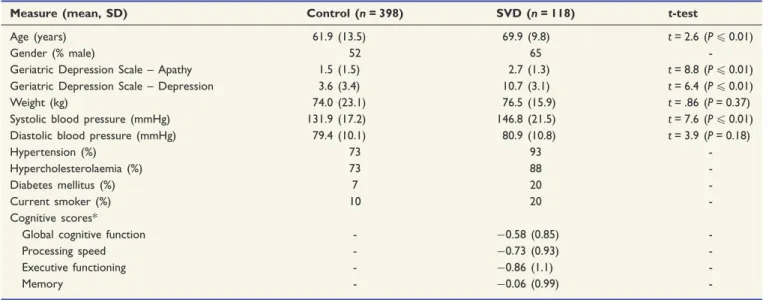
Table 1 Descriptive statistics of demographic and clinical variables for participants with SVD and controls
Measure (mean, SD) Control (n= 398) SVD (n= 118) t-test
Age (years) 61.9 (13.5) 69.9 (9.8) t= 2.6 (P40.01)
Gender (% male) 52 65
-Geriatric Depression Scale – Apathy 1.5 (1.5) 2.7 (1.3) t= 8.8 (P40.01)
Geriatric Depression Scale – Depression 3.6 (3.4) 10.7 (3.1) t= 6.4 (P40.01)
Weight (kg) 74.0 (23.1) 76.5 (15.9) t= .86 (P= 0.37)
Systolic blood pressure (mmHg) 131.9 (17.2) 146.8 (21.5) t= 7.6 (P40.01)
Diastolic blood pressure (mmHg) 79.4 (10.1) 80.9 (10.8) t= 3.9 (P= 0.18)
Hypertension (%) 73 93
-Hypercholesterolaemia (%) 73 88
-Diabetes mellitus (%) 7 20
-Current smoker (%) 10 20
-Cognitive scores*
Global cognitive function - 0.58 (0.85)
-Processing speed - 0.73 (0.93)
-Executive functioning - 0.86 (1.1)
-Memory - 0.06 (0.99)
Diffusion tensor imaging
Images were acquired from a 1.5 T General Electric Signa HDxt (General Electric), maximum gradient amplitude 33 mTm 1, using a proprietary head coil. All sequences had whole brain coverage and the total acquisition time was 45 min. Axial single shot spin echo planar images were acquired (repetition time/echo time = 15 600/93.4 ms, field of view = 240240 mm2, matrix = 9696, 55 slices of 2.5 mm with no slice gap). Eight volumes without diffusion weighting (b = 0 smm 2) were followed by 25 diffusion-sensitized images with gradients applied in non-collinear direc-tions (b = 1000 smm2). These acquisitions were repeated to obtain four more unweighted volumes and the negative of the 25 gradients.
Images were realigned to remove eddy current distortions using FMRIB’s Linear Image Registration Tool (FLIRT) (Jenkinson and Smith, 2001), part of the FMRIB Software Library (FSL) (Smithet al., 2004; Jenkinsonet al., 2012), ver-sion 5.0. Slices with signal loss due to motion were identified using an in-house intensity-based program and excluded from further analysis. Most diffusion tensor images (111/118) were identified as requiring some degree of correction by excluding diffusion-weighted image slices with excessive patient move-ment prior to computation of the DTI. In most cases slice exclusion was minimal (median 8.5/2750 slices, interquartile range = 14.75 slices). The maximum number of excluded slices was 133 over 50 diffusion-weighted image volumes (i.e. there are 55 slices per diffusion-weighted gradient direction image meaning that 4.83% of slices for that individual were excluded). All corrected and uncorrected diffusion tensor images were visually inspected prior to further analysis. Diffusion-weighted volumes with opposite gradients were geo-metrically averaged to eliminate gradient cross-terms (Neeman
et al., 1991). The eight b = 0 smm 2images were co-registered and the average taken to provide a T2-weighted echo planar image, which we term the ‘b0’ image.
A mask which excluded non-brain tissue was created from the b0 image using FSL’s Brain Extraction Tool (Smith, 2002). Masks were inspected and manually corrected where necessary.
Diffusion tensors were fitted using the least squares method (Basseret al., 1994) for each voxel of the brain mask by FSL’s ‘dtifit’ program. The tensor was decomposed to create maps of fractional anisotropy and mean diffusivity. When disease-related white matter changes are present you would expect to observe reduced fractional anisotropy, and increased mean diffusivity values. Prior to voxel-wise analysis the fractional anisotropy images underwent binary erosion with a 331 voxel kernel.
Voxel-based analysis
Analysis of fractional anisotropy and mean diffusivity maps was conducted using a voxel-based analysis methodology described by Schwarz and colleagues (2014). This combines the use of an advanced registration technique implemented in the Advanced Normalization Tools (ANTS) software (Klein
et al., 2009; Avantset al., 2011) with an iteratively generated unbiased group-template to allow valid voxel-wise compari-sons (Schwarzet al., 2014), similar to the voxel-based morph-ometry technique (VBM) (Ashburner and Friston, 2000).
To create a standard fractional anisotropy template with min-imal bias for use in the voxel-based analysis we adopted an iterative procedure with three stages. Linear registrations were implemented in FSL FLIRT using default settings (12 parameter affine transformation computed using the correlation ratio cost function). First, a representative subject fractional anisotropy image was linearly registered to the FMRIB58 fractional anisot-ropy template provided with FSL. All images were linearly regis-tered to this initial target and the mean average of these images used to create an initial group template. The template was updated with two further iterations of the above steps. The final mean average image was used to initialize a non-linear template generated using ANTS via the buildtemplateparallel.sh script (Avantset al., 2011). As a final step, a linear transform-ation was computed between the non-linear group template and the FMRIB58 template. The above transformations bringing fractional anisotropy and mean diffusivity maps in subject space to the FMRIB58 template analysis space (via the group template) were concatenated and the combined transform applied in a single step to minimize interpolation errors. Following previously used methodology, the voxel-based ana-lysis of all subject images were smoothed using a Gaussian kernel (= 1 mm) and analysis was restricted to voxels with an average fractional anisotropy 50.2 representing regions of mostly white matter (Schwarzet al., 2014).
In order to include whole white matter summary measures of fractional anisotropy and mean diffusivity in the structural equation model (see below) we extracted the median fractional anisotropy and mean diffusivity for all non-zero white matter voxels included within the voxel-based analysis mask using the ‘fslstats’ function in FSL.
Statistical analysis
We compared firstly the severity of apathy and depression be-tween the SVD cases and controls and then thresholded the scale using median splits to identify high and low scoring par-ticipants, allowing us to establish the degree of overlap be-tween apathy and depression in the group of patients with SVD. To confirm validity of the apathy subscale in our sample, we conducted a confirmatory factor analysis based on the six factor model (Adams et al., 2004).
Structural equation model
Structural equation modelling is a statistical technique which enables the testing of a theoretical model that includes a set of directional (regression) and non-directional relationships (covariances). This allows the quantification of the independ-ent contributions of predictor variables on outcome variables, while accounting for dependencies between them. Here we used multilevel structural equation modelling to investigate the relationship between white matter microstructure and apathy, depression, global cognitive impairment and the final outcome variable, quality of life; this was done within the SVD sample. However, we also wanted to identify any independent contributions of apathy, depression and global cognitive func-tioning on quality of life. Overall, this allows us to estimate the independent contributions of both apathy and depression on quality of life while taking into account relationships with white matter changes and any inter-relationships between the other observed variables, age and IQ included, within the co-variation matrix.
For the structural equation modelling analysis, key variables and covariates were included in an initial hypothesized model (Fig. 1) and then pathways were dropped in a systematic fash-ion to create alternate models for comparison, revealing the most parsimonious model (Supplementary material). Models were fitted to raw data using full information maximum like-lihood to account for data missing at random and alternative models were compared using chi-square likelihood ratio test of comparative model fit, comparative fit index (CFI), and root mean square error of approximation (RMSEA), along with a number of other parameters (Supplementary material). Structural equation modelling and factor analysis was per-formed in the statistical modelling software MPLUS version 5 (Muthe´n and Muthe´n, 2012).
Voxel-based analysis of diffusion images
To identify which regions of white matter may be specifically associated with either apathy or depression, statistical analysis was performed using the randomize program in FSL (Winkler
et al., 2014) with 10 000 permutations. A general linear model was used to test positive and negative linear associations between fractional anisotropy/mean diffusivity and (i) geriatric depression scale-apathy score; and (ii) geriatric depression scale-depression, controlling for global cognitive impairment age and gender (dummy coded) as covariates of no interest in a single model (a single general linear model was con-structed for fractional anisotropy and mean diffusivity separ-ately). Multiple comparisons were adjusted for using threshold-free cluster enhancement (TFCE; H = 2, E = 0.5, C = 6) and corrected P-values were thresholded at P40.01 and their spatial distribution compared with standard white matter atlases (Harvard-Oxford Cortical and Subcortical Atlas and Johns Hopkins University DTI-based white-matter atlases) (Mori et al., 2005; Hua et al., 2008).
Results
Rates of depression and apathy in
patients with small vessel disease and
controls
Demographic characteristics of the patients with SVD and control populations are shown in Table 1. The cases with SVD had higher apathy (0–6) and depression (0–24) scores than the controls; apathy mean [standard deviation (SD)] 2.7 (1.3) versus; 1.5 (1.5); P40.001; depression mean (SD) 10.7 (3.1) versus 3.6 (3.4) P40.001. Using a median cut-off of 3 for the apathy scale, and 10 for depres-sion, 62/120 (52%) of participants with SVD had high apathy and 67/120 (56%) had high depression. There was considerable dissociation between apathy and depres-sion, with 41/120 (34%) of the patients with SVD report-ing co-occurrreport-ing apathy and depression, but with an additional 47/120 (39%) experiencing either apathy or de-pression in isolation.
Confirmatory factor analysis of the
Geriatric Depression Scale
The confirmatory factor analysis was conducted in MPLUS. The literature has suggested a number of differ-ent factor structures for the Geriatric Depression Scale, with four factors being most common, with apathy/ social withdrawal consistently occurring as a single factor (Kim et al., 2012). Our results suggested a six factor structure, consistent with that found by Adams and colleagues (2004), with adequate model fit statistics [2(58) = 75, CFI = 0.96 RMSEA = 0.05]. Therefore, both depression (0–24) and apathy variables (0–6) were included in the structural equation model, as described in the ‘Materials and methods’ section.
Structural equation model of the
relationships between white matter
microstructure and clinical measures
in SVD
Our initial model had adequate model fit statistics [2(2) = 3.8, P= 0.15; CFI = 0.99; RMSEA = 0.08, 90% confidence interval (CI) = 0.0–0.22] and found that while reduction in white matter microstructure (median fractional anisotropy) was a significant predictor of greater apathy scores (standardized coefficient = 0.38, P40.001) and more cognitive impairment (standardized coefficient = 0.45,
P40.001), it was not significantly related to symptoms of depression (standardized coefficient = 0.16, P= 0.07). There was also a significant direct effect of median frac-tional anisotropy on quality of life (standardized coeffi-cient = 0.23, P= 0.01). The model also revealed that apathy (standardized coefficient = 0.22, P= 0.01) and de-pression (standardized coefficient = 0.39,P40.001) both
Figure 1 Hypothesized model examining the relationship between median fractional anisotropy/mean diffusivity, apathy, depression, cognitive impairment and quality of life. FA = fractional anisotropy; MD = median diffusivity. NART errors and age are regressed onto all independent variables.
independently predicted a poorer quality of life, while cog-nitive impairment (standardized coefficient = 0.12,P= 0.19) did not. Apathy, depression and cognitive impairment were all significantly correlated with each other (all P40.05).
We then constrained non-significant pathways to a value of zero in order to test a number of alternative nested models (Supplementary material). This resulted in a series of models with adequate model fit (Supplementary material) but with Model 3 having fit indices comparable to our hypothesized model, to which it was identical, but with the non-significant path from cognitive impairment to quality of life being con-strained to zero [2(3) = 5.4, P= 0.15; CFI = 0.98; RMSEA = 0.08, 90% CI = 0.0–0.22; Fig. 2]. A 2difference test revealed no significant difference between the hypothe-sized model and Model 3 [2(1) = 1.6,P= 0.21], and there-fore Model 3 was accepted as the most parsimonious.
We repeated the final model substituting the median fractional anisotropy value with median mean diffusivity, and found consistent results [2(2) = 3.6, P= 0.16; CFI = 0.99; RMSEA = 0.08, 90% CI = 0.0–0.22]. Again we found that apathy (standardized coefficient = 0.33, P40.001) and cognitive functioning (standardized coefficient = 0.37, P40.001), but not depression (standardized coefficient = 0.10, P= 0.52; Fig. 2), were significantly associated with median mean diffusivity (Fig. 2). The correlations between all the key variables included in the models can be found in Table 2.
Structural equation models of the
effect of cognitive domains on the
relationship between white matter
and apathy
To ensure the relationships between white matter changes, apathy and depression were not mediated by specific cognitive domains we conducted a post hoc ana-lysis using executive functioning, memory and processing
speed performance instead of global cognitive function-ing. The executive function [2 (2) = 2.9, P= 0.23; CFI = 0.99; RMSEA = 0.06, 90% CI = 0.0–0.20], long-term memory [2(2) = 2.7, P= 0.25; CFI = 0.99; RMSEA = 0.06, 90% CI = 0.0–0.20] and processing speed models [2(2) = 2.8, P= 0.25; CFI = 0.99; RMSEA = 0.06, 90% CI = 0.0–0.20] all had adequate model fit statistics, and reliably showed that regardless of the cognitive measure used, a reduced median frac-tional anisotropy significantly predicts more apathy, poorer cognitive function and a worse quality of life, but that depression was not significantly related to white matter changes (see Fig. 3 for full models). These models are consistent with the previous analyses, con-firming that the relationship between white matter changes and apathy is maintained when accounting indi-vidually for each of the following cognitive domains: ex-ecutive functioning, memory and processing speed. These models were repeated using the median mean diffusivity value and showed consistent results (Fig. 3).
White matter correlates of apathy
and depression
Consistent with the structural equation model, when controlling for depression, apathy was found to be signifi-cantly related to widespread changes in both fractional an-isotropy and mean diffusivity (Fig. 4). Fractional anisotropy and mean diffusivity associations with apathy were primarily in anterior brain regions, but also areas within the parietal and temporal lobes. This included sig-nificant results within the bilateral anterior cingulum, corpus callosum, fornix, uncinate/inferior fronto-occipital fasciculus and the anterior thalamic radiation, including portions of the anterior limbs of the external capsule. In contrast, when controlling for apathy, we found no signifi-cant relationship between our white matter parameters and symptoms of depression. The analysis was also repeated
Figure 2 Final model depicting significant pathways between median fractional anisotropy/median diffusivity and apathy, global cognition and quality of life in patients with SVD.FA = fractional anisotropy; MD = median diffusivity. NART errors and age are regressed onto all independent variables. Dark lines represent significant paths (P40.05), while grey lines are non-significant paths. Note: the non-significant path between Global Cognitive Impairment and Quality of Life was dropped to acquire best model fit. The values presented are standardizedbcoefficients.
Figure 3 Post hoc structural equation model in v estigating the possible mediating effect of ex ecutiv e functioning and memor y o n the relationship betw een apath y and median fractional anisotr op y. FA = fractional anisotr op y; MD = median diffusivity . NAR T err ors and age are regr essed onto all independent variables. Dark lines repr esent significa nt paths ( P 4 0.05), while gr e y lines are non-significant paths. Note: the non-significant path betw een Global Cognitiv e Impairment and Quality of life was dr opped to acquir e best model fit. The values pre sented are standard ized b coefficients.
using the whole Geriatric Depression Scale and again there were no significant findings. These findings strongly suggest the involvement of limbic-cortical-thalamic-striatal circuits in apathy in people with SVD. As the regions associated with apathy in this analysis have previously been associated with processing speed (Tuladhar et al., 2015), we repeated the analysis specifically controlling for this cognitive domain, finding consistent results (Supplementary material).
Discussion
In this study, we first determined the presence of apathy and depression in SVD compared to a large sample of healthy controls, and investigated the overlap between apathy and depression in SVD, before determining the re-lationship between white matter microstructure and apathy, depression and quality of life. Finally, we investigated re-gional associations between white matter microstructure and apathy to identify possible mechanisms that may underlie these symptoms.
These results demonstrate that both apathy and depres-sion are increased in patients with SVD, but that frequently they do not occur together in the same patient. We found that while 34% of patients with SVD displayed both symp-toms concurrently, 39% had dissociated sympsymp-toms of either depression or apathy. Of particular significance is our finding that 18% who are not depressed had high
levels of apathy as this may frequently be missed in the clinical setting.
Despite evidence that apathy is a common neuropsychi-atric symptom following stroke (Hackettet al., 2014), there are little data on its impact on patients’ quality of life. Previous studies in SVD have reported that both cognitive functioning (Brookes et al., 2014b) and depression (Brookes et al., 2013) following stroke are associated with a poorer quality of life. Here we demonstrate that both apathy and depression each have their own independ-ent contributions to the quality of life in SVD. In our model, cognitive impairment impacted indirectly on quality of life through its association with apathy and depression. This serves to highlight the importance of the assessment of emotional neuropsychiatric symptoms such as apathy and depression in patients with SVD. We also found a direct path between white matter damage and quality of life. This effect is most likely mediated by one or more factors not included in the modelling because of model size constraints, for example, physical disability or motor problems.
It has been suggested that the increased prevalence of apathy and depression in SVD may be caused by white matter damage to the cortico-subcortical pathways that connect brain regions important for regulating emotion (Taylor et al., 2013). DTI of these patients revealed that white matter microstructural changes in SVD are associated with increased apathy, but not depressive symptoms. Spatial analysis identified a number of subcortical regions
Figure 4 Areas of reduced fractional anisotropy/median diffusivity associated with apathy in patients with SVD, controlling for age gender and cognitive functioning.Images displayed using neurological convention. AC = anterior cingulum; ATR = Anterior Thalamic Radiation; CC = corpus callosum; FA = fractional anisotropy; IFOF = inferior fronto-occipital fasciculus; MD = mean diffusivity; PC = posterior cingulum; UF = uncinate fasciculus.
commonly thought to be affected by SVD pathology as underlying the association between white matter changes and apathy (Prins et al., 2005). This included the anterior cingulum, fornix, uncinate fasciculus, inferior frontal oc-cipital fasciculus and the body and genu of the corpus callosum.
The anterior cingulate cortex,
goal-directed behaviour and apathy
The cingulum is a large white matter tract underlying the cingulate cortex (Shackmanet al., 2011), which has been linked to both affect regulation and cognitive control, and has been associated with apathy in both structural (Apostolova et al., 2007; Stantonet al., 2013) and func-tional MRI studies (Marshall et al., 2007; Alexopoulos
et al., 2013). Consistent with our current findings, reduc-tions in cingulum white matter integrity have also been associated with apathy in patients with dementia and in normal ageing (Cacciari et al., 2010; Kim et al., 2011; Hahn et al., 2013; Spalletta et al., 2013). In particular, lesions, including strokes, involving the anterior cingulate cortex can lead to profound apathy often referred to as abulia (Cohen et al., 1999; Grunsfeld and Login, 2006). It has now been suggested that in conjunction with the ventral striatal reward systems and frontal regions im-portant for task initiation, the anterior cingulate cortex has a role in the selection and maintenance task perform-ance (Holroyd and Yeung, 2012). Given that, in addition to the cingulum, we also found significant associations between apathy and areas of both the internal and exter-nal capsules and diffuse regions of frontal white matter, one hypothesis is that apathy in SVD is caused by dis-ruption of the connections between frontal-striatal cir-cuits, including the anterior cingulate cortex, which select and initiate goal-directed behaviour based on pre-vious reward information.
The fornix, episodic memory and
apathy
There was a significant association between apathy and white matter structural changes to the fornix and the an-terior thalamic radiation. Reduction in fornix white matter integrity has typically been associated with episodic memory (Metzler-Baddeley et al., 2012). This suggests that an ability to access autobiographical information about times, places and their associated emotions or reward value may be important in apathy. However, our structural equation model revealed that performance on memory tasks was not related to apathy scores. It is pos-sible that the relationship between apathy and memory may be isolated to only affective or reward related memories, and future studies may be able to investigate this using experimental paradigms rather than more generic neuro-psychological tests.
The uncinate fasciculus, mnemonic
associations and apathy
Consistent with studies in Alzheimer’s disease (Hahnet al., 2013), progressive supranuclear palsy (Agostaet al., 2014) and normal ageing (Groolet al., 2014), we found a signifi-cant relationship between apathy and the uncinate fascic-ulus. The uncinate fasciculus is considered to be important for a range of cognitive functions such as episodic memory (Metzler-Baddeleyet al., 2012), linguistic ability (Papagno
et al., 2011) and social-emotional ability (Hornbergeret al., 2011). It has recently been demonstrated that apathy in those with late-life depression is significantly associated with fractional anisotropy changes in the left uncinate fas-ciculus and that reduced structural integrity in this region may mediate treatment response (Yuen et al., 2014).
There was also a significant association between apathy and the inferior fronto-occipital fasciculus, both at its
Table 2 Pearson’s correlations between key variables included in the structural equation model
Median FA
Median MD
Depression Apathy Cognitive function Processing speed Executive function Memory Quality of life Age Median FA Median MD 0.90** Depression (GDS 0–24) 0.12 0.02 Apathy (GDS 0–6) 0.38** 0.34** 0.51** Cognitive function 0.43** 0.37** 0.23* 0.32** Processing speed 0.23** 0.34** 0.10 0.20* 0.85** Executive function 0.23** 0.16# 0.02 0.01 0.28** 0.23* Memory 0.15 0.10 0.10 0.10 0.18* 0.18* 0.68** Quality of life 0.41** 0.38 0.65** 0.58** 0.35** 0.26* 0.11 0.01 Age 0.06 0.01 0.12 0.14 0.09 0.10 0.18* 0.10 0.05 NART 0.21* 0.15 0.14 0.06 0.66** 0.45** 0.10 0.13 0.03 0.14
GDS = Geriatric Depression Scale; FA = fractional anisotropy; MD = median diffusivity; NART = National Adult Reading Test; Cognitive function = global cognitive composite score;
#
anterior termination in the frontal operculum, but also more posteriorly (Fig. 3). The inferior fronto-occipital fas-ciculus projects from the frontal operculum and proceeds posteriorly passing though the insular to posterior temporal lobe and finally the occipital lobe (Martino et al., 2010). There is little evidence for a direct role for the inferior fronto-occipital fasciculus in apathy. However, Stanton and colleagues (2013) reported that a reduced insula volume, as well as atrophy in the cingulate region, was associated with high levels of apathy in patients with neu-rodegenerative disorders.
Apathy or depression in small
vessel disease?
Given that our results are not consistent with the previous finding in SVD that the white matter tracts implicated here have a role in depressive symptoms, it is essential to discuss why this may be. First, it is important to state that based on this evidence alone, we are not suggesting that people with SVD do not suffer from symptoms of depression. Rather, these findings suggest that depression in SVD may to some extent be secondary to motivational loss and cognitive impairment, which are the more direct con-sequence of white matter damage. Second, while our results indicate that there is no direct association between white matter integrity and depression, our data do suggest that depression still has a significant impact on functional out-comes in patients with SVD.
Our results show a high level of correspondence with the only other paper that has investigated regional associations between white matter and depression in SVD (van Uden
et al., 2015). In that study, depressive symptoms were mea-sured using the Centre for Epidemiologic Studies Depression Scale (CES-D; Radloff, 1977). The CES-D does contain some questions that tap into apathy, including questions such as ‘I could not get going’ and ‘I talked less than normal’, which may have driven the significant asso-ciations between depression and white matter microstruc-ture in this study.
Conclusions, limitations and future
directions
Based on the results of this study we concluded that apathy is a significant symptom in people with SVD and is asso-ciated with damage to white matter tracts which connect regions in the frontal lobe with both subcortical structures and the temporal lobe. Taking evidence from a wide range of literature we have inferred that apathy may be caused by damage to a number of networks responsible for the inte-gration of reward, mnemonic associations and the selection and initiation of goal-directed behaviour. Future research should now focus on probing these regions with behav-ioural and cognitive tasks designed to elicit reward-related behaviour in combination with functional MRI. Adam
and colleagues (2013) have shown that a reward-based decision-making task is sensitive to treatment of profound apathy with a dopamine agonist in a patient with bilateral basal–ganglia lesions as a result of ischaemic stroke. While Kohno et al. (2010) demonstrated the potential benefit of Ropinirole, another dopamine receptor agonist, to reduce apathy in a patient who had suffered an ischaemic lesion to the prefrontal cortex.
This is the first study to show specific relationships be-tween white matter pathology and apathy in people with SVD. We have also supported this by using structural equa-tion modelling to demonstrate that both apathy and depres-sion can be dissociated in SVD and have independent associations with quality of life. However, future research would benefit from taking a more detailed approach when assessing apathy. For instance, by using the Lille Apathy Rating Scale (Sockeel et al., 2006), which divides apathy into subdomains enabling the development of patient specific profiles, also determining whether the current re-sults can be replicated. Furthermore, although we carefully selected our voxel-based analysis methodology (Avants
et al., 2011) to reduce common problems with voxel-based methods (Ashburner and Friston, 2000; Bookstein, 2001), the localization of apathy within the white matter should be interpreted with caution and replicated in an independent sample of patients with SVD.
It is important to note that despite our findings that apathy is significantly related to white matter microstruc-ture in SVD, even when controlling for cognitive function-ing, some regions do overlap with those reported to be associated with cognitive functioning. This is particularly the case for processing speed, which has also been shown to be related to diffuse white matter changes particularly in frontal regions (Tuladhar et al., 2015).
This study has a number of clinical implications. Firstly, our findings that apathy and depression are dissociable in SVD, and that they both impact significantly on patient quality of life, suggests a more routine assessment of apathy is warranted. Secondly, an ability to accurately dis-tinguish between apathy and depression, whether they occur together or in isolation from each other, may allow the development of distinct treatment approaches for each. For instance, while antidepressant treatments such as sero-tonin re-uptake inhibitors (SSRIs) may be effective for symptoms of depression (Salzman et al., 2002), they may not be effective at treating apathy and its specific contribu-tion to a person’s funccontribu-tional outcome. While there is lim-ited literature on effective treatments for apathy, a number of dopaminergic compounds have been suggested (Kohno
et al., 2010; Rosenberget al., 2013; Thoboiset al., 2013). In addition, evidence suggests that the presence of symp-toms of apathy reduces the effectiveness of treatments for depression (Chaturvedi and Sarmukaddam, 1986; Ho¨ltta¨
et al., 2012). It is currently unclear whether this is only due to a reduced engagement with the treatment regime or if neurobiological differences between those who have both symptoms of apathy and depression versus those with
depression alone may play a part. A clear delineation of divergent properties of apathy and depression, both at the behavioural level and the neurobiological level, may allow the development of treatment plans for apathy itself or treatments for post-stroke depression that take into account the presence or absence of apathy.
Funding
The SCANS study was supported by a Wellcome Trust grant (081589). Recruitment to the SCANS study was sup-ported by the English National Institute of Health Research Clinical Stroke Research Network. Matthew Hollocks is supported by a Stroke Association/British Heart Foundation Project Grant (TSA/BHF Prog 2010/01).
Andrew Lawrence is supported by an Alzheimer’s
Research UK project grant (ARUK-PG2013-2). Masud Husain is supported by a Wellcome Trust Principal Research Fellowship. Hugh Markus is supported by an NIHR Senior Investigator award and by the Cambridge University Hospitals Department of Health’s NIHR Comprehensive Biomedical Research Centre.
Supplementary material
Supplementary material is available at Brainonline.References
Adam R, Leff A, Sinha N, Turner C, Bays P, Draganski B, et al. Dopamine reverses reward insensitivity in apathy following globus pallidus lesions. Cortex 2013; 49: 1292–303.
Adams KB, Matto HC, Sanders S. Confirmatory factor analysis of the geriatric depression scale. Gerontologist 2004; 44: 818–26. Agosta F, Galantucci S, Svetel M, Lukic´ MJ, Copetti M, Davidovic K,
et al. Clinical, cognitive, and behavioural correlates of white matter damage in progressive supranuclear palsy. J Neurol 2014; 261: 913–24.
Alexopoulos GS, Hoptman MJ, Yuen G, Kanellopoulos DK, Seirup J, Lim KO, et al. Functional connectivity in apathy of late-life depres-sion: a preliminary study. J Affect Disord 2013; 149: 398–405. Apostolova LG, Akopyan GG, Partiali N, Steiner CA, Dutton RA,
Hayashi KM, et al. Structural correlates of apathy in Alzheimer’s disease. Dement Geriatr Cogn Disord 2007; 24: 91–7.
Ashburner J, Friston KJ. Voxel-based morphometry—the methods. Neuroimage 2000; 11: 805–21.
Avants BB, Tustison NJ, Song G, Cook PA, Klein A, Gee JC. A re-producible evaluation of ANTs similarity metric performance in brain image registration. Neuroimage 2011; 54: 2033–44. Basser PJ, Mattiello J, LeBihan D. Estimation of the effective
self-diffusion tensor from the NMR spin echo. J Magn Reson B 1994; 103: 247–54.
Bookstein FL. ‘Voxel-based morphometry’ should not be used with imperfectly registered images. Neuroimage 2001; 14: 1454–62. Brookes RL, Hannesdottir K, Lawrence R, Morris RG, Markus HS.
Brief Memory and Executive Test: evaluation of a new screening test for cognitive impairment due to small vessel disease [Internet]. Age Ageing 2012; 41: 212–8.
Brookes RL, Herbert V, Lawrence AJ, Morris RG, Markus HS. Depression in small-vessel disease relates to white matter ultrastruc-tural damage, not disability. Neurology 2014a; 83: 1417–23. Brookes RL, Herbert V, Paul S, Hannesdottir K, Markus HS, Morris
RG. Executive dysfunction, awareness deficits and quality of life in patients with cerebral small vessel disease: a structural equation model. Neuropsychology 2014b; 28: 247–53.
Brookes RL, Hollocks MJ, Khan U, Morris RG, Markus HS. The Brief Memory and Executive Test (BMET) for detecting vascular cognitive impairment in small vessel disease: a validation study. BMC Med 2015; 13: 290.
Brookes RL, Willis T a, Patel B, Morris RG, Markus HS. Depressive symptoms as a predictor of quality of life in cerebral small vessel disease, acting independently of disability; a study in both sporadic small vessel disease and CADASIL. Int J Stroke 2013; 8: 510–7. Cacciari C, Moraschi M, Di Paola M, Cherubini A, Orfei MD, Giove
F, et al. White matter microstructure and apathy level in amnestic mild cognitive impairment. J Alzheimers Dis 2010; 20: 501–7. Chaturvedi SK, Sarmukaddam SB. Prediction of outcome in depression
by negative symptoms. Acta Psychiatr Scand 1986; 74: 183–6. Cohen RA, Kaplan RF, Zuffante P, Moser DJ, Jenkins MA, Salloway
S, et al. Alteration of intention and self-initiated action associated with bilateral anterior cingulotomy. J Neuropsychiatry Clin Neurosci 1999; 11: 444–53.
Fazekas F, Chawluk JB, Zimmerma A, June M. MR signal abnorm-alities at 1.5 T in Alzheimer ’s dementia and normal aging defi-ciency. AJR Am J Roentgenol 1987; 149: 351–6.
Grool AM, Geerlings MI, Sigurdsson S, Eiriksdottir G, Jonsson PV., Garcia ME, et al. Structural MRI correlates of apathy symptoms in older persons without dementia: AGES-Reykjavik Study. Neurology 2014; 82: 1628–35.
Grunsfeld AA, Login IS. Abulia following penetrating brain injury during endoscopic sinus surgery with disruption of the anterior cin-gulate circuit: case report. BMC Neurol 2006; 6: 4.
Hackett ML, Ko¨hler S, O’Brien JT, Mead GE. Neuropsychiatric out-comes of stroke. Lancet Neurol 2014; 13: 525–34.
Hahn C, Lim HK, Won WY, Ahn KJ, Jung WS, Lee CU. Apathy and white matter integrity in Alzheimer’s disease: a whole brain analysis with tract-based spatial statistics. PLoS One 2013; 8
Holroyd CB, Yeung N. Motivation of extended behaviors by anterior cingulate cortex. Trends Cogn Sci 2012; 16: 122–8.
Ho¨ltta¨ EH, Laakkonen ML, Laurila J V., Strandberg TE, Tilvis RS, Pitka¨la¨ KH. Apathy: prevalence, associated factors, and prognostic value among frail, older inpatients. J Am Med Dir Assoc 2012; 13: 541–5.
Hornberger M, Geng J, Hodges JR. Convergent grey and white matter evidence of orbitofrontal cortex changes related to disinhibition in behavioural variant frontotemporal dementia. Brain 2011; 134: 2502–12.
Hua K, Zhang J, Wakana S, Jiang H, Li X, Reich DS, et al. Tract probability maps in stereotaxic spaces: analyses of white matter anatomy and tract-specific quantification. Neuroimage 2008; 39: 336–47.
Jenkinson M, Beckmann CF, Behrens TEJ, Woolrich MW, Smith SM. FSL. Neuroimage 2012; 62: 782–90.
Jenkinson M, Smith S. A global optimisation method for robust affine registration of brain images. Med Image Anal 2001; 5: 143–56. Kim G, DeCoster J, Huang C-H, Bryant AN. A meta-analysis of the
factor structure of the Geriatric Depression Scale (GDS): the effects of language. Int Psychogeriatr 2012: 1–11.
Kim HJ, Kang SJ, Kim C, Kim GH, Jeon S, Lee JM, et al. The effects of small vessel disease and amyloid burden on neuropsychiatric symptoms: a study among patients with subcortical vascular cogni-tive impairments. Neurobiol Aging 2013; 34: 1913–20.
Kim JW, Lee DY, Choo IH, Seo EH, Kim SG, Park SY, et al. Microstructural alteration of the anterior cingulum is associated with apathy in Alzheimer disease. Am J Geriatr Psychiatry 2011; 19: 644–53.
Kirsch-Darrow L, Marsiske M, Okun MS, Bauer R, Bowers D. Apathy and depression: separate factors in Parkinson’s disease. J Int Neuropsychol Soc 2011; 17: 1058–66.
Klein A, Andersson J, Ardekani BA, Ashburner J, Avants B, Chiang M-C, et al. Evaluation of 14 nonlinear deformation algorithms applied to human brain MRI registration. Neuroimage 2009; 46: 786–802.
Kohno N, Abe S, Toyoda G, Oguro H, Bokura H, Yamaguchi S. Successful treatment of post-stroke apathy by the dopamine receptor agonist ropinirole. J Clin Neurosci 2010; 17: 804–6.
Landes AM, Sperry SD, Strauss ME. Prevalence of apathy, dysphoria, and depression in relation to dementia severity in Alzheimer’s dis-ease. J Neuropsychiatry Clin Neurosci 2005; 17: 342–9.
Lawrence AJ, Patel B, Morris RG, MacKinnon AD, Rich PM, Barrick TR, et al. Mechanisms of cognitive impairment in cerebral small vessel disease: multimodal MRI results from the St George’s cognition and neuroimaging in stroke (SCANS) study. PLoS One 2013; 8: e61014. Levy R, Czernecki V. Apathy and the basal ganglia. J Neurol 2006; 253 Levy R, Dubois B. Apathy and the functional anatomy of the prefrontal
cortex-basal ganglia circuits. Cereb Cortex 2006; 16: 916–28. Makin SDJ, Turpin S, Dennis MS, Wardlaw JM. Cognitive
impair-ment after lacunar stroke: systematic review and meta-analysis of incidence, prevalence and comparison with other stroke subtypes. J Neurol Neurosurg Psychiatry 2013; 84: 893–900.
Marin RS, Biedrzycki RC, Firinciogullari S. Reliability and validity of the apathy evaluation scale. Psychiatry Res 1991; 38: 143–62. Marin RS, Firinciogullari S, Biedrzycki RC. The sources of
conver-gence between measures of apathy and depression. J Affect Disord 1993; 28: 7–14.
Marshall GA, Monserratt L, Harwood D, Mandelkern M, Cummings JL, Sultzer DL. Positron emission tomography metabolic correlates of apathy in Alzheimer disease. Arch Neurol 2007; 64: 1015–20. Martino J, Brogna C, Robles SG, Vergani F, Duffau H. Anatomic
dissection of the inferior fronto-occipital fasciculus revisited in the lights of brain stimulation data. Cortex 2010; 46: 691–9. Metzler-Baddeley C, Hunt S, Jones DK, Leemans A, Aggleton JP,
O’Sullivan MJ. Temporal association tracts and the breakdown of episodic memory in mild cognitive impairment. Neurology 2012; 79: 2233–40.
Mitchell AJ, Bird V, Rizzo M, Meader N. Diagnostic validity and added value of the Geriatric Depression Scale for depression in pri-mary care: a meta-analysis of GDS30 and GDS15. J Affect Disord 2010; 125: 10–17.
Moretti R, Cavressi M, Tomietto P. Gait and apathy as relevant symp-toms of subcortical vascular dementia. Am J Alzheimers Dis Other Demen 2015; 30: 390–9.
Mori S, Wakana S, Nagae-Poetscher LM, Van Zijl PCM. MRI atlas of human white matter. Amsterdam, The Netherlands: Elsevier; 2005. Muthe´n LK, Muthe´n BO. Mplus user’s guide. Los Angeles, CA:
Muthe´n & Muthe´n; 2012.
Neeman M, Freyer JP, Sillerud LO. A simple method for obtaining cross-term-free images for diffusion anisotropy studies in NMR microimaging. Magn Reson Med 1991; 21: 138–43.
O’Brien JT, Firbank MJ, Krishnan MS, van Straaten ECW, van der Flier WM, Petrovic K, et al. White matter hyperintensities rather than lacunar infarcts are associated with depressive symptoms in older people: the LADIS study. Am J Geriatr Psychiatry 2006; 14: 834–41.
Papagno C, Miracapillo C, Casarotti A, Romero Lauro LJ, Castellano A, Falini A, et al. What is the role of the uncinate fasciculus? Surgical removal and proper name retrieval. Brain 2011; 134: 405–14. Prins ND, Van Dijk EJ, Den Heijer T, Vermeer SE, Jolles J, Koudstaal
PJ, et al. Cerebral small-vessel disease and decline in information processing speed, executive function and memory. Brain 2005; 128: 2034–41.
Radloff LS. The CES-D scale: a self-report depression scale for re-search in the general population. Appl Psychol Meas 1977; 1: 385–401.
Robert P, Onyike CU, Leentjens AFG, Dujardin K, Aalten P, Starkstein S, et al. Proposed diagnostic criteria for apathy in Alzheimer’s disease and other neuropsychiatric disorders. Eur Psychiatry 2009; 24: 98–104.
Rosenberg PB, Lanctoˆt KL, Drye LT, Herrmann N, Scherer RW, Bachman DL, et al. Safety and efficacy of methylphenidate for apathy in Alzheimer’s disease: a randomized, placebo-controlled trial. J Clin Psychiatry 2013; 74: 810–6.
Salzman C, Wong E, Wright BC. Drug and ECT treatment of depres-sion in the elderly, 1996-2001: a literature review. Biol Psychiatry 2002. p. 265–84.
Schwarz CG, Reid RI, Gunter JL, Senjem ML, Przybelski SA, Zuk SM, et al. Improved DTI registration allows voxel-based analysis that outperforms tract-based spatial statistics. Neuroimage 2014; 94: 65–78.
Shackman AJ, Salomons TV, Slagter HA, Fox AS, Winter JJ, Davidson RJ. The integration of negative affect, pain and cognitive control in the cingulate cortex. Nat Rev Neurosci 2011; 12: 154–67. Smith EE, Schneider JA, Wardlaw JM, Greenberg SM. Cerebral
micro-infarcts: the invisible lesions. Lancet Neurol 2012; 11: 272–82. Smith SM. Fast robust automated brain extraction. Hum Brain Mapp
2002; 17: 143–55.
Smith SM, Jenkinson M, Woolrich MW, Beckmann CF, Behrens TEJ, Johansen-Berg H, et al. Advances in functional and structural MR image analysis and implementation as FSL. Neuroimage 2004; 23 (Suppl 1): S208–19.
Sneed JR, Rindskopf D, Steffens DC, Krishnan KRR, Roose SP. The vascular depression subtype: evidence of internal validity. Biol Psychiatry 2008; 64: 491–7.
Sockeel P, Dujardin K, Devos D, Dene`ve C, Deste´e A, Defebvre L. The Lille apathy rating scale (LARS), a new instrument for detecting and quantifying apathy: validation in Parkinson’s disease. J Neurol Neurosurg Psychiatry 2006; 77: 579–84.
Spalletta G, Fagioli S, Caltagirone C, Piras F. Brain microstructure of subclinical apathy phenomenology in healthy individuals. Hum Brain Mapp 2013; 34: 3193–203.
Stanton BR, Leigh PN, Howard RJ, Barker GJ, Brown RG. Behavioural and emotional symptoms of apathy are associated with distinct patterns of brain atrophy in neurodegenerative dis-orders. J Neurol 2013; 260: 2481–90.
Starkstein SE, Ingram L, Garau ML, Mizrahi R. On the overlap be-tween apathy and depression in dementia. J Neurol Neurosurg Psychiatry 2005; 76: 1070–4.
Tang WK, Chen YK, Lu JY, Chu WCW, Mok VCT, Ungvari GS, et al. White matter hyperintensities in post-stroke depression: a case control study. J Neurol Neurosurg Psychiatry 2010; 81: 1312–15.
Taylor WD, Aizenstein HJ, Alexopoulos GS. The vascular depression hypothesis: mechanisms linking vascular disease with depression [Internet]. Mol Psychiatry 2013; 18: 963–74.
Thobois S, Lhomme´e E, Klinger H, Ardouin C, Schmitt E, Bichon A, et al. Parkinsonian apathy responds to dopaminergic stimulation of D2/D3 receptors with piribedil. Brain 2013; 136: 1568–77. Tuladhar AM, van Norden AGW, de Laat KF, Zwiers MP, van Dijk
EJ, Norris DG, et al. White matter integrity in small vessel disease is related to cognition [Internet]. Neuroimage Clin 2015; 7: 518–24. Uher R, Payne JL, Pavlova B, Perlis RH. Major depressive disorder in
DSM-5: implications for clinical practice and research of changes from DSM-IV. Depress Anxiety 2013; 31: 459–71.
van der Mast RC, Vinkers DJ, Stek ML, Bek MC, Westendorp RGJ, Gussekloo J, et al. Vascular disease and apathy in old age. The Leiden 85-plus Study. Int J Geriatr Psychiatry 2008; 23: 266–71. van Norden AGW, De Laat KF, Van Dijk EJ, van Uden IWM, Van
Oudheusden LJB, Gons RAR, et al. Diffusion tensor imaging and cognition in cerebral small vessel disease. The RUN DMC study. Biochim Biophys Acta Mol Basis Dis 2012; 1822: 401–7. van Uden IWM, Tuladhar AM, de Laat KF, van Norden AGW, Norris
symptoms in cerebral small vessel disease: The RUN DMC study. Am J Geriatr Psychiatry 2015; 23: 525–35.
Wen MC, Steffens DC, Chen MK, Zainal NH. Diffusion tensor ima-ging studies in late-life depression: systematic review and meta-analysis [Internet]. Int J Geriatr Psychiatry 2014; 29: 1173–84. White CL, McClure LA, Wallace PM, Braimah J, Liskay A, Roldan A,
et al. The correlates and course of depression in patients with lacu-nar stroke: results from the secondary prevention of small subcor-tical strokes (SPS3) study. Cerebrovasc Dis 2011; 32: 354–60. Williams LS, Weinberger M, Harris LE, Clark DO, Biller J.
Development of a stroke-specific quality of life scale. Stroke 1999; 30: 1362–9.
Winkler AM, Ridgway GR, Webster MA, Smith SM, Nichols TE. Permutation inference for the general linear model. Neuroimage 2014; 92: 381–97.
Yesavage JA, Brink TL, Rose TL, Lum O, Huang V, Adey M, et al. Development and validation of a geriatric depression screening scale: a preliminary report [Internet]. J Psychiatr Res 1983; 17: 37–49. Yuen GS, Gunning FM, Woods E, Klimstra SA, Hoptman MJ,
Alexopoulos GS. Neuroanatomical correlates of apathy in late-life depression and antidepressant treatment response. J Affect Disord 2014; 166: 179–86.