R E S E A R C H
Open Access
Failure rates of mini-implants placed in the
infrazygomatic region
Flavio Uribe
1*, Rana Mehr
2, Ajay Mathur
3, Nandakumar Janakiraman
4and Veerasathpurush Allareddy
5Abstract
Background:The purpose of this pilot study was to evaluate the failure rates of mini-implants placed in the infrazygomatic region and to evaluate factors that affect their stability.
Methods:A retrospective cohort study of 30 consecutive patients (55 mini-implants) who had infrazygomatic mini-implants at a University Clinic were evaluated for failure rates. Patient, mini-implant, orthodontic, surgical, and mini-implant maintenance factors were evaluated by univariate logistic regression models for association to failure rates.
Results:A 21.8 % failure rate of mini-implants placed in the infazygomatic region was observed. None of the predictor variables were significantly associated with higher or lower odds for failed implants.
Conclusions:Failure rates for infrazygomatic mini-implants were slightly higher than those reported in other maxilla-mandibular osseous locations. No predictor variables were found to be associated to the failure rates.
Keywords:Infrazygomatic mini-implants; Failure rate; Stability
Background
Mini-implants have become a tool to address anchorage needs in the modern orthodontic practice. They have been widely utilized for anchorage reinforcement and placed within and outside of the dentoalveolar region. Possible insertion sites for mini-implants in the maxilla include the area below the nasal spine [1], the palate [2], the alveolar process [3–5], and the infrazygomatic (IZ) crest [6]. Insertion sites other than the alveolar process allow for more versatility of orthodontic tooth movements since the roots do not interfere with tooth displacement. Specifically, the IZ crest of the maxilla is one of these anatomical sites distant from the dentoal-veolar region, which allows unobstructed tooth move-ment, decreasing the chance of root contact.
The IZ region has important osseous characteristics such as the presence of thicker cortical bone, which al-lows good primary stability [7]. In fact, this region in partially edentulous patients is considered to have the best bone quality in the maxilla [8]. IZ mini-implants
have been successfully used to provide skeletal anchor-age for en-masse anterior retraction and intrusion of the maxillary posterior teeth [6, 9, 10].
The success and failure rate of mini-implants have been studied extensively, especially for mini-implants placed in tooth-bearing regions. Success has been de-fined when mini-implants are maintained in bone until the end of treatment or intentional removal, regardless of [11]. On the other hand, failure is considered as se-vere clinical mobility of a mini-implant that results in its inability to act as a stationary anchor, which requires removal or replacement, or loss of a mini-implant less than 8 months after placement [12–14]. Factors affect-ing the success and failure rate of mini-implants have been divided into different categories. These categories are patient, mini-implant, orthodontic, surgical, and mini-implant maintenance factors [11, 12, 15, 16].
There is lack of evidence in the literature specifically investigating the failure rate of mini-implants placed in the IZ region. Therefore, the objective of this study was to evaluate failure rates of mini-implants placed in the IZ crest of the maxilla and investigate the factors affect-ing this unfavorable outcome.
* Correspondence:[email protected]
1Division of Orthodontics, Department of Craniofacial Sciences, Charles
Burstone Professor, University of Connecticut School of Dental Medicine, 263 Farmington Avenue, Farmington, CT 06030, USA
Full list of author information is available at the end of the article
Methods
A retrospective pilot chart review of patients that had received IZ mini-implants for orthodontic treatment from July 2007 to November 2013 was conducted at the University of Connecticut after IRB approval (IRB 13-146-3). Inclusion criteria were all patients that had received a mini-implant placed in the IZ region for use as temporary anchorage device and that had complete records. The database of the orthodontic clinic was used to search for these patients. Exclusion criteria included patients with chart notes that did not record status of the mini-implants throughout treatment. In the database search for patients that had received infrazygomatic mini-implants in our in-stitution during the specific time period, we found a total of 40 subjects. Of these, 10 subjects were excluded from the study due to lack of complete records such as incom-plete chart notes or photographs not taken at 3 months interval in the digital record.
Chart notes and photographic images of the digital charts of the patients were analyzed to evaluate the dependent and independent variables. The primary out-come was mini-implant failure. Independent variables associated to mini-implant success were as follows: pa-tient-, implant-, orthodontic-, surgical-, and mini-implant maintenance-related factors. These independent variables were evaluated as predictors of mini-implant failure.
Data from a total of 30 consecutive patients (mean age 22.2 ± 11 years) who had 55 IZ mini-implants placed and met the inclusion criteria was collected (Table 1). Four different types of mini-implants [Lomas (Mondeal, Tuttligen, Germany), Imtec (Unitek 3M, Monrovia, California), Aarhus (Medicon, Tuttligen, Germany), Dual Top (RMO, Denver, Colorado)] were used and analyzed. The brand selection for each patient was based on the availability of the mini-implant system and mini-implant in the clinic at the time of placement. The surgical pro-cedure included local anesthesia followed by a small tissue punch. A pilot hole was placed with a manual driver for 22 of the 55 mini-implants. The same manual driver was used for the pilot hole regardless of the brand of the mini-implant. Placement of the mini-implants was performed with the specific driver designed for that par-ticular system by the manufacturer. The mini-implants were placed by two types of operators, an experienced clinician (more than 50 mini-implants placed, FU) and by residents under direct supervision of this operator. Nine orthodontic residents placed the mini-implants during this time period, all of whom had minimal ex-perience in implant placement (less than 10 mini-implants placed). All mini-mini-implants were placed at an approximate angle of 40° to 70° to maxillary occlusal plane in the IZ area by palpating the “key ridge” above the first permanent molar (Fig. 1) [7, 10].
After placement, initial stability of the mini-implant was checked by the operator who ensured there were no signs of mobility. The mini-implants were used for the retraction, distalization, and intrusion purposes. Com-bination of these movements such as intrusion– retrac-tion and intrusion–distalization was also recorded. The mini-implants were loaded with these types of forces for an average of 13.67 ± 6.79 months. The primary outcome variable of interest was implant failure of the mini-implants.
Failure was defined as a mini-implant that had to be removed or had fallen out after placement. The effect of oral hygiene on survival of the mini-implants was assessed. The sample was divided into three groups de-pending on the patients’ oral hygiene: good, fair, or poor, and was determined based on the photographic images and notes from the record. Vertical facial pat-tern was evaluated based on the mandibular plane angle (MPA) and Frankfurt/mandibular plane angle (FMA).
Statistical analysis
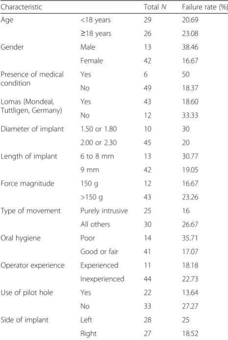
The outcome of interest was“failure”of implants. This was modeled as a binomial variable (Failure—yes/no). Simple Table 1Failure rates by characteristics of patients
Characteristic TotalN Failure rate (%)
Age <18 years 29 20.69
≥18 years 26 23.08
Gender Male 13 38.46
Female 42 16.67
Presence of medical condition
Yes 6 50
No 49 18.37
Lomas (Mondeal, Tuttligen, Germany)
Yes 43 18.60
No 12 33.33
Diameter of implant 1.50 or 1.80 10 30
2.00 or 2.30 45 20
Length of implant 6 to 8 mm 13 30.77
9 mm 42 19.05
Force magnitude 150 g 12 16.67
>150 g 43 23.26
Type of movement Purely intrusive 25 16
All others 30 26.67
Oral hygiene Poor 14 35.71
Good or fair 41 17.07
Operator experience Experienced 11 18.18
Inexperienced 44 22.73
Use of pilot hole Yes 22 13.64
No 33 27.27
Side of implant Left 28 25
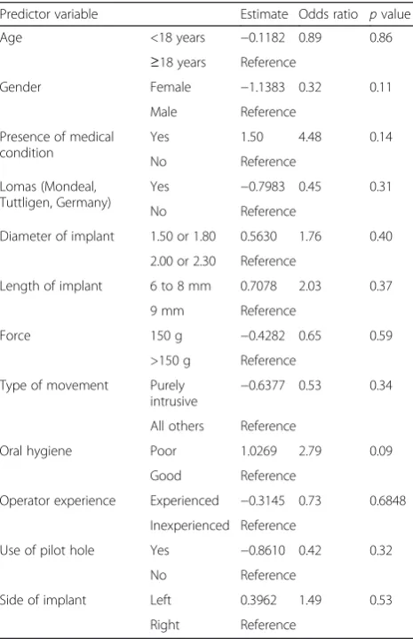
descriptive statistics were used to summarize the esti-mates of failure rates across different levels of predictor variables. The predictor variables examined included age, gender, presence of medical conditions, mini-implant brand (Lomas versus others), diameter of implant, length of implant, amount of force used, type of movement, oral hygiene, operator experience, use of pilot hole, and side of infrazygomatic implant. Univariate logistic regression analyses were used to examine the independent associ-ation of each predictor variable with the outcome (fail-ure of implants). The regression models were fit using Generalized Estimating Equations method. The effect of clustering of outcomes within patients was adjusted in the models. An exchangeable correlation matrix was specified. Odds ratio for each characteristic that resulted in a failed mini-implant was calculated. Each individual implant was the unit of analysis. All tests were two sided and apvalue of <0.05 was deemed to be statistically sig-nificant. All statistical analyses were conducted using SPSS Version 22.0 (IBM Corp, New York City, NY) and SAS Version 9.3 (SAS Institute, Cary, NC).
Results
Table 1 describes all the patient-, implant-, orthodon-tic-, surgical-, and implant maintenance-related charac-teristics of the patient sample and mini-implants placed. Of the 30 patients (55 mini-implants) that received IZ mini-implants, majority were female, with no significant medical condition. Approximately 80 % of the mini-implants were Lomas (Mondeal, Tuttligen, Germany) with a 2 × 9 mm dimension. All mini-implants were placed in unattached gingiva almost evenly distributed between left and right sides and immediately loaded. Most patients maintained good or fair oral hygiene and no infection
developed around any of the mini-implants. A loading force of 200 g for intrusion of posterior teeth purposes was used on the majority of mini-implants. The average MPA and FMA angles were 39.9° ± 6.60° and 31.3° ± 6.36°, respectively.
Over the course of treatment, 21.8° % of the mini-implants failed. Overall, failure rates were higher among those aged≥18 years, males, with medical conditions, use of non-Lomas implants, implants with length of 6 to 8 mm (compared to 9 mm), implants with 1.5/1.8 mm diameter (compared to 2 or 2.3 mm diameter), use of force greater than 150 g, with poor oral hygiene, when placed by inex-perienced operators, and left-sided implants. Purely intru-sive movements had lower failure rates when compared to all other combinations of movements. Failure rates were lower when pilot holes were used. The results of the uni-variate logistic regression analyses are summarized in Table 2. The estimates from the regression models indi-cated that none of the predictor variables were significantly associated with higher or lower odds for failed implants. Fig. 1Mini-implant placed in the IZ region of the maxilla
Table 2Estimates from univariate regression analysis (independent association between predictor variables and failed implants)
Predictor variable Estimate Odds ratio pvalue
Age <18 years −0.1182 0.89 0.86
≥18 years Reference
Gender Female −1.1383 0.32 0.11
Male Reference
Presence of medical condition
Yes 1.50 4.48 0.14
No Reference
Lomas (Mondeal, Tuttligen, Germany)
Yes −0.7983 0.45 0.31
No Reference
Diameter of implant 1.50 or 1.80 0.5630 1.76 0.40
2.00 or 2.30 Reference
Length of implant 6 to 8 mm 0.7078 2.03 0.37
9 mm Reference
Force 150 g −0.4282 0.65 0.59
>150 g Reference
Type of movement Purely
intrusive −
0.6377 0.53 0.34
All others Reference
Oral hygiene Poor 1.0269 2.79 0.09
Good Reference
Operator experience Experienced −0.3145 0.73 0.6848
Inexperienced Reference
Use of pilot hole Yes −0.8610 0.42 0.32
No Reference
Side of implant Left 0.3962 1.49 0.53
Discussion
A recent meta-analysis reported that the average overall success rate of mini-implants to be approximately 86 % [14]. This analysis included studies for mini-implants placed in different maxillomandibular locations. How-ever, the vast majority of the studies reporting on mini-implant failure rate have predominantly focused on those placed in interradicular sites [3, 4, 11, 17, 18]. The findings of our study show that IZ mini-implants have slightly lower success rate (78.2 %) than that of the average mini-implant. This is in contrast to Liou et al.’s [6] findings who reported 100 % success of mini-implants placed in this region.
The reason for the different results in success rates with our study may be attributed to the size of the mini-implants. In their study, the length of the mini-implants was 17 mm. Additionally, the success rate on that study was based on a limited time period of 9 months com-pared to our study where mini-implants were loaded for an average of 13 months. Furthermore, mini-implant mobility, recorded as displacement, was reported in Liou’s study in 44 % of the patients. Thus, failure could have been evidenced at a later time point for these pa-tients. Finally, although the mini-implants in our study were either placed by an experienced operator, or super-vised by an experienced operator who had placed more than 50 mini-implants, our experience in placement of the IZ mini-implants developed through the duration of the study. It is possible that the perfect success rate reported in Liou’s study might be related to experienced operators with more than 50 mini-implants placed in this specific region.
One important variable for the different success rates of mini-implants is skeletal facial pattern. Moon et al. [19] found similar success rates (77 %) to those of our study for mini-implants placed interdentally in patients with high Frankfurt-mandibular plane angle (FMA). This skeletal type was prevalent in the majority of our patients where the average FMA and mandibular plane angle (MPA) was 31.3° and 39.9°, respectively. This finding is also in agreement with a study by Miyawaki et al. [3] who also reported that mini-implants placed in patients with high MPA had lower success rates (72.7 %). Indeed, it has been found that patients with an increased vertical skeletal pattern have reduced cor-tical bone thickness, which may affect primary stability of the mini-implants [20]. However, it is unknown if this reduced cortical bone thickness is also present in the infrazygomatic region.
An evaluation of patient-, mini-implant-, orthodon-tic-, surgical-, and mini-implant maintenance-related factors that could affect the stability of mini-implant was performed. Among all these factors, none were as-sociated with greater odds of failure. Poor oral hygiene
showed a trend to be associated to failure rates. Al-though this is an expected finding, there is controversy of the role of oral hygiene in mini-implant failure. Sharma et al. [21] reported that poor oral hygiene and inflammation were associated to mini-implant failure. On the other hand, Park et al. [11] found that oral hy-giene played no role, but local inflammation around the mini-implants did.
Perhaps the type of mucosa surrounding the mini-implant may play a more important role in the inflamma-tory reaction and thus the success of the mini-implant. It has been reported that nonkeratanized gingiva may be a risk factor for mini-implant failure. Viwattanatipa et al. [22] found low survival rates of mini-implants placed in the infrazygomatic region or vestibular area (46 % after 1 year). In this study, all the mini-implants were placed on nonkeratanized tissue which could be less resistant to the effects of plaque and thus compromise mini-implant sta-bility. Possibly a longer mini-implant that approximates the attached gingiva may reduce the potential for the development of an inflammatory process.
Although there were some mini-implants that became mobile, some of these did not fail. This is consistent with the findings of Liou et al. [6] who specifically eval-uated IZ mini-implants and found this type of screws have some degree of mobility without failure. However, we observed that mobility appeared to be closely re-lated to failure.
One surprising finding was the fact that operator ex-perience was unrelated to mini-implant failure. Since this type of mini-implant placement has more tech-nical difficulty, it was expected that non-experienced operators would have more failures. This nonsignifi-cant finding in the regression analysis may be related to the fact that these mini-implants, although placed by residents, were still supervised by the experienced operator.
One important factor that could contribute to the failure rate is the angle of placement and the direction of loading force in mini-implants placed in the IZ re-gion. In fact, Perillo et al. [23] found in a recent study using a finite element analysis that the insertion angle of the mini-implant and the direction of force have a significant influence in the stress on the bone. This par-ameter was not evaluated in the present study, as it would have needed to be examined in a prospective na-ture. Moreover, recording the direction of the force vector may be difficult as it may vary as treatment pro-gresses based on the biomechanical needs.
clinic database may have not accounted patients where the IZ mini-implant was placed and removed immediately due to inadequate primary stability, thus underestimating the true failure rate. Although there is a possibility for this, based on the authors’experience, inadequate primary sta-bility of the mini-implants in this IZ region has rarely been observed. Regardless of these limitations, this study provided data for expected success rates in mini-implants placed in the IZ region, which from a biomechanical per-spective, provide significant versatility for orthodontic tooth movements difficult to achieve from anchorage drawn from mini-implants placed in interradicular sites.
The present study was designed to be a pilot explora-tive study of a multitude of patient- and provider-related factors associated with failure of IZ mini-implants. A multitude of patient- and provider-related factors could influence outcomes (in this case—failure of IZ mini-implants) and the precise role of each variable on the outcome is difficult to elucidate with a small sample size. This is particularly true when there are variations in the distribution of covariates. The current study was designed to be a pilot project and we identified a mix of patient-related factors that are associated with IZ mini-implant failures. We intend to use results from the present study to design a future prospective study to identify factors associated with failure of IZ implants. Our study included 30 patients (55 mini-implants). These patients were selected based on a chart review over a 6-year time period. Our unit of analysis was each individual mini-implant. In effect, our sample size was 55. This number is still inadequate and the present study may be underpowered considering the number of variables we included in the regression models. It is difficult to increase the sample sizes for such single center studies owing to the fact that very few patients elect to have IZ mini-implants and rela-tively few number of orthodontists place the IZ mini-implants. The solution will be to increase sample sizes by conducting multi-center studies where we can cap-ture an adequate number of patients that are also het-erogeneous in terms of covariate distribution. The present study results will aid in designing better con-trolled multi-center prospective studies. Therein lies the importance of the present study.
Conclusions
– Mini-implants placed in the IZ region had a 21.8 % failure rate. This failure rate is slightly higher than that reported for mini-implants placed
interradicularly.
– Patient, mini-implant, orthodontic, surgical, and mini-implant maintenance factors were not predictive of failure rates.
Competing interests
The authors declare that they have no competing interests.
Authors’contributions
FU is the principal investigator who designed study, analyzed data, and wrote manuscript. RM is the study coordinator in the initial phases of the study who obtained IRB approval for the study and initiated data collection. AM is the study coordinator for the majority of the study who gathered, compiled, and analyzed data and did the initial draft of the manuscript. NJ collaborated in compiling and analyzing data and contributed to writing the manuscript. VA did the statistical analysis prior and after data was collected. All authors read and approved the final manuscript.
Author details
1
Division of Orthodontics, Department of Craniofacial Sciences, Charles Burstone Professor, University of Connecticut School of Dental Medicine, 263 Farmington Avenue, Farmington, CT 06030, USA.2Private Practice, Houston, TX, USA.3Private Practice, Mumbai, India.4Division of Orthodontics, Department of Craniofacial Sciences, University of Connecticut School of Dental Medicine, Farmington, CT, USA.5Department of Orthodontics, College of Dentistry, The University of Iowa, Iowa, IA, USA.
Received: 28 May 2015 Accepted: 31 August 2015
References
1. Kim TW, Kim H, Lee SJ. Correction of deep overbite and gummy smile by using a mini-implant with a segmented wire in a growing Class II Division 2 patient. Am J Orthod Dentofac Orthop. 2006;130(5):676–85.
2. Karagkiolidou A, Ludwig B, Pazera P, Gkantidis N, Pandis N, Katsaros C. Survival of palatal miniscrews used for orthodontic appliance anchorage: a retrospective cohort study. Am J Orthod Dentofac Orthop.
2013;143(6):767–72.
3. Miyawaki S, Koyama I, Inoue M, Mishima K, Sugahara T, Takano-Yamamoto T. Factors associated with the stability of titanium screws placed in the posterior region for orthodontic anchorage. Am J Orthod Dentofac Orthop. 2003;124(4):373–8.
4. Motoyoshi M, Uemura M, Ono A, Okazaki K, Shigeeda T, Shimizu N. Factors affecting the long-term stability of orthodontic mini-implants. Am J Orthod Dentofac Orthop. 2010;137(5):588 e1–5. discussion−9.
5. Foot R, Dalci O, Gonzales C, Tarraf NE, Darendeliler MA. The short-term skeleto-dental effects of a new spring for the intrusion of maxillary posterior teeth in open bite patients. Prog Orthod. 2014;15:56.
6. Liou EJ, Pai BC, Lin JC. Do miniscrews remain stationary under orthodontic forces? Am J Orthod Dentofac Orthop. 2004;126(1):42–7.
7. Liou EJ, Chen PH, Wang YC, Lin JC. A computed tomographic image study on the thickness of the infrazygomatic crest of the maxilla and its clinical implications for miniscrew insertion. Am J Orthod Dentofac Orthop. 2007;131(3):352–6.
8. Melsen B, Petersen JK, Costa A. Zygoma ligatures: an alternative form of maxillary anchorage. J Clin Orthod. 1998;32(3):154–8.
9. Kuroda S, Katayama A, Takano-Yamamoto T. Severe anterior open-bite case treated using titanium screw anchorage. Angle Orthod. 2004;74(4):558–67. 10. Liou EJ, Lin JC. Appliances, mechanics, and treatment strategies toward
orthognathic-like treatment results. In: Nanda R, editor. Temporary anchorage in orthodontics. St. Louis: Mosby; 2009.
11. Park HS, Jeong SH, Kwon OW. Factors affecting the clinical success of screw implants used as orthodontic anchorage. Am J Orthod Dentofac Orthop. 2006;130(1):18–25.
12. Antoszewska J, Papadopoulos MA, Park HS, Ludwig B. Five-year experience with orthodontic miniscrew implants: a retrospective investigation of factors influencing success rates. Am J Orthod Dentofac Orthop. 2009;136(2):158 e1–10. discussion−9.
13. Baek SH, Kim BM, Kyung SH, Lim JK, Kim YH. Success rate and risk factors associated with mini-implants reinstalled in the maxilla. Angle Orthod. 2008;78(5):895–901.
15. Chugh T, Ganeshkar SV, Revankar AV, Jain AK. Quantitative assessment of interradicular bone density in the maxilla and mandible: implications in clinical orthodontics. Prog Orthod. 2013;14:38.
16. Kalra S, Tripathi T, Rai P, Kanase A. Evaluation of orthodontic mini-implant placement: a CBCT study. Prog Orthod. 2014;15:61.
17. Kuroda S, Sugawara Y, Deguchi T, Kyung HM, Takano-Yamamoto T. Clinical use of miniscrew implants as orthodontic anchorage: success rates and postoperative discomfort. Am J Orthod Dentofac Orthop. 2007;131(1):9–15.
18. Lee SJ, Ahn SJ, Lee JW, Kim SH, Kim TW. Survival analysis of orthodontic mini-implants. Am J Orthod Dentofac Orthop. 2010;137(2):194–9. 19. Moon CH, Park HK, Nam JS, Im JS, Baek SH. Relationship between vertical
skeletal pattern and success rate of orthodontic mini-implants. Am J Orthod Dentofac Orthop. 2010;138(1):51–7.
20. Ozdemir F, Tozlu M, Germec-Cakan D. Cortical bone thickness of the alveolar process measured with cone-beam computed tomography in patients with different facial types. Am J Orthod Dentofac Orthop. 2013;143(2):190–6.
21. Sharma P, Valiathan A, Sivakumar A. Success rate of microimplants in a university orthodontic clinic. ISRN Surg. 2011;2011:982671.
22. Viwattanatipa N, Thanakitcharu S, Uttraravichien A, Pitiphat W. Survival analyses of surgical miniscrews as orthodontic anchorage. Am J Orthod Dentofac Orthop. 2009;136(1):29–36.
23. Perillo L, Jamilian A, Shafieyoon A, Karimi H, Cozzani M. Finite element analysis of miniscrew placement in mandibular alveolar bone with varied angulations. Eur J Orthod. 2015;37(1):56–9.
Submit your manuscript to a
journal and benefi t from:
7Convenient online submission
7Rigorous peer review
7Immediate publication on acceptance
7Open access: articles freely available online 7High visibility within the fi eld
7Retaining the copyright to your article