1535-9778/05/$08.00⫹0 doi:10.1128/EC.4.9.1503–1512.2005
Copyright © 2005, American Society for Microbiology. All Rights Reserved.
MINIREVIEW
Septin Function in Yeast Model Systems and Pathogenic Fungi
Lois M. Douglas,
1Francisco J. Alvarez,
2Cheryl McCreary,
1and James B. Konopka
1*
Department of Molecular Genetics and Microbiology1and Graduate Program in Genetics,2
State University of New York, Stony Brook, New York 11794-5222
The septins were first discovered in the budding yeast
Sac-charomyces cerevisiaeand were named for their role in
cytoki-nesis and septum formation (69). Septins are now known to be highly conserved in fungi and animals, although absent in plants and many protozoans (e.g., Plasmodium fasciculatum
and Dictyostelium discoideum). The septin proteins are
char-acterized by presence of a distinct type of GTPase domain and by their ability to form filaments. TheS. cerevisiaeseptin pro-teins form a series of 10-nm filaments that assemble into a ring on the inner surface of the plasma membrane at the bud neck. Septin rings are thought to function as a scaffold to recruit proteins to the bud neck and to act as a boundary domain to restrict diffusion during budding and cytokinesis (30, 39, 66, 68). However, septins are now implicated in a broad range of dynamic membrane events. InS. cerevisiae, septins have been found to also play a role in conjugation and sporulation. More-over, analyses of septin function in other organisms, including fungi that undergo different patterns of growth and differen-tiation, are revealing new aspects of septin function. Therefore, we will provide an overview of the current understanding of the relatively well-studied roles of septins duringS. cerevisiae bud-ding and then use this as a context to review studies of septin function during other developmental pathways inS. cerevisiae
and other fungi. The other fungi will include the model fission yeastSchizosaccharomyces pombeand two opportunistic fungal pathogens: the multimorphic Candida albicans and the fila-mentous fungusAspergillus nidulans.
SEPTIN PROTEINS FORM FILAMENTS
TheS. cerevisiaegenome encodes seven septin proteins, five
of which associate during vegetative growth to form a ring at the bud neck (Cdc3, Cdc10, Cdc11, Cdc12, and Shs1/Sep7). Two other septins are only expressed during sporulation (Spr3 and Spr28). A common structural feature of all septins from yeast to man is the presence of a GTPase domain (Fig. 1) (21, 82). This domain is not highly homologous to the well-known Ras family of GTPases but does contain the characteristic motifs conserved in other GTPase domains (65). (However, as described below, not all septins appear to function as GTPases.) The GTPase domain is flanked on the N-terminal side by a short basic rich region and on the C-terminal side by
a conserved domain that is unique to the septin protein family. The region of basic amino acids is thought to mediate binding to phosphoinositide containing membranes, similar to what has been shown for a basic domain in the mammalian septin H5 (12, 115). MostS. cerevisiaeseptins also contain a C-terminal coil domain, although it is absent in Cdc10. The coiled-coil region appears to act together with the adjacent conserved domain to bind to other septins (106).
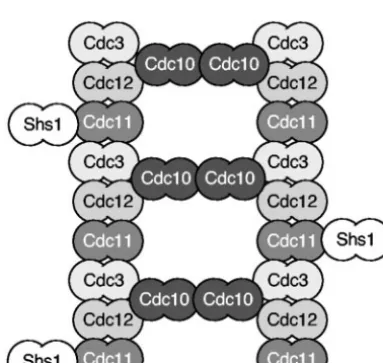
Septin proteins assemble to form a ring at the bud neck that correlates with the position of 10-nm filaments seen by elec-tron microscopy (9, 33, 89). The 10-nm filaments are thought to be composed of septins, since purified septin proteins bind each other and form similar types of filaments in vitro (33, 106). Recent studies indicate that the five septin proteins pro-duced in vegetative cells form a pentameric complex with the orientation shown in Fig. 2 (1, 106). Based on the ability of purified septin proteins to interact in vitro and their relative roles in septin ring formation in vivo, Cdc3, Cdc11, and Cdc12 are thought to form the main filament polymer, and Cdc10 is proposed to promote cross-linking. Shs1/Sep 7 is not essential for filament formation (10, 79) but plays a role in stabilizing the structure and is a target for regulation by phosphorylation (26). The septin proteins are indicated as dimers in Fig. 2 to signify that the individual septin proteins also interact in a homotypic fashion (106). Previous analysis of septin complexes purified from yeast showed a stoichiometry of 2:2:1:2 for Cdc3, -10, -11, and -12 (33), but other studies suggested that this might be due to loss of some subunits during purification (106). Studies on other fungi have identified general similarities for septin filament assembly. For example, theS. pombeorthologs of Cdc3 (Spn1), Cdc10 (Spn2), Cdc11 (Spn3), and Cdc12 (Spn4) interact with each other in a manner similar to that described above forS. cerevisiaeseptins (1). Although there are some differences, such as the absence of an ortholog of Shs1/ Sep7 inS. pombe, the relative importance of the septins for ring formation in vivo seems conserved. The orthologs of Cdc3 (Spn1) and Cdc12 (Spn4) play a central role in septin ring formation, whereas the Cdc10 ortholog (Spn2) was less impor-tant (1). Similarly, inC. albicansCdc3 and Cdc12 are essential but Cdc10 is not (110). However, theC. albicansortholog of Cdc11 was also not required for septin ring formation (al-though the rings were partially defective). The ability of C.
albicans cdc11⌬mutants to form septin rings is not compatible
with the model ofS. cerevisiaeseptin filament formation shown in Fig. 2. One possibility is that Cdc3 and Cdc12 can form filaments in vivo and that the other septins help to stabilize the filaments or recruit other proteins to do so. In this regard it is * Corresponding author. Mailing address: Department of Molecular
Genetics and Microbiology, State University of New York, Stony Brook, NY 11794-5222. Phone: (631) 632-8715. Fax: (631) 632-9797. E-mail: [email protected].
1503
on September 8, 2020 by guest
http://ec.asm.org/
interesting that the microsporidiumEncephalitozoon cuniculi, an obligate intracellular parasite representing a very divergent branch of fungi with a compact genome, appears to contain only three septin homologs (57). Multicellular organisms can also get by on a small complement of septins; theCaenorhabditis elegans
genome appears to contain only three septin genes (20).
REGULATION OF SEPTIN RING ASSEMBLY AND DISASSEMBLY DURING THE
S. CEREVISIAECELL CYCLE
The dynamic localization of septins during the cell cycle has made the septins both an interesting target for analysis of cell-cycle regulation and a useful landmark for monitoring cell
cycle progression. Examples of septin localization during bud-ding are shown in Fig. 3 forC. albicanscells to demonstrate that the patterns are identical to those observed inS. cerevisiae
and are thus likely to be conserved in other budding yeasts.
InS. cerevisiae, a septin ring forms at the future site of bud
formation about 15 min before the bud is observed to emerge (32, 42, 59, 69). As budding progresses, the septin ring extends into the daughter cell and forms an hourglass-shaped structure that is often described as a collar. The collar then changes from a fluid state, wherein the septin subunits are interchangeable, to a frozen state in which the septin subunits are stable during S, G2, and M phase, as judged by FRAP (fluorescence recovery
after photobleaching) (14, 26). Ultrastructural studies suggest the possibility that septin filaments may be more compact in the frozen state (89). Interestingly, the septins transiently be-come fluid again when the septin ring splits at the start of actomyosin ring contraction during cytokinesis (26, 67). The split septin rings then become frozen again and then later disassemble in order to reassemble at the next bud site in G1.
Thus, the alternating periods of fluid and frozen states are thought to permit the septin ring to alter its shape to match the changes in neck morphology during bud emergence and cyto-kinesis. The frozen state is thought to allow septins to form a stable structure that can act as a scaffold or as a barrier do-main, as will be described in the following sections.
A key aspect of septin function is that ring formation is regulated during the cell cycle by assembly and disassembly rather than by synthesis and degradation. A large number of genes have been implicated in influencing septin regulation (30, 39, 68). Therefore, we will summarize below three aspects of septin regulation that have been relatively well studied: regulation by the Rho-family GTPase Cdc42, phosphorylation, and GTP binding. Some of the other proteins that bind septins and may regulate their function include Bni5, Iqg1, and Hof1 (18, 30, 40, 64).
Septin ring assembly.Septins are thought to initially form a patch at the future bud site that subsequently coalesces into a ring (14, 68, 106, 107). The role of the patch is not well defined, however, since it was discovered in mutants that are defective in forming septin rings. This has suggested to others that the patch may be an abnormal structure and not a true interme-diate in septin ring assembly (40). Either way, Cdc42 plays an important role in septin ring formation, in addition to its other roles in promoting actin localization and polarized cell growth. Analysis of specific Cdc42 point mutations showed that Cdc42 FIG. 1. Comparison ofS. cerevisiaeseptin proteins. Diagrams of
theS. cerevisiaeseptin protein structures are shown.■, Region rich in basic amino acids that is implicated in binding to phosphinositide-containing membranes; u, GTPase domain; 䊐, conserved domain characteristic of septins; p, coiled-coil domain. The length of each septin is listed after the protein diagram. aa, Amino acids.
FIG. 2. Model for septin filament assembly. Recent students pre-dict that septin proteins assemble into filaments based on a repeating structure of the five mitotic septins that assemble into a pentameric complex (1, 106). Septin proteins are indicated as dimers to signify that the septin proteins associate with themselves.
FIG. 3. Examples of septin ring structures during budding. Fluo-rescence microscope photographs of C. albicans producing Cdc10-GFP. Representative cells are shown as examples of ring formation (A), septin collar at junction between mother and daughter cell (B), ring splitting (C), and cytokinesis and ring disassembly (D).
on September 8, 2020 by guest
http://ec.asm.org/
carries out a special role in septin ring formation that is inde-pendent of its role in actin polarization (38). In particular, these studies indicated that cycles of Cdc42 GTPase activity are needed for proper septin ring formation. Cdc42 GTPase activity was also implicated by the phenotypes of mutants lack-ing the GTPase-activatlack-ing proteins (GAPs) for Cdc42 (rga1⌬,
rga2⌬, andbem3⌬) (14). Several possibilities have been sug-gested for the role of Cdc42 in septin assembly, including a direct role for Cdc42 in shuttling septin subunits to the ring (36). Alternatively, the Cdc42 GAPs themselves may act as effectors to promote septin organization, as was suggested by colocalization of the Cdc42 GAPs and septins and by the abil-ity of overproduced GAPs to suppress septin mutant pheno-types (14). It is also possible that there is a downstream effector of Cdc42 that has not yet been discovered, since in mammalian cells the Borg proteins, which are downstream effectors of Cdc42, regulate septin assembly (50). In addition, it has also been proposed that the primary role of Cdc42 is to recruit the PAK kinase Cla4 to the future bud site and activate it to phosphorylate septins, as will be described next.
At least six protein kinases are thought to contribute to septin regulation. Cla4, a PAK kinase family member that binds to Cdc42, interacts with the septins and phosphorylates Cdc3 and 10 in vitro and possibly other septins in vivo (107). Septin phosphorylation by Cla4 appears to play a direct role in stabilizing septin filaments, since it coincides with the change to a frozen septin structure in vivo and because the septins remain fluid incla4⌬cells (107).cla4⌬mutants form aberrant septin structures, including rings that move away from the bud neck. The Gin4 protein kinase also localizes to the bud neck and is thought to regulate septin function by phosphorylating at least one septin, Shs1/Sep7 (10, 26, 70). Interestingly,gin4⌬
cells do not form a septin ring; instead, the septins are present as a series of bars across the bud neck that run parallel to the mother-daughter axis (39, 70). Similar bars are observed at the neck of mating pheromone-induced shmoos that are arrested in G1 by the pheromone signal pathway. These results have
suggested that Gin4 may stabilize the ring by promoting lateral association of septin filaments. Septins are also phosphorylated by the cyclin-dependent kinase Cdc28 to influence septin ring disassembly, as will be discussed below. Elm1, Cdc5, and Yck2 protein kinases have also been implicated in septin regulation, although the specific mechanisms are not clear, since they have other roles in morphogenesis.
GTP has long been implicated in septin function, given that all septins contain a similar GTPase domain. Analysis of septin proteins purified from yeast and other organisms confirmed that septins bind GTP and hydrolyze it in vitro (31, 33). Anal-ysis of individual recombinant septin proteins showed that Cdc10 and Cdc12 display GTPase activity, but not Cdc3 or Cdc11 (106). In fact, Cdc3 and Cdc11 may not bind GTP. Cdc10 and Cdc12 mutants that block GTP binding caused a temperature-sensitive phenotype in vivo and block septin fila-ment formation in vitro, indicating an important role for GTP binding as a prerequisite for filament formation (106). Muta-tions that prevent GTP binding did not impair the ability of septins to associate with each other into a pentameric complex, suggesting the role of GTP binding is to promote association of these complexes into filaments. GTP binding was also found to promote filament formation for septins from other organisms
(80). In contrast,S. cerevisiaeseptin mutants that block GTP hydrolysis did not affect septin filament assembly or disassem-bly, in vivo or in vitro (106). In addition, most GTP bound to septins does not appear to get hydrolyzed during the cell cycle (108). Thus, GTP binding to septins may play a structural role similar to the binding of GTP to␣-tubulin rather than the role of GTP binding and hydrolysis in signaling by Ras (106, 108).
Septin disassembly.The mechanisms that trigger septin ring disassembly at the end of the cell cycle are not as well under-stood as those that promote assembly. One likely mechanism is the dephosphorylation of septins by the PP2A phosphatase bound to Rts1, a B⬘regulatory subunit. PPA2RTS1is thought to
directly dephosphorylate the septins in part because it localizes to the bud neck after the Tem1 GTPase promotes spindle breakdown and septin ring splitting (25). Also, septin rings are stabilized inrts1⌬mutants (25). Interestingly, phosphorylation of Cdc3 by Cln-activated Cdc28 cyclin-dependent kinase in G1
phase appears to play a role in septin disassembly (102). cdc3-S1S2mutants that lack the target sites for Cln-Cdc28 show a delay in septin ring disassembly. Other mechanisms may con-tribute in addition to phosphorylation/dephosphorylation. For example, Cdc42 localizes to the septin rings late in the cell cycle and may potentially affect septin filament disassembly. Septin modification by SUMO (Smt3), the small ubiquitin-like protein, was also implicated in disassembly (51). However, this now seems unlikely, since mutation of the E3 attachment fac-tor (siz1⌬) blocked sumoylation of the septins but did not cause a delay in septin ring disassembly (52). Nonetheless, the observation that bud neck proteins other than the septins are SUMO modified inC. albicanssuggests a conserved role for SUMO at the bud neck (75).
SEPTIN RINGS ACT AS A SCAFFOLD TO RECRUIT PROTEINS TO THE BUD NECK
The distinctive pattern of septin localization has facilitated the identification of about 40 proteins that associate, directly or indirectly, with the septins at the bud neck (18, 39). Thus, the septin filaments at the bud neck are thought to act as a scaffold for anchoring proteins. However, the septin scaffold is not a simple hitching post since proteins can be recruited to specific subdomains of the ring, and subsets of proteins are found to bind or dissociate from the ring at different stages of the cell cycle (18, 39). For example, Bni4 initially binds in G1
to the mother cell side of the ring and then is later found throughout the ring (22). In contrast, Hsl1 binds to the daugh-ter cell side (4, 71). Given the large number of septin-binding proteins, we will review below only three well-studied examples of protein networks that bind to the septins, including com-plexes involved in bud site selection, chitin ring deposition, and a cell cycle checkpoint.
Bud site selection. The process of bud site selection is an interesting example of how distinct types of protein networks can be recruited to the septins to specify the future site of cell polarization and bud formation. Each new bud inS. cerevisiae
is positioned relative to the previous bud site in a specific manner. Haploidaor␣cells bud in an axial manner adjacent to the previous division site, and diploid a/␣ cells bud in a bipolar pattern near the poles (16, 55). A central role of septin proteins in this process is indicated by the random budding
on September 8, 2020 by guest
http://ec.asm.org/
pattern of septin mutants (15, 93). Axial budding requires recruitment to the bud neck of the Bud3, Bud4, and Axl2/ Bud10 proteins (11, 15, 93). It has been suggested by several groups that Bud3 and Bud4 may remain at the cortex after septin disassembly to define the position of the septin ring in the next cell cycle (11, 85). Bipolar budding is specified in part by persistent markers involving Bud8 at the pole distal to the birth scar and Bud9p at the proximal pole, along with the interacting integral membrane proteins Rax1 and Rax2 (55). Correct localization of Bud9 depends upon the septin proteins and the actin cytoskeleton, while proper targeting of Bud8p relies on other factors including actin and the formin Bni1 (16, 45, 55, 94). Proteins needed both for proper axial and bipolar budding are also recruited to septin rings, including the Bud5 guanine-nucleotide exchange factor (GEF) and the Bud2 GAP, that act on the Bud1/Rsr1 Ras-like GTPase (56, 72, 87).
Chitin ring formation.The formation of a chitin ring in the cell wall at the bud site provides an important example of how some proteins are recruited to the septin ring by intermediary proteins that bind to the septins. Just prior to bud emergence and coincident with formation of a septin ring at the incipient bud site in G1, chitin deposition occurs in the cell wall to form
a ring. The chitin ring remains throughout the cell cycle, ulti-mately marking the cortical site of division as the bud scar. Synthesis depends upon activity of Chs3 (chitin synthase III) and its localization to the proper site. Chs3 is tethered to the septin ring by a hierarchy of proteins involving the binding of Chs3 to Chs4, which then binds to Bni4 (22). Bni4 binds Cdc10 and is therefore thought to directly anchor this protein com-plex to the septin ring in vivo. Bni4 also binds to the Glc7 serine-threonine phosphatase and recruits it to the septin ring, which also facilitates interaction of Bni4 with septin proteins (63). The recruitment of Bni4 to the septin ring in G1is also
interesting, because it indicates that the septins can act as a scaffold while they are in a fluid state.
Cell cycle checkpoints/sensors.Septins organize protein net-works that sense the normal progression of bud morphogenesis and spindle position (18). The role of the septins in the spindle checkpoint has not been well defined (13, 18), so this section will focus on the bud morphogenesis checkpoint because it provides an interesting example of how proteins can be se-quentially recruited to the septin ring (58, 66). Perturbation of bud morphogenesis was discovered to activate a checkpoint by stabilizing the Swe1 protein kinase, which then phosphorylates the cyclin-dependent kinase Cdc28, leading to arrest of cell division in G2(4, 66, 71, 78). Swe1 also regulates cell size inS.
cerevisiae, similar to the function of its ortholog Wee1 inS.
pombe(48, 58). Thus, Swe1 plays a key role in sensing the
progression of bud morphogenesis (48, 58, 66, 90, 91). If bud morphogenesis proceeds normally during the cell cycle, com-ponents of the morphogenesis/size checkpoint are sequentially localized to the septin ring to promote phosphorylation of Swe1 and consequently its degradation. The first protein to be recruited is the Hsl1 protein kinase, which binds to the daugh-ter side of the septin collar, and then recruits Hsl7 and Swe1 (4, 19, 71, 96). Binding of Hsl1 to the Cdc11 and Cdc12 septins activates its protein kinase activity, but Hsl1 does not appear to phosphorylate Swe1 directly (44). Instead, two other protein kinases, Cla4 and Cdc5, are targeted sequentially to the bud neck and directly phosphorylate Swe1 and appear to contribute
to its degradation via a ubiquitin-dependent pathway (92). Abnormal morphogenesis prevents the proper function of this regulatory module, leading to Swe1 stabilization and cell cycle arrest in G2. The Swe1-mediated delay in G2contributes to the
elongated bud morphology characteristic of septin mutants in
S. cerevisiae. Recent studies suggest that the checkpoint may
sense either the shape of the neck region or the transition of the septin ring from a fluid to a frozen state at the bud neck (which may only occur in cells undergoing proper morphogen-esis) (66, 104).
SEPTIN RINGS FORM A BARRIER DURING BUDDING AND CYTOKINESIS
Bud neck barrier.Septins also contribute to cell polarization by mediating a barrier function that blocks diffusion between the daughter and mother cells. One line of evidence for this came from analysis of the integral membrane protein Ist2p, whose mRNA was discovered to localize to the bud tip (101). Interestingly, green fluorescent protein (GFP)-tagged Ist2 in the plasma membrane of the daughter cell was not able to diffuse across the bud neck to the mother cell. The asymmetric localization of Ist2p depends on the septins: GFP-Ist2p dif-fused into the mother cell whencdc12-6cells were shifted to the restrictive temperature to disrupt septin rings (101). Sep-tins are peripheral membrane proteins, making it possible that the septin ring acts directly as a physical barrier to diffusion of integral membrane proteins, such as Ist2. However, septin bar-rier function is probably more complex, since septin rings are also required to maintain the polarization to the bud of actin patches, as well as exocyst and polarisome components such as Sec3p, Sec5p, and Spa2p (3). Septins were also reported to act as a boundary to restrict to the bud Lte1, the GEF for the TEM1 GTPase, that promotes exit from mitosis (13). In septin mutants Lte1 can diffuse into the mother cell, where it activates Tem1 and thereby promotes premature exit from mitosis prior to proper nuclear division.
Cytokinesis barrier.Later in the cell cycle the septin ring splits and acts as a boundary to trap between the rings some of the proteins involved in cytokinesis and septum formation (25). During cytokinesis, the polarisome component Spa2p, exocyst component Sec3p, and chitin synthase II (Chs2p) localize be-tween the split septin rings, as does the actomyosin ring. Cy-tokinesis then proceeds by actomyosin ring contraction and the fusion of secretory vesicles to form the septum. However, shifting
acdc12-6septin mutant to the restrictive temperature to disrupt
septin rings revealed that Spa2, Sec3, and Chs2 were rapidly lost from the bud neck, indicating that their diffusion was normally constrained by the split septin rings. In contrast, the actomyosin ring was maintained at the bud neck after the temperature shift. Although the septins were not required for actomyosin ring main-tenance during cytokinesis or its subsequent contraction, septins were required for proper abscission (25).
SEPTIN FUNCTION DURING ALTERNATE DEVELOPMENTAL PATHWAYS INS. CEREVISIAE
(MATING AND SPORULATION)
Septin function during mating.S. cerevisiaecells conjugate when cells of the opposite mating type (a and␣) signal each
on September 8, 2020 by guest
http://ec.asm.org/
other with secreted mating pheromones (27, 28). Mating pher-omone-stimulated cells arrest in G1 phase and then undergo
polarized morphogenesis to form a conjugation bridge that connects the mating cells. Pheromone-stimulated cells are dis-tinct from budding cells in that they form an acute projection of growth, which is commonly referred to as a shmoo. Inter-estingly, septins localize to the neck of shmoos even though these pheromone-arrested cells are not progressing through the cell cycle and do not undergo cytokinesis at this site (32, 59). However, several observations suggest that septins carry out distinct roles during mating. For one, septins at the shmoo neck do not form a tight ring as seen during budding and instead form a more diffuse ring that appears to be composed of a series of bars that run parallel to the long axis of the cell (70). These septin bars are similar in appearance to the septins in budding cells that lack Gin4 (gin4⌬). Also, the septin bars function differently than rings, since Gin4 does not localize with septins in mating cells, even though it is present (70).
Septins are thought to act as a scaffold during mating be-cause they are required to localize Afr1 (35, 62) and Glc7 (8) to the shmoo neck, and they are required for the proper chitin localization at this position. Afr1 may bind directly to the septins, since it interacts with Cdc12 in the Two-Hybrid Assay. Afr1 is required for the proper formation of an acute projec-tion of shmoo morphogenesis (23, 35). Localized chitin depo-sition at the neck of the mating projection also requires the function of septins and Afr1 (62). Interestingly, the C terminus of Afr1 is highly similar to the C-terminal region of Bni4 that binds to Glc7 and recruits it to bud necks, suggesting that Afr1 plays an analogous role in recruiting the Glc7 phosphatase to the neck of shmoos.
The septins may also act as a barrier at the neck of shmoos to prevent diffusion of factors away from the tip. Polarization of mating pheromone receptor signaling to the shmoo tip is thought to help stimulate highly polarized growth of mating projections to form the conjugation bridge (49). Receptor po-larization results from a combination of the removal of old receptors by ligand-induced endocytosis, along with the target-ing of new receptors to the shmoo tip by the secretory pathway. The septins may help to polarize pheromone signaling by act-ing as a barrier to prevent diffusion away from the tip of receptors and possibly other pheromone pathway components. The localization of Afr1 to the septin ring at the shmoo neck is thought to further narrow the active zone of pheromone sig-naling to the shmoo tip, since Afr1 can negatively regulate receptor signaling (23, 35). Consistent with this model, muta-tion of eitherAFR1or the septins results in cells that form a broad shmoo tip with a more diffuse localization of receptors. It is also possible that septins could play a role as a barrier during cell fusion and zygote formation by creating a special zone in the conjugation bridge. However, septin mutants do not display a significant mating defect.
Septin proteins display unique localization during sporula-tion.InS. cerevisiae, nutrient limitation stimulates diploid a/␣ cells to undergo DNA replication and meiosis that results in development of four haploid spores within a sac-like ascus derived from the mother cell. This process is quite different from budding in that there is no equivalent of the bud neck or septation site. Instead, the spore membranes result from de novo synthesis of membrane around the postmeiotic nuclei.
Septins were first implicated in sporulation because transcrip-tion ofCDC3andCDC10is increased more than tenfold, and expression of the sporulation-specific septins, SPR3 and
SPR28, is highly induced late in meiosis at the time of spore
development (17, 24, 29, 86).
Septins are not observed at the plasma membrane of sporu-lating cells and are instead first observed in meiosis II when chromosomes divide into four nuclear lobes. Cdc3, Cdc11, Spr3, and Spr28 were detected in ring-like structures surround-ing each of the four spindle pole bodies (24, 29). However, these rings are distinct from those seen during budding. They initially form at the leading edge of prospore membrane and then achieve a broader localization. As the prospore mem-brane elongates and expands along the outer surface of each nucleus, septins are detected in sheets that appear as pairs of bars in optical sections (24, 29, 99). Upon closure of the pro-spore membranes around each nucleus, the septins then dis-play a generally uniform distribution around each nascent spore. The septins appear to colocalize with each other in the prospore membranes, and the localization of Cdc3 and Cdc11 was less intense inspr3⌬cells, suggesting the septins form a complex (29). However, Spr3 did not detectably localize at bud necks when expression ofSPR3was engineered in vegetative cells. Further work will be required to determine the regula-tion of septin protein interacregula-tions during sporularegula-tion.
Septins appear to act as a scaffold during sporulation. The Glc7 phosphatase is recruited to the septins by Gip1, a target-ing subunit, which is only expressed in sporulattarget-ing cells (99). Interestingly, Gip1 and Glc7 are essential for the organization of septin complexes, although it is unclear whether this is due to a direct dephosphorylation of septin proteins (99). Thus, an interesting similarity between budding and sporulating cells is that they each have a unique protein that targets Glc7 to the septins; Bni4 in budding cells and Gip1 in sporulating cells. This similarity may also extend to mating cells since Afr1 shows similarity to the domain of Bni4 that binds Glc7 and thus may recruit Glc7 to the septins in mating cells. Septin localization to the spore periphery after prospore membrane closure has been proposed to act as a scaffold for recruitment of factors that contribute to spore wall formation within the lumen of the prospore double membrane (29).
Although these studies implicate septins in spore formation, septin mutant strains display no significant sporulation pheno-type.spr3⌬,cdc10⌬, and double mutantspr3⌬spr28⌬strains exhibited little or no reduction in sporulation efficiency (24, 29, 99). It has been proposed that the lack of severe sporulation defects upon septin gene deletion may be due to “functional redundancy” among the septin proteins (24).
SEPTIN FUNCTION INC. ALBICANS,S. POMBE,
ANDA. NIDULANS
The essential role of the septins has lead to their study in other fungi, includingS. pombe,C. albicans, andA. nidulans. A common feature of these organisms is that the septins assem-ble into a ring that functions in cytokinesis. However, the study of these other fungi is also providing new insights into how septins are regulated during different cell cycle programs asS.
pombedivides by fission,C. albicans grows in different
mor-phologies ranging from buds to filamentous hyphae, and A.
on September 8, 2020 by guest
http://ec.asm.org/
nidulansis a filamentous fungus. In addition, these studies are revealing new aspects of septin function in developmental pathways not seen inS. cerevisiae, such as the formation of chlamydospores inC. albicansand asexual conidiospores inA.
nidulans. Studies onC. albicansandA. nidulansare also
sig-nificant in that they are expected to help to identify mecha-nisms underlying the ability of fungal pathogens to grow inva-sively in human hosts.
Septins inS. pombe.S. pombeundergoes a very distinct type of cell division fromS. cerevisiae in that it divides by fission rather than by budding. Consistent with this, septins are not localized at early stages in the cell cycle and do not appear to contribute to morphogenesis (1, 6, 103). Later in the cell cycle at anaphase, the septins form a ring around the inner surface of the plasma membrane at the medial region of the cell, marking the future site of cytokinesis. The general behavior of the septins at this stage appears to be similar to what happens in budding yeast cells in that assembly and disassembly of the septin ring is regulated during the cell cycle. However, septins are not needed for cytokinesis; deletion of septins appears to cause primarily a delay in cell separation (1). The localization of septins to the septation site late in anaphase is similar to what was observed in animal cells (60), suggestingS. pombe
may be a good model system for understanding septin regula-tion in animals.
TheS. pombegenome contains seven genes that are
homol-ogous to the septins inS. cerevisiae. Four of the septins (Spn1, Spn2, Spn3, and Spn4) function during vegetative growth and appear to be orthologous to the key septins in S. cerevisiae
(Cdc3, -10, -11, and -12, respectively) (1). For example, Spn1 and Spn4 are required to form a septin ring in vivo just as Cdc3 and Cdc12 are essential forS. cerevisiae. Additional analysis of the localization of septins in vivo, together with biochemical studies of the ability of different septin proteins to interact, suggests that theS. pombeseptins associate in a manner similar to theS. cerevisiaeseptins (1, 6, 103) (See section above on septin proteins forming filaments.)S. pombedoes not appear to contain an ortholog of Shs1/Sep7, which is a target for regulation by phosphorylation inS. cerevisiae, indicating that future studies will likely identify significant differences in the regulation of S. pombe septins by phosphorylation. The re-maining three S. pombe septin genes (spn5, -6, and -7) are induced during sporulation (77), but their role in this process has not been reported.
Although initial studies identified many similarities in septin function between S. pombe and S. cerevisiae, there are also several significant differences. For example, the anillin ho-molog Mid2 colocalizes with septins in S. pombeand is also required for septin ring formation (6, 103). In contrast, the similar septin-binding proteins Bud4 inS. cerevisiaeand Int1 in
C. albicanscolocalize with septins in their respective
organ-isms, but they are not required for ring formation. However, recent studies have implicated Bud4 in contributing to proper septin organization in S. cerevisiae (37). (It should also be noted that the low level of sequence identity of Mid2 with these other proteins makes it unclear that their functions are or-thologous.) Mid2 also influences septin ring disassembly, since overproduction of Mid2 leads to a delay in septin ring disas-sembly (6, 103). This indicates that Mid2 degradation, which is regulated by the Skp1/Cdc53/F-box (SCF)-dependent
proteol-ysis, facilitates septin disassembly in S. pombe (6, 103). An-other difference between these organisms is that S. pombe
septins are not involved in recruiting the ortholog of Gin4 (Cdr2), which inS. pombefunctions independently of the sep-tins (83). Thus, further differences in septin regulation are likely to be found in future studies ofS. pombe.
Septins inC. albicans.In response to different environmen-tal stimuliC. albicanswill form either rounded buds, elongated chains of cells known as pseudohyphae, or long filamentous cells with parallel cell walls known as hyphae (97). In addition, certain low-nutrient conditions induce C. albicans to form chlamydospores, which are thought to be asexual resting spores.C. albicanshas not been observed to undergo meiotic spore formation. Studies on the regulation ofC. albicans mor-phogenesis are important for understanding the mechanisms of fungal pathogenesis, since the ability to switch between budding and hyphal cells has been linked to virulence. The budding and hyphal growth phases differ in the production of virulence factors, the ability to grow invasively, and the ability to escape the immune system in human hosts (7, 112).
The budding phase inC. albicansshows general similarity to
S. cerevisiaewith some key differences. Five septins form the
septin ring at the bud neck that are orthologous to Cdc3, -10, -11, and -12 and Shs1/Sep7 (34, 54, 98, 110). Deletion analysis indicates that the overall contribution of most septins is
simi-lar;CDC3andCDC12are essential, andSHS1/SEP7plays a
relatively minor role (110). Deletion ofCDC10 and CDC11
resulted in defects in cytokinesis and spindle orientation but, in contrast toS. cerevisiae, these septins were not essential for growth, even at 42°C. The observation thatcdc11⌬cells still form a septin ring, even at elevated temperatures, contradicts current models that propose an essential role for Cdc11 in filament formation (1, 106). This suggests either that the cur-rent models are wrong or thatC. albicansseptins are organized in a different manner or are stabilized in vivo by different factors. The higher thermostability ofC. albicansseptins may relate to this organism being adapted for growth at 37°, the temperature of its host. Septins act as a scaffold to recruit proteins to the bud neck, including theC. albicanshomologs of Hsl1, Gin4, Int1, and Bni4 (34, 73, 105, 113). However, the role of the bud neck proteins may be altered. For example, C.
albicanslacks a homolog of Hsl7 that inS. cerevisiaebinds to
Hsl1 and is important for its function. Septins are not detect-ably modified by SUMO (Smt3) as they are inS. cerevisiae, but they do act as a scaffold to recruit SUMO-modified proteins, suggesting a role for SUMO in regulation of bud neck proteins (75).
The role of septins in pseudohyphae is thought to be similar to budding, since in both cases a septin ring forms at the junction with the mother cell. In contrast, hyphal cells form the septin ring about 10 m away from the junction with the mother cell (see below). Thus, GFP-tagged septins can be used to help distinguish between pseudohyphal and hyphal cells (34, 97, 98) and can also be used to analyze septin structures inC.
albicanscells taken from infected mice (41).
C. albicanshyphae are commonly seen at sites of infection,
and their formation can be induced in vitro at 37°C by various stimuli, such as serum. Three types of septin localization were observed in hyphae:
(i) A classic septin ring forms in the initial protrusion of
on September 8, 2020 by guest
http://ec.asm.org/
hyphal growth (known as a germ tube) about 10 to 15m from the mother cell and also at subsequent sites of cell division as the hypha elongates (34, 98, 110). This ring functions similarly to the bud neck ring to promote septum formation. Hyphae are distinct from budding cells in that the previous septin rings are not fully disassembled, particularly on the mother cell side, after septation. An interesting possibility is that the subapical cells arrest at a stage prior to the signal for septin ring disas-sembly, since these subapical cells lag before beginning a new cell cycle.
(ii) A diffuse ring of septins was detected at the junction between the mother cell and the germ tube (98, 110). This basal septin band is regulated differently from the septin rings at sites of cytokinesis because it still forms in agin4⌬strain, whereas septin rings do not (113). It is not clear that the basal septin band acts as a scaffold, since Gin4 and Int1 do not localize to this region, but the basal band may have other functions as described below. The basal septin band appears to be similar to the septin localization in pheromone-induced shmoos inS. cerevisiae(70). Thus, shmoo formation may be a good model for aspects of germ tube formation in that both processes can occur in G1, whereas budding initiates as cells
enter the S phase of a new cell cycle.
(iii) A faint cap of septins was also detected at the leading edge of growth in germ tubes and hyphae (98, 110). Interest-ingly, this localization coincides with an ergosterol-rich region of the plasma membrane at hyphal tips that was identified by filipin staining (74). A similar ergosterol rich domain was also seen at shmoo tips in S. cerevisiae and at hyphal tips in A.
nidulans(2, 88). There may be a general connection between
septins and the organization of ergosterol in the plasma mem-brane, since filipin staining coincides with septin rings during cytokinesis inC. albicans hyphae and inS. pombe(74, 100). Thus, septins may play a role in cell polarization by facilitating the organization of specific plasma membrane domains, such as sterol-rich lipid rafts.
C. albicans cdc10⌬andcdc11⌬mutants displayed
abnormal-ities in hyphal growth in addition to defects in septum forma-tion that suggest that septins in the basal band and at the hyphal tip contribute to proper morphogenesis (110). Both
cdc10⌬andcdc11⌬mutants form hyphae that are more curved
than the relatively straight hyphae formed by wild-type cells. The sites of extreme bending or curvature are associated with altered Calcofluor staining of cell walls, suggesting that the septins may play a direct role in promoting even hyphal growth.
Thecdc10⌬andcdc11⌬mutants were also defective in
select-ing sites of secondary germ tube formation. Wild-type cells typically initiate a second germ tube at a distal site so that the two germ tubes form at an angle of⬎90° apart on the mother cell. In contrast,cdc10⌬andcdc11⌬mutants often formed a secondary germ tube adjacent to the initial hypha and in some cases from within the hypha. This may be related to the defects in bud site selection seen for septin mutants inC. albicansand
S. cerevisiae. Although the septin mutants form hyphae, they
were defective in invasive growth, both in vitro in agar and in vivo in a mouse model ofCandidainfection (109). Thecdc10⌬
andcdc11⌬mutants grew to high levels in kidneys of infected
mice but did not cause a disseminated infection, as did the wild type. Instead, the septin mutants formed large clumps of fungal cells that were surrounded by lymphocytes. Thus, it may not be
necessary to completely block hyphal formation to have signif-icant effects on preventing the spread ofC. albicansinfections. Analysis of chlamydospore formation identified a novel pat-tern of septin localization. Chlamydospores are large thick-walled cells whose role in infection is unclear, but they act as a resting form in other species. During chlamydospore morpho-genesis, cells switch to filamentous growth and then develop elongated suspensor cells that in turn give rise to chlamydo-spores. The cdc10⌬ and cdc11⌬ mutants were defective in forming chlamydospores, primarily because of a failure to un-dergo septation (73). Interestingly, analysis of GFP-tagged sep-tin proteins in chlamydospores revealed that, after septation, the septins are present throughout the plasma membrane in a series of filamentous structures that run parallel to the axis of the chlamydospore-suspensor cell junction. This peripheral lo-calization of septins is reminiscent of septin lolo-calization in meiotic spores inS. cerevisiae and suggests that septins may play a role in the formation of the specialized cell walls ob-served in these cell types.
Septins inA. nidulans. A. nidulans provides an interesting system for comparative analysis of septins, since it undergoes distinct patterns of cell division characteristic of many filamen-tous fungi and can also form aerial hyphae that produce asex-ual conidiospores (46). These studies also have application to fungal pathogenesis, since A. nidulans is an opportunistic pathogen, and it is related to the most common filamentous fungal pathogen of humans,Aspergillus fumigatis.
Five septin genes,aspAto-E, were identified inA. nidulans
(82). Comparison of the predicted protein sequences indicates that four septins—AspA, -B, -C, and -D—correspond to the key septins inS. cerevisiaeand appear to be orthologs of Cdc11, -3, -12, and -10. As predicted for an ortholog ofCDC3,aspBis an essential gene (111). The remaining septin-like protein, AspE, does not show strong similarity to any of theS. cerevisiae
septins, but all five septins are expressed during vegetative growth (82). There do not appear to be any sporulation-spe-cific septins.
A unique aspect of septin localization inA. nidulansis that the timing of septin ring formation varies, depending on the type of cell division. Germination of a spore results in the growth of a germ tube that elongates without septation until a critical size is reached. After this size threshold is passed the next mitosis triggers formation of the first septum (46, 47, 114). Comparison of the timing of septin localization and nuclear division indicates that septins localize postmitotically (111). Thus,A. nidulansis similar toS. pombein forming a septin ring late in the cell cycle. After the septin ring splits, the ring on the apical side persists and may act as a marker of cell polarity. In contrast, the timing of septin localization was different during formation of a secondary germ tube from the conidiospore or when branching occurs and a filament emerges from a subapi-cal cell within a hypha. In these latter cases, a septin ring was observed before mitosis, similar to the timing of septin ring formation inS. cerevisiae. AnaspBtemperature-sensitive mu-tant demonstrated hyperbranching when shifted to the re-stricted temperature, suggesting septins might also recruit components that regulate induction of mitotic events (111). It will be interesting to determine how cell cycle regulation of septin localization is controlled in these different cell types. Another interesting question is how the sites of branching and
on September 8, 2020 by guest
http://ec.asm.org/
secondary germ tubes are selected, since the bud genes are not conserved.
Septins also localize to cell junctions during the formation of the conidiophore, the specialized structure that bears asexual conidiospores (81, 111). During this developmental process, the tip of an aerial hypha becomes swollen and then gives rise to two layers of small bud-like cells, the metulae and phialides. The conidiospores themselves emerge from the second layer (the phialides) in a budding process. Immunofluorescence lo-calized AspB to a diffuse band at the base of each forming metula, which disappeared as the structure elongated (111). Additional AspB rings were observed at the interfaces of metu-lae and developing phialides, which similarly were no longer seen as the phialides matured, and septins were also present at the interface of each phialide and emerging conidiospore. An
aspBtemperature-sensitive mutant exhibited a lack of metulae formation when shifted to the restrictive temperature, suggest-ing that AspB may function as a scaffold or barrier to direct specific components to sites within the developing conidio-phore (111).A. nidulansseptins are therefore suggested to act as an “organizational scaffold” that targets specific components to designated sites at different points in development.
EXPANDING ROLES OF SEPTINS: FROM YEAST TO HUMANS
The yeast bud neck septins have provided a valuable model for the ability of septins to form filaments, act as scaffolds, and create diffusion barriers. Analysis of septins in other develop-mental pathways, and in fungi with distinct modes of cell divi-sion, has identified many common aspects of septin function. The study of septins in these other contexts is also identifying many critical differences that are broadening our knowledge of different types of septin organization and functions. A further example of this is the new insight into septin function coming from the analysis of septins in animal cells. Many septin func-tions are conserved in animal cells, such as the ability of GTP binding to promote septin filament formation, the role of sep-tins in cytokinesis, and a likely role for sepsep-tins in a diffusion barrier at the cleavage furrow of dividing mammalian cells (60, 95). Studies on fungal septins may therefore prove to be a good model for understanding the role of septins in human disease; septin mutations have been implicated in cancer, neurodegen-erative conditions, and other pathologies (43). Similarly, the studies on animal cells are also helping to identify novel septin functions that are not seen in budding yeast, such as a role in regulated secretion (5, 76). In animal cells it is also interesting that actin, together with the adaptor protein anillin, acts as a template for septin filament assembly (61). The relationship between actin and septins in fungal cells is not well defined, but an anillin homolog (Mid2) promotes septin organization inS.
pombe(6, 103) and several recent studies inS. cerevisiaealso
indicate that actin influences septin assembly (53, 84). Thus, the synergism of research from yeast to humans will aid future studies on the septin family of proteins.
ACKNOWLEDGMENTS
We thank Aaron Neiman and Doug Kellogg for helpful comments and suggestions on the manuscript.
Recent studies of septins in our lab have been supported by grant RO1 AI47837 from the National Institutes of Health (J.B.K.).
REFERENCES
1.An, H., J. L. Morrell, J. L. Jennings, A. J. Link, and K. L. Gould.2004. Requirements of fission yeast septins for complex formation, localization, and function. Mol. Biol. Cell15:5551–5564.
2.Bagnat, M., and K. Simons.2002. Cell surface polarization during yeast mating. Proc. Natl. Acad. Sci. USA99:14183–14188.
3.Barral, Y., V. Mermall, M. S. Mooseker, and M. Snyder.2000. Compart-mentalization of the cell cortex by septins is required for maintenance of cell polarity in yeast. Mol. Cell5:841–851.
4.Barral, Y., M. Parra, S. Bidlingmaier, and M. Snyder.1999. Nim1-related kinases coordinate cell cycle progression with the organization of the pe-ripheral cytoskeleton in yeast. Genes Dev.13:176–187.
5.Beites, C. L., H. Xie, R. Bowser, and W. S. Trimble.1999. The septin CDCrel-1 binds syntaxin and inhibits exocytosis. Nat. Neurosci.2:434–439. 6.Berlin, A., A. Paoletti, and F. Chang.2003. Mid2p stabilizes septin rings
during cytokinesis in fission yeast. J. Cell Biol.160:1083–1092.
7.Berman, J., and P. E. Sudbery.2002.Candida albicans: a molecular revo-lution built on lessons from budding yeast. Nat. Rev. Genet.3:918–930. 8.Bloecher, A., and K. Tatchell.2000. Dynamic localization of protein
phos-phatase type 1 in the mitotic cell cycle ofSaccharomyces cerevisiae.J. Cell Biol.149:125–140.
9.Byers, B., and L. Goetsch.1976. A highly ordered ring of membrane-associated filaments in budding yeast. J. Cell Biol.69:717–721.
10.Carroll, C. W., R. Altman, D. Schieltz, J. R. Yates, and D. Kellogg.1998. The septins are required for the mitosis-specific activation of the Gin4 kinase. J. Cell Biol.143:709–717.
11.Casamayor, A., and M. Snyder.2002. Bud-site selection and cell polarity in budding yeast. Curr. Opin. Microbiol.5:179–186.
12.Casamayor, A., and M. Snyder.2003. Molecular dissection of a yeast septin: distinct domains are required for septin interaction, localization, and func-tion. Mol. Cell. Biol.23:2762–2777.
13.Castillon, G. A., N. R. Adames, C. H. Rosello, H. S. Seidel, M. S. Longtine, J. A. Cooper, and R. A. Heil-Chapdelaine.2003. Septins have a dual role in controlling mitotic exit in budding yeast. Curr. Biol.13:654–658. 14.Caviston, J. P., M. Longtine, J. R. Pringle, and E. Bi.2003. The role of
Cdc42p GTPase-activating proteins in assembly of the septin ring in yeast. Mol. Biol. Cell14:4051–4066.
15.Chant, J., M. Mischke, E. Mitchell, I. Herskowitz, and J. R. Pringle.1995. Role of Bud3p in producing the axial budding pattern of yeast. J. Cell Biol. 129:767–778.
16.Chant, J., and J. R. Pringle.1995. Patterns of bud-site selection in the yeast Saccharomyces cerevisiae.J. Cell Biol.129:751–765.
17.Chu, S., J. DeRisi, M. Eisen, J. Mulholland, D. Botstein, P. O. Brown, and I. Herskowitz.1998. The transcriptional program of sporulation in budding yeast. Science282:699–705.
18.Cid, V. J., J. Jimenez, M. Molina, M. Sanchez, C. Nombela, and J. W. Thorner.2002. Orchestrating the cell cycle in yeast: sequential localization of key mitotic regulators at the spindle pole and the bud neck. Microbiology 148:2647–2659.
19.Cid, V. J., M. J. Shulewitz, K. L. McDonald, and J. Thorner.2001. Dynamic localization of the Swe1 regulator Hsl7 during theSaccharomyces cerevisiae cell cycle. Mol. Biol. Cell12:1645–1669.
20.Consortium, C. E. S.1998. Genome sequence of the nematode Caenorhab-ditis elegans: a platform for investigating biology. Science282:2012–2018. 21.Cooper, J. A., and D. P. Kiehart. 1996. Septins may form a ubiquitous
family of cytoskeletal filaments. J. Cell Biol.134:1345–1348.
22.DeMarini, D. J., A. E. Adams, H. Fares, C. De Virgilio, G. Valle, J. S. Chuang, and J. R. Pringle.1997. A septin-based hierarchy of proteins required for localized deposition of chitin in theSaccharomyces cerevisiae cell wall. J. Cell Biol.139:75–93.
23.DeMattei, C. R., C. Davis, and J. B. Konopka.2000. Point mutations within a highly conserved region of theSaccharomyces cerevisiae AFR1gene cause a defect in both the pheromone signaling and morphogenesis functions. Genetics155:43–55.
24.De Virgilio, C., D. J. DeMarini, and J. R. Pringle.1996.SPR28, a sixth member of the septin gene family inSaccharomyces cerevisiaethat is ex-pressed specifically in sporulating cells. Microbiology142:2897–2905. 25.Dobbelaere, J., and Y. Barral.2004. Spatial coordination of cytokinetic
events by compartmentalization of the cell cortex. Science305:393–396. 26.Dobbelaere, J., M. S. Gentry, R. L. Hallberg, and Y. Barral.2003.
Phos-phorylation-dependent regulation of septin dynamics during the cell cycle. Dev. Cell4:345–357.
27.Dohlman, H. G., and J. W. Thorner.2001. Regulation of G protein-initiated signal transduction in yeast: paradigms and principles. Annu. Rev. Bio-chem.70:703–754.
28.Elion, E. A. 2000. Pheromone response, mating, and cell biology. Curr. Opin. Microbiol.3:573–581.
29.Fares, H., L. Goetsch, and J. R. Pringle.1996. Identification of a
on September 8, 2020 by guest
http://ec.asm.org/
mentally regulated septin and involvement of the septins in spore formation inSaccharomyces cerevisiae.J. Cell Biol.132:399–411.
30.Faty, M., M. Fink, and Y. Barral.2002. Septins: a ring to part mother and daughter. Curr. Genet.41:123–131.
31.Field, C. M., O. Al-Awar, J. Rosenblatt, M. L. Wong, B. Alberts, and T. J. Mitchison.1996. A purifiedDrosophilaseptin complex forms filaments and exhibits GTPase activity. J. Cell Biol.133:605–616.
32.Ford, S. K., and J. R. Pringle.1991. Cellular morphogenesis in the Sac-charomyces cerevisiaecell cycle: localization of theCDC11gene product and the timing of events at the budding site. Dev. Genet.12:281–292. 33.Frazier, J. A., M. L. Wong, M. S. Longtine, J. R. Pringle, M. Mann, T. J.
Mitchison, and C. Field.1998. Polymerization of purified yeast septins: evidence that organized filament arrays may not be required for septin function. J. Cell Biol.143:737–749.
34.Gale, C., M. Gerami-Nejad, M. McClellan, S. Vandoninck, M. S. Longtine, and J. Berman.2001.Candida albicansInt1p interacts with the septin ring in yeast and hyphal cells. Mol. Biol. Cell12:3538–3549.
35.Giot, L., and J. B. Konopka.1997. Functional analysis of the interaction between Afr1p and the Cdc12p septin, two proteins involved in pheromone-induced morphogenesis. Mol. Biol. Cell8:987–998.
36.Gladfelter, A. S., I. Bose, T. R. Zyla, E. S. Bardes, and D. J. Lew.2002. Septin ring assembly involves cycles of GTP loading and hydrolysis by Cdc42p. J. Cell Biol.156:315–326.
37.Gladfelter, A. S., L. Kozubowski, T. R. Zyla, and D. J. Lew.2005. Interplay between septin organization, cell cycle, and cell shape in yeast. J. Cell Sci. 118:1617–1628.
38.Gladfelter, A. S., J. J. Moskow, T. R. Zyla, and D. J. Lew.2001. Isolation and characterization of effector-loop mutants of CDC42 in yeast. Mol. Biol. Cell12:1239–1255.
39.Gladfelter, A. S., J. R. Pringle, and D. J. Lew.2001. The septin cortex at the yeast mother-bud neck. Curr. Opin. Microbiol.4:681–689.
40.Gladfelter, A. S., T. R. Zyla, and D. J. Lew.2004. Genetic interactions among regulators of septin organization. Eukaryot. Cell3:847–854. 41.Gonzalez-Novo, A., J. Jimenez, M. J. Garcia, I. Rios-Serrano, J. Pla, A.
Jimenez, and M. Sanchez-Perez.2004. Dynamics of CaCdc10, a septin of Candida albicans, in living cells and during infection. Int. Microbiol.7:105– 112.
42.Haarer, B. K., and J. R. Pringle.1987. Immunofluorescence localization of theSaccharomyces cerevisiae CDC12gene product to the vicinity of the 10-nm filaments in the mother-bud neck. Mol. Cell. Biol.7:3678–3687. 43.Hall, P. A., and S. E. Russell.2004. The pathobiology of the septin gene
family. J. Pathol.204:489–505.
44.Hanrahan, J., and M. Snyder.2003. Cytoskeletal activation of a checkpoint kinase. Mol. Cell12:663–673.
45.Harkins, H. A., N. Page, L. R. Schenkman, C. De Virgilio, S. Shaw, H. Bussey, and J. R. Pringle.2001. Bud8p and Bud9p, proteins that may mark the sites for bipolar budding in yeast. Mol. Biol. Cell12:2497–2518. 46.Harris, S. D., and M. Momany.2004. Polarity in filamentous fungi: moving
beyond the yeast paradigm. Fungal Genet. Biol.41:391–400.
47.Harris, S. D., J. L. Morrell, and J. E. Hamer.1994. Identification and characterization of Aspergillus nidulansmutants defective in cytokinesis. Genetics136:517–532.
48.Harvey, S. L., and D. R. Kellogg.2003. Conservation of mechanisms con-trolling entry into mitosis: budding yeast wee1 delays entry into mitosis and is required for cell size control. Curr. Biol.13:264–275.
49.Jackson, C. L., J. B. Konopka, and L. H. Hartwell.1991.Saccharomyces cerevisiae␣-pheromone receptors activate a novel signal transduction path-way for mating partner discrimination. Cell67:389–402.
50.Joberty, G., R. R. Perlungher, P. J. Sheffield, M. Kinoshita, M. Noda, T. Haystead, and I. G. Macara.2001. Borg proteins control septin organiza-tion and are negatively regulated by Cdc42. Nat. Cell Biol.3:861–866. 51.Johnson, E. S., and G. Blobel.1999. Cell cycle-regulated attachment of the
ubiquitin-related protein SUMO to the yeast septins. J. Cell Biol.147:981– 994.
52.Johnson, E. S., and A. A. Gupta.2001. An E3-like factor that promotes SUMO conjugation to the yeast septins. Cell106:735–744.
53.Kadota, J., T. Yamamoto, S. Yoshiuchi, E. Bi, and K. Tanaka.2004. Septin ring assembly requires concerted action of polarisome components, a PAK kinase Cla4p, and the actin cytoskeleton inSaccharomyces cerevisiae.Mol. Biol. Cell15:5329–5345.
54.Kaneko, A., T. Umeyama, N. Hanaoka, B. C. Monk, Y. Uehara, and M. Niimi.2004. Tandem affinity purification of theCandida albicansseptin protein complex. Yeast21:1025–1033.
55.Kang, P. J., E. Angerman, K. Nakashima, J. R. Pringle, and H. O. Park. 2004. Interactions among Rax1p, Rax2p, Bud8p, and Bud9p in marking cortical sites for bipolar bud-site selection in yeast. Mol. Biol. Cell15:5145– 5157.
56.Kang, P. J., A. Sanson, B. Lee, and H. O. Park.2001. A GDP/GTP ex-change factor involved in linking a spatial landmark to cell polarity. Science 292:1376–1378.
57.Katinka, M. D., S. Duprat, E. Cornillot, G. Metenier, F. Thomarat, G. Prensier, V. Barbe, E. Peyretaillade, P. Brottier, P. Wincker, F. Delbac, H.
El Alaoui, P. Peyret, W. Saurin, M. Gouy, J. Weissenbach, and C. P. Vivares.2001. Genome sequence and gene compaction of the eukaryote parasiteEncephalitozoon cuniculi.Nature414:450–453.
58.Kellogg, D. R.2003. Wee1-dependent mechanisms required for coordina-tion of cell growth and cell division. J. Cell Sci.116:4883–4890. 59.Kim, H. B., B. K. Haarer, and J. R. Pringle.1991. Cellular morphogenesis
in theSaccharomyces cerevisiaecell cycle: localization of theCDC3gene product and the timing of events at the budding site. J. Cell Biol.112:535– 544.
60.Kinoshita, M.2003. Assembly of mammalian septins. J. Biochem.134:491– 496.
61.Kinoshita, M., C. M. Field, M. L. Coughlin, A. F. Straight, and T. J. Mitchison.2002. Self- and actin-templated assembly of mammalian septins. Dev. Cell3:791–802.
62.Konopka, J. B., C. DeMattei, and C. Davis.1995.AFR1promotes polarized apical morphogenesis inSaccharomyces cerevisiae.Mol. Cell. Biol.15:723– 730.
63.Kozubowski, L., H. Panek, A. Rosenthal, A. Bloecher, D. J. DeMarini, and K. Tatchell.2003. A Bni4-Glc7 phosphatase complex that recruits chitin synthase to the site of bud emergence. Mol. Biol. Cell14:26–39. 64.Lee, P. R., S. Song, H. S. Ro, C. J. Park, J. Lippincott, R. Li, J. R. Pringle,
C. De Virgilio, M. S. Longtine, and K. S. Lee. 2002. Bni5p, a septin-interacting protein, is required for normal septin function and cytokinesis in Saccharomyces cerevisiae.Mol. Cell. Biol.22:6906–6920.
65.Leipe, D. D., Y. I. Wolf, E. V. Koonin, and L. Aravind.2002. Classification and evolution of P-loop GTPases and related ATPases. J. Mol. Biol.317: 41–72.
66.Lew, D. J.2003. The morphogenesis checkpoint: how yeast cells watch their figures. Curr. Opin. Cell Biol.15:648–653.
67.Lippincott, J., K. B. Shannon, W. Shou, R. J. Deshaies, and R. Li.2001. The Tem1 small GTPase controls actomyosin and septin dynamics during cyto-kinesis. J. Cell Sci.114:1379–1386.
68.Longtine, M. S., and E. Bi.2003. Regulation of septin organization and function in yeast. Trends Cell Biol.13:403–409.
69.Longtine, M. S., D. J. DeMarini, M. L. Valencik, O. S. Al-Awar, H. Fares, C. De Virgilio, and J. R. Pringle.1996. The septins: roles in cytokinesis and other processes. Curr. Opin. Cell Biol.8:106–119.
70.Longtine, M. S., H. Fares, and J. R. Pringle.1998. Role of the yeast Gin4p protein kinase in septin assembly and the relationship between septin as-sembly and septin function. J. Cell Biol.143:719–736.
71.Longtine, M. S., C. L. Theesfeld, J. N. McMillan, E. Weaver, J. R. Pringle, and D. J. Lew.2000. Septin-dependent assembly of a cell cycle-regulatory module inSaccharomyces cerevisiae.Mol. Cell. Biol.20:4049–4061. 72.Marston, A. L., T. Chen, M. C. Yang, P. Belhumeur, and J. Chant.2001. A
localized GTPase exchange factor, Bud5, determines the orientation of division axes in yeast. Curr. Biol.11:803–807.
73.Martin, S. W., L. M. Douglas, and J. B. Konopka.2005. Cell cycle dynamics and quorum sensing inCandida albicanschlamydospores are distinct from budding and hyphal cells. Eukaryot. Cell4:1191–1202.
74.Martin, S. W., and J. B. Konopka.2004. Lipid raft polarization contributes to hyphal growth inCandida albicans.Eukaryot. Cell3:675–684. 75.Martin, S. W., and J. B. Konopka.2004. SUMO modification of
septin-interacting proteins inCandida albicans.J. Biol. Chem.279:40861–40867. 76.Martinez, C., and J. Ware.2004. Mammalian septin function in hemostasis
and beyond. Exp. Biol. Med.229:1111–1119.
77.Mata, J., R. Lyne, G. Burns, and J. Bahler.2002. The transcriptional program of meiosis and sporulation in fission yeast. Nat. Genet.32:143–147. 78.McMillan, J. N., R. A. Sia, and D. J. Lew.1998. A morphogenesis check-point monitors the actin cytoskeleton in yeast. J. Cell Biol.142:1487–1499. 79.Mino, A., K. Tanaka, T. Kamei, M. Umikawa, T. Fujiwara, and Y. Takai. 1998. Shs1p: a novel member of septin that interacts with spa2p, involved in polarized growth inSaccharomyces cerevisiae.Biochem. Biophys. Res. Com-mun.251:732–736.
80.Mitchison, T. J., and C. M. Field.2002. Cytoskeleton: what does GTP do for septins? Curr. Biol.12:R788–R790.
81.Momany, M.2002. Polarity in filamentous fungi: establishment, mainte-nance and new axes. Curr. Opin. Microbiol.5:580–585.
82.Momany, M., J. Zhao, R. Lindsey, and P. J. Westfall.2001. Characteriza-tion of theAspergillus nidulansseptin (asp) gene family. Genetics157:969– 977.
83.Morrell, J. L., C. B. Nichols, and K. L. Gould.2004. The GIN4 family kinase, Cdr2p, acts independently of septins in fission yeast. J. Cell Sci. 117:5293–5302.
84.Norden, C., D. Liakopoulos, and Y. Barral.2004. Dissection of septin actin interactions using actin overexpression inSaccharomyces cerevisiae.Mol. Microbiol.53:469–483.
85.Osman, M. A., J. B. Konopka, and R. A. Cerione.2002. Iqg1p links spatial and secretion landmarks to polarity and cytokinesis. J. Cell Biol.159:601– 611.
86.Ozsarac, N., M. Bhattacharyya, I. Dawes, and M. Clancy.1995. TheSPR3 gene encodes a sporulation-specific homologue of the yeastCDC3/10/11/12
on September 8, 2020 by guest
http://ec.asm.org/
family of bud neck microfilaments and is regulated byABFI.Gene164: 157–162.
87.Park, H. O., A. Sanson, and I. Herskowitz.1999. Localization of bud2p, a GTPase-activating protein necessary for programming cell polarity in yeast to the presumptive bud site. Genes Dev.13:1912–1917.
88.Pearson, C. L., K. Xu, K. E. Sharpless, and S. D. Harris.2004. MesA, a novel fungal protein required for the stabilization of polarity axes in As-pergillus nidulans.Mol. Biol. Cell15:3658–3672.
89.Rodal, A. A., L. Kozubowski, B. L. Goode, D. G. Drubin, and J. H. Hartwig. 2005. Actin and septin ultrastructures at the budding yeast cell cortex. Mol. Biol. Cell16:372–384.
90.Rupes, I.2002. Checking cell size in yeast. Trends Genet.18:479–485. 91.Rupes, I., B. A. Webb, A. Mak, and P. G. Young.2001. G2/M arrest caused
by actin disruption is a manifestation of the cell size checkpoint in fission yeast. Mol. Biol. Cell12:3892–3903.
92.Sakchaisri, K., S. Asano, L. R. Yu, M. J. Shulewitz, C. J. Park, J. E. Park, Y. W. Cho, T. D. Veenstra, J. Thorner, and K. S. Lee.2004. Coupling morphogenesis to mitotic entry. Proc. Natl. Acad. Sci. USA101:4124–4129. 93.Sanders, S. L., and I. Herskowitz.1996. The Bud4 protein of yeast, required for axial budding, is localized to the mother/bud neck in a cell cycle-dependent manner. J. Cell Biol.134:413–427.
94.Schenkman, L. R., C. Caruso, N. Page, and J. R. Pringle.2002. The role of cell cycle-regulated expression in the localization of spatial landmark pro-teins in yeast. J. Cell Biol.156:829–841.
95.Schmidt, K., and B. J. Nichols.2004. A barrier to lateral diffusion in the cleavage furrow of dividing mammalian cells. Curr. Biol.14:1002–1006. 96.Shulewitz, M. J., C. J. Inouye, and J. Thorner.1999. Hsl7 localizes to a
septin ring and serves as an adapter in a regulatory pathway that relieves tyrosine phosphorylation of Cdc28 protein kinase inSaccharomyces cerevi-siae.Mol. Cell. Biol.19:7123–7137.
97.Sudbery, P., N. Gow, and J. Berman.2004. The distinct morphogenic states ofCandida albicans.Trends. Microbiol.12:317–324.
98.Sudbery, P. E. 2001. The germ tubes ofCandida albicanshyphae and pseudohyphae show different patterns of septin ring localization. Mol. Mi-crobiol.41:19–31.
99.Tachikawa, H., A. Bloecher, K. Tatchell, and A. M. Neiman. 2001. A Gip1p-Glc7p phosphatase complex regulates septin organization and spore wall formation. J. Cell Biol.155:797–808.
100.Takeda, T., T. Kawate, and F. Chang.2004. Organization of a sterol-rich membrane domain by cdc15p during cytokinesis in fission yeast. Nat. Cell Biol.6:1142–1144.
101.Takizawa, P. A., J. L. DeRisi, J. E. Wilhelm, and R. D. Vale.2000. Plasma membrane compartmentalization in yeast by messenger RNA transport and a septin diffusion barrier. Science290:341–344.
102.Tang, C. S., and S. I. Reed.2002. Phosphorylation of the septin Cdc3 in G1
by the Cdc28 kinase is essential for efficient septin ring disassembly. Cell Cycle1:42–49.
103.Tasto, J. J., J. L. Morrell, and K. L. Gould.2003. An anillin homologue, Mid2p, acts during fission yeast cytokinesis to organize the septin ring and promote cell separation. J. Cell Biol.160:1093–1103.
104.Theesfeld, C. L., T. R. Zyla, E. G. Bardes, and D. J. Lew.2003. A monitor for bud emergence in the yeast morphogenesis checkpoint. Mol. Biol. Cell 14:3280–3291.
105.Umeyama, T., A. Kaneko, Y. Nagai, N. Hanaoka, K. Tanabe, Y. Takano, M. Niimi, and Y. Uehara. 2005.Candida albicansprotein kinase CaHsl1p regulates cell elongation and virulence. Mol. Microbiol.55:381–395. 106.Versele, M., B. Gullbrand, M. J. Shulewitz, V. J. Cid, S. Bahmanyar, R. E.
Chen, P. Barth, T. Alber, and J. Thorner.2004. Protein-protein interactions governing septin heteropentamer assembly and septin filament organiza-tion inSaccharomyces cerevisiae.Mol. Biol. Cell5:4568–4583.
107.Versele, M., and J. Thorner.2004. Septin collar formation in budding yeast requires GTP binding and direct phosphorylation by the PAK, Cla4. J. Cell Biol.164:701–715.
108.Vrabioiu, A. M., S. A. Gerber, S. P. Gygi, C. M. Field, and T. J. Mitchison. 2004. The majority of theSaccharomyces cerevisiaeseptin complexes do not exchange guanine nucleotides. J. Biol. Chem.279:3111–3118.
109.Warenda, A. J., S. Kauffman, T. P. Sherrill, J. M. Becker, and J. B. Konopka.2003.Candida albicansseptin mutants are defective for invasive growth and virulence. Infect. Immun.71:4045–4051.
110.Warenda, A. J., and J. B. Konopka. 2002. Septin function inCandida albicansmorphogenesis. Mol. Biol. Cell13:2732–2746.
111.Westfall, P. J., and M. Momany.2002.Aspergillus nidulansseptin AspB plays pre- and postmitotic roles in septum, branch, and conidiophore de-velopment. Mol. Biol. Cell13:110–118.
112.Whiteway, M., and U. Oberholzer.2004.Candidamorphogenesis and host-pathogen interactions. Curr. Opin. Microbiol.7:350–357.
113.Wightman, R., S. Bates, P. Amornrrattanapan, and P. Sudbery.2004. In Candida albicans, the Nim1 kinases Gin4 and Hsl1 negatively regulate pseudohypha formation and Gin4 also controls septin organization. J. Cell Biol.164:581–591.
114.Wolkow, T. D., S. D. Harris, and J. E. Hamer.1996. Cytokinesis in Aspergil-lus nidulansis controlled by cell size, nuclear positioning and mitosis. J. Cell Sci.109(Pt. 8):2179–2188.
115.Zhang, J., C. Kong, H. Xie, P. S. McPherson, S. Grinstein, and W. S. Trimble.1999. Phosphatidylinositol polyphosphate binding to the mamma-lian septin H5 is modulated by GTP. Curr. Biol.9:1458–1467.