(UDC: 618.19-089:519.87)
Multi-scale modeling and computational surgery: application to breast
conservative therapy
M. Garbey1,2, D. Thanoon1, R.Salmon1, B. Bass3
1Department of Computer Science, University of Houston, Houston, TX 77204, USA
2 The Methodist Institute for Technology Innovation and Education · 3The Methodist Hospital Research Institute, Houston, TX , USA
Abstract
After a lumpectomy, cosmetic defect occurs up to 30% of the time and have negative impact on the quality of life of the patient Clough et al. [1998], Veiga et al. [2010]. The complex interplay between mechanical forces due to gravity, the breast tissue distribution, and the internal stress generated by the healing process play dominant roles in determining the success or failure of the surgery. The purpose of this project is to develop a mathematical model for assessing and predicting the effect of lumpectomies on postsurgical cosmesis - see M.Garbey et al. [2010] chapter 1 and 15. We will present an image based domain decomposition method coupling the mechanical model of the breast tissue with a model simulating the slow healing process.
Keywords: multi-scale modeling, breast conservative therapy
1. Introduction & motivation
According to the world health organization - http://www.who.int/cancer/en/ - breast cancer is the top cancer in women both in the developed and the developing world: It is estimated that 519 000 women died in 2004 due to breast cancer.
The treatment of early breast stage carcinoma will usually involve mastectomy (complete breast removal) or alternatively lumpectomy (only tumor removal with a partial mastectomy), more commonly referenced as Breast Conserving Surgery (BCS). Lumpectomy followed by radiotherapy is labeled as Breast Conserving Therapy (BCT). The goal of BCT is to achieve local control of the cancer as well as to preserve a breast that satisfies the woman’s cosmetic concerns.
M. Garbey et al.: Multi-scale modeling and computational surgery: application to breast conservative therapy 82
Such model should go beyond the immediate mechanical impact of tissue removal. Our hypothesis is that the complex interplay between mechanical forces due to gravity, the constitutive law of breast tissue distribution, and the internal stress generated by the healing process play a dominant role in determining the cosmetic success or failure of BCT. In order to correctly simulate the impact of tissue removal on breast deformation we build a multiscale model where the healing process and the mechanical aspect are coupled.
Multiscale model have successfully been achieved in several biomedical fields . Cancer tumor growth is a major example of such an application. [Ribba et al., 2006a,b] developed a multiscale model that take into account the coupling between a microscopic model of the molecular pathways involved in the process of tumor angiogenesis, a macroscopic model of tumor growth and adjuvant therapy. Another relevant example is the modeling of the dynamic interaction of hemodynamics and vascular response to injury [M.Garbey et al., 2010]. Domain Decomposition (DD) is a natural paradigm to implement a multiscale model where local environmental conditions described by continuous mechanic are coupled to tissue plasticity described at the cellular level. We will use a modular approach for the software design that promote the agile development of the model to fit clinical trial data.
This paper offers a proof of concept in two space dimensions. Let us first present briefly the patient specific tissue deformation component of that computational framework. We refer to the companion paper [D.Thanoon and B.Bass, 2011] for an extensive study in 3 D.
2. Image base mechanical model
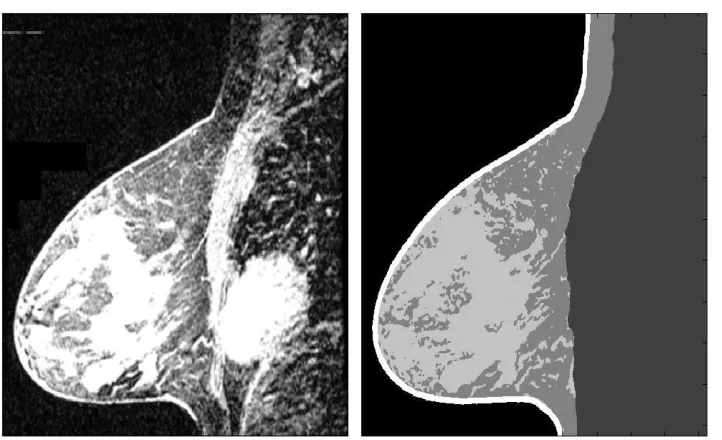
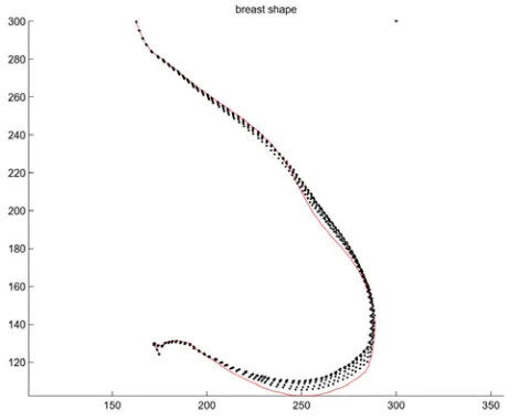
We build first the mechanical framework starting from medical images (MRI data) of patients. The breast contour was successfully segmented using the Lankton et al method [Lankton and Tannenbaum, 2008]. In order to take into account the heterogeneity of the breast tissue, as in [Tanner et al., 2006], we implemented an image classification method. We decompose the breast into three region: dense tissue, fat and fibrous tissue and skin, see Fig. 1.
Fig. 1. Comparision of the initial picture and the segmented one (dense tissue, fat and fibrous tissue, skin)
Based on previous work [Samani et al., 2001], [Chung, 2008], [Rajagopal, 2007], [Ozan, 2008], the mechanical model chosen to simulate breast tissue was the Neo-Hookean hyperelastic model. Hyperelastic material is well adapted to simulate soft tissue deformation. The stress-strain relationship is derived from a strain density energy function denoted W. For the Neo-Hookean material:
2 1
( 3) ( 1)
2 2
k
w I J (1)
W is the strain energy per unit of volume, I1 the first deviatoric strain invariant, μ the initial
shear modulus of the material, k the bulk modulus and J the determinant of the elastic deformation gradient. The material parameter for the Neo-Hookean material are μ and k and represent respectively the stiffness and the compressibility of the material. In isotropic linear elastic material the shear modulus μ and the bulk K can be linked to the Young modulus E and the Poisson ratio of a material by the following equations:
, 2(1 )
E
and 3(1 2 ) E K
(2)
Instead of separating different domain of computation with different material property, heterogeneity of the tissue was directly simulated by imposing a variable Young modulus E(x,y) in the domain.
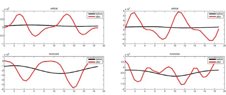
BCT surgical operation removes the breast tumor and surrounding tissue in order to satisfy a negative margin. The numerical simulation shows that the void created by the lumpectomy induces significant mechanical disturbances see Fig. 4. Our hypothesis is that the strain energy plays a key role in the wound healing process [J.D.Murray, 2003]
M. Garbey et al.: Multi-scale modeling and computational surgery: application to breast conservative therapy 84
Fig. 2. Plot of the strain energy function at the wound resection boundary before and after surgery for different gravity direction
3. Multiscale modeling
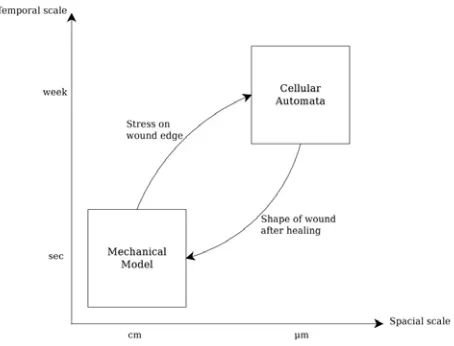
Healing is a process that takes weeks while the soft tissue deformation under a gravity load is in comparison instantaneous. The wound healing process involves complex phenomena engaging biological, chemical and mechanical interactions. This unfolds a two side interaction: on the one hand mechanical stress affects cell mitosis, extracellular matrix production and cell migration underlining tissue production and plasticity; on the other hand new tissue generation impact the breast tissue mechanics and therefore changes the stress distribution inside the breast. We will neglect here the effect of radiotherapy that does affect indeed both the healing history and the tissue mechanical properties [Dormand et al., 2005]. For simplicity we consider one direction and amplitude of gravity, relating to the standing position of the patient. In principle we should take into account the dynamic through the various daily activities such as walking, kneeling or supine position etc... [Gefen and Dilmoney, 2007]. Our multiscale computational model is summarized in Fig. 3.We start from an unloaded initial position of the breast. Thanks to the mechanical model we compute the loaded shape and its stress distribution. The stress is mapped back onto the unloaded shape. This unloaded shape serves as the reference geometry for the healing model that advances the wound edge. From the wound edge progression we output a new unloaded breast shape and compute again the loaded shape and its new stress distribution. We iterate the cycle of Fig. 3 until, eventually, the wound closure.
The simplicity of our modular approach allows us to couple an off the shelf commercial code for the tissue mechanic with an in-house Cellular Automata (CA) code for tissue plasticity. The two models communicate through the location of the wound edge and the spatial distribution of whatever mechanical properties we would like to retain. The unloaded shape serves as the reference stage for mapping. In principle both models can run on adequate separate computer architectures and can be developed independently!
Fig. 3. Computational model
the grid is considered either empty or occupied by a single cell. In principle the ”diameter” of our hexagonal automata is of order of a cell size h = O(10)μm. The 2D computational domain should contain ~ 106 to 107 CA sites. We considered that cells can undergo two types of
transformations: cell division (mitosis), and cell motion. The time step settled in the CA model corresponds roughly to the time scale of a cell cycle, i.e. between 10 to 20 hours. The CA algorithm can be decomposed into four operations that are applied sequentially at each time step.
3.1 Cell division at the wound edge:
Cells that are located at the wound edge can divide with probability pedge. The new daughter cell should occupy one of the free neighbor sites. This cell division might be controlled by some form of contact inhibition [Drasdo, 2005].
Cell Motility: Cell migrations has equal probability to move into one of the 6 directions of the CA grid, provided that the site is free. Rather than using a Monte Carlo method we compute directly the new probability distribution of cells denoted Ui[0,1] using the diffusion operator
t
.At the end of the motility step we select the level set of the probability distribution that insure conservation of ”mass”. More precisely. we select the sites where U U o with
[0,1] o
U such that the total number of cells is conserved.
3.2 Cell division inside the active layer:
We suppose that otherwise cells divide into an active layer next to the wound edge where the concentration of growth factor is high. Let us suppose for simplicity that its dimensions is fixed a priori to .The probability of cell division inside this region denoted plauerwill be either constant (one way coupling), or function of the strain energy, W, provided by the mechanical model (two way coupling):
0(1 ( )) layer
p p F W
M. Garbey et al.: Multi-scale modeling and computational surgery: application to breast conservative therapy 86
3.3 Cell redistribution:
We work with a fix hexagonal grid. In principle the cell distribution should minimize some mechanical energy. We compute the path that link the cell that goes under division to the closest empty site. This site is next to the wound edge. We shift then the cell along that path in order to accommodate the space needed to the new cell. We introduce also some noise in the computation of the optimum path to simulate the fact that cell redistribution may fit a local optimum.
This cell redistribution can be simulated again thanks to the diffusion operator followed by the level set allocation that respect the new number of cells. Overall we obtain a CA scheme that scales well on a parallel system and can accommodate a very large number of cells.
4. Implementation with Distributed Computing
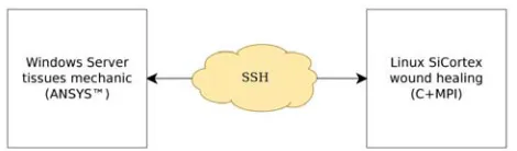
We developed a parallel version of the CA described previously using MPI 2.2 for message passing. This allow us to increase the resolution of our CA in order to reach a realistic order of magnitude for the number of cells. The scheme for our distributed architecture is the following see Figure 4: the main time stepping loop run on a four core PC’ server running at 2 GHz. On the same PC the mechanical model is launched through the finite element modeling software ANSYSTM. The CA code is called after each mechanical simulation and run on a large multicore Sicortex system that has 1434 cores running at 600 Mhz. The CA simulation still takes the vast majority of the total elapse time. Each call to the CA procedure is actually decomposed into three steps. First the input data is sent to the parallel machine through the SCP protocol. Then the code itself is run on this machine through a SSH connection and finally the results are retrieve in the same way after completion of the code.
Fig. 4. A Simple Heterogeneous Distributed Computing Implementation
5. Results
Let us first present the result for the one way coupling where the mitosis rate is fixed and independent of the mechanical stress. In Fig. 5 (left) we show the wound closure history of such a simulation at the end of each cycle. We observe that a relatively low motility parameter value in our CA still allow the wound to keep a roughly circular shape. Further we observe an acceleration of the wound closure as the curvature of the edge increases. This is similar to the model of Javierre et Al ? based on the level set technique, except that the result emerges from the CA’s choice of basic rules and is not imposed priori.
that the symmetry is lost for this configuration, suggesting that more complicated behavior is expected thanks to gravity.
This observation is closer to clinical experience than the previous one. Although these simulations are done for illustration purpose only, they can highlight some phenomenological behavior of the lumpectomy healing process. Beside thanks to parallel computing, we were able to run realistic number of cells of the order of 16K for a two dimensional slice.
Fig. 5. Closing of the wound computed from the CA model: (left) with a fix rate of mitosis;(right) with the rate of mitosis depending on strain energy.
Overall we may compute from that multiscale simulation the surgery outcome after complete healing that indeed may indicate an increased cosmetic defect due to some tissue lost - see Figure 6.
6. Conclusion
We have presented a multiscale model that shows the feasibility of a general computational framework for lumpectomy modeling. This simulation presents the advantage of coupling the biological phenomena with mechanical proper ties.
M. Garbey et al.: Multi-scale modeling and computational surgery: application to breast conservative therapy 88
DD is an essential paradigm not only for simulation but also for refining our model in the future, as a function of clinical trials. Each model component of our BCT framework deals with its own space and/or time scale, its own anatomic representation and is easy to modify for improvement. We strongly believe that this method might be a key to understand the complexity of surgery outcome and to produce some relevant numerical predictions for surgery outcome.
Thanks We would like to thanks the UH IT Reseach Computing Center for its invaluable technical help in distributed computing.
Извод
Моделирање навише скалаикомпјутерскахирургија: применанаконзервативну
терапијудојке
M. Garbey1,2, D. Thanoon1, R.Salmon1, B. Bass3
1Department of Computer Science, University of Houston, Houston, TX 77204, USA
2 The Methodist Institute for Technology Innovation and Education · 3The Methodist Hospital Research Institute, Houston, TX , USA
Резиме
Послелумпектомије, јављајусекозметичкесметњедо 30% иимајунегативанутицајна квалитетживотапацијента, [Clough et al. 1998], [Veiga et al. 2010]. Сложенаинтеракција између механичких сила услед гравитације, расподеле ткива дојке, као и унутрашњи напонгенерисанпроцесомзарастањаиграјудоминантнуулогууодређивањууспехаили неуспеха операције. Сврха овог рада је да се развије математички модел за птврду и предвиђањеефекталумпектомијенапостоперативнукозметику - погледати [M.Garbey et al. 2010] главе 1 и 15. У раду је описан на имиџингу засновани метод декомпозиције домена, спрежућимеханичкимоделткивадојкесамоделомспорогпроцесазарастања.
Кључнеречи: Моделирањенавишескала, конзервативнатерапијадојке
References
J.H. Chung. Modelling Mammographic Mechanics. PhD thesis, Auckland Bioengineering Institute, 2008.
K. Clough, J. Cuminet, A. Fitoussi, C. Nos, and V. Mosseri. Cosmetic sequelae after conservative treatment for breast cancer: classiffication and results of surgical correction. Ann Plast Surg., 41:471–481, 1998.
E.L. Dormand, P. E Banwell, and T. EE Goodacre. Radiotherapy and wound healing. Blackwell Publishing Ltd and Medicalhelplines.com Inc, International Wound Journal, 2, 2005. Dirk Drasdo. Coarse graining in simulated cell populations. Advances in Complex Systems
(ACS), 8(02):319–363, 2005.
A. Gefen and B. Dilmoney. Mechanics of the normal woman’s breast. Technology and Health Care, 15:259–271, 2007.
J.D.Murray. Mathematical Biology: II Spatial Modles and Biomedical Applications. Third Edition Springer New York, 2003.
S. Lankton and A. Tannenbaum. Localizing region-based active contours. IEEE Trans Image Process., 17(11):2029–2039, 2008.
M.Garbey, B. Bass, M. De Matelin, C. Collet, and R. Tran Son Tay. Computational Surgery and Dual Training. Springer Verlag, 2010.
C. Ozan. Mechanical Modeling of Brain and Breast Tissue. PhD thesis, Georgia Institute of Technology, 2008.
V. Rajagopal. Modelling Breast Tissue Mechanics Under Gravity Loading. PhD thesis, Auckland Bioengineering Institute, 2007.
B. Ribba, T. Colin, and S. Schnell. A multiscale mathematical model of cancer, and its use in analyzing irradiation therapies. Theor Biol Med Model., 3, 2006a.
B. Ribba, O. Saut, T. Colin, D. Bresch, E. Grenier, and JP. Boissel. A multiscale mathematical model of avascular tumor growth to investigate the therapeutic benefit of antiinvasive agents. J. Theor. Biol., 243:523–541, 2006b.
A. Samani, Bishop J., Yaffe M.J., and Plewes D.B. Biomechanical 3d finite element modeling of the human breast using mri data, medical imaging. IEEE Trans Med Imaging., 20:271– 279, 2001.
C. Tanner, J. A. Schnabel, M. O. Leach, D. R. Hose, D. L. G. Hill, and D. J. Hawkes. Factors influencing the accuracy of biomechanical breast models. American Association of Physicists in Medicine, 33:1758–1769, 2006.