R E S E A R C H A R T I C L E
Open Access
Tdp-43 cryptic exons are highly variable
between cell types
Yun Ha Jeong
1,5†, Jonathan P. Ling
1†, Sophie Z. Lin
1†, Aneesh N. Donde
1,2, Kerstin E. Braunstein
1, Elisa Majounie
4,6,
Bryan J. Traynor
3,4, Katherine D. LaClair
1, Thomas E. Lloyd
2,3and Philip C. Wong
1,2*Abstract
Background:TDP-43 proteinopathy is a prominent pathological feature that occurs in a number of human diseases including amyotrophic lateral sclerosis (ALS), frontotemporal dementia (FTD), and inclusion body myositis (IBM). Our recent finding that TDP-43 represses nonconserved cryptic exons led us to ask whether cell type-specific cryptic exons could exist to impact unique molecular pathways in brain or muscle.
Methods:In the present work, we investigated TDP-43’s function in various mouse tissues to model disease pathogenesis. We generated mice to conditionally delete TDP-43 in excitatory neurons or skeletal myocytes and identified the cell type-specific cryptic exons associated with TDP-43 loss of function.
Results:Comparative analysis of nonconserved cryptic exons in various mouse cell types revealed that only some cryptic exons were common amongst stem cells, neurons, and myocytes; the majority of these nonconserved cryptic exons were cell type-specific.
Conclusions:Our results suggest that in human disease, TDP-43 loss of function may impair cell type-specific pathways.
Keywords:TDP-43–Nonconserved cryptic exons, Bioinformatics, Amyotrophic lateral sclerosis, Frontotemporal dementia, Inclusion body myositis
Background
Recent genetic evidence has established the linkage be-tween the neurological disorders amyotrophic lateral sclerosis (ALS) and frontotemporal dementia (FTD) [1–5]. The key pathological feature that is shared between ALS and FTD is the cytoplasmic aggregation and nuclear clearance of an RNA binding protein called transactive response DNA binding protein 43 kDa (TDP-43, TARDBP) [6]. Since the discovery of TDP-43, a number of other human diseases have also been characterized with TDP-43 pathology [7–12]. Of particular interest, however, is the pathogenesis of inclusion body myositis (IBM), which is believed to be primarily myogenic rather than neurogenic [13, 14]. To understand the mechanisms of
disease pathogenesis that will inform appropriate thera-peutic strategies, it will be critical to determine whether the pathways affected by TDP-43 proteinopathy differ between neurons and myocytes.
We have recently found that TDP-43 plays a major role in repressing nonconserved cryptic exons [15]. These cryptic exons are regions of the genome that are normally skipped by the spliceosome due to the pres-ence of adjacent UG microsatellite repeats, the consen-sus binding site of TDP-43. When TDP-43 function is lost, these cryptic exons become activated and often lead to nonsense-mediated decay (NMD) of the associated mRNA. In our previous report [15], we utilized an in vitro inducible stem cell model of TDP-43 deletion. However, we have yet to establish the cell type-specific cryptic exons that arise in vivo. Here, we generated conditional Tdp-43 knockout mice to specifically delete Tdp-43 in excitatory neurons and skeletal myocytes. We found that Tdp-43 cryptic exons are highly variable between cell types and that many distinct pathways are
* Correspondence:[email protected] †Equal contributors
1
Departments of Pathology, Johns Hopkins University School of Medicine, Baltimore, MD 21205, USA
2Departments of Neuroscience, Johns Hopkins University School of Medicine, Baltimore, MD 21205, USA
Full list of author information is available at the end of the article
altered—novel findings that have mechanistic and thera-peutic implications for human diseases exhibiting TDP-43 proteinopathy.
Methods
Mouse breeding strategy
We crossbred our conditional Tardbp knockout mice (TardbpF/+) withCamKIIa-Cretransgenic mice to obtain a cohort of CamKIIa-Cre;TardbpF/+mice which were subse-quently crossbred to TardbpF/+ mice to generate the final cohort: CamKIIa-Cre;Tardbp+/+, CamKIIa-Cre;TardbpF/+ and CamKIIa-Cre;TardbpF/F mice. A similar strategy was applied when crossbreeding the MLC-Cre driver line to TardbpF/+ mice. All mouse experiments were approved by the Johns Hopkins University Animal Care and Use Committee.
Histology and immunohistochemistry
For the CamKIIa-Cre line, wildtype and floxed mice were anaesthetized and perfused with 4% paraformalde-hyde. Brains were embedded into paraffin, cut into 10μm sections and stained according to standard proto-cols. For the MLC-Cre line, wildtype and floxed mice were anaesthetized and sacrificed by decapitation. Muscle tissue was then rapidly dissected and flash frozen in liquid nitrogen cooled isopentane. Frozen cryosec-tions were cut at 10μm thickness and stained according to standard protocols. Immunoreactivity was visualized using the Vectastain ABC Kit and diaminobenzidine per-oxidase substrate (Vector Laboratories). Images were ob-tained using Olyumpus BX53 microscope.
Immunoblot analysis
For the CamKIIa-Cre line, wildtype and floxed mice were anaesthetized and sacrificed by decapitation. Brain tissue was then rapidly dissected and manually homoge-nized in RIPA buffer (Sigma) containing an EDTA-free protease inhibitor cocktail (Thermo Scientific). For the MLC-Cre line, wildtype and floxed mice were also anaesthetized and sacrificed by decapitation. Muscle tissue was snap frozen in isopentane cooled with liquid nitrogen, manually ground into a powder, and then homogenized in RIPA buffer with protease inhibitor cocktail. Protein concentration was determined using the BCA assay (Pierce). Proteins were resolved using the NuPAGE 4-12% Bis-Tris Gel (Novex) with NuPAGE MES SDS Running Buffer (Novex), and transferred to PVDF membrane (Millipore) with NuPAGE Transfer Buffer (Invitrogen).
The following antibodies were used for protein blots, immunofluorescence, and immunohistochemical analyses: rabbit anti-TDP-43 (Proteintech 10782-2-AP and 12892-1-AP), NeuN monoclonal antibody (Chemicon), anti-GAPDH monoclonal antibody (Sigma), Alexa Fluor
488-conjugated Donkey anti-Guinea Pig IgG (H + L) antibody (Jackson ImmunoResearch), Alexa Fluor 594- and 647-conjugated Donkey anti-goat and anti-rabbit IgG (H + L) antibodies (Life Tech.).
RNA extraction, RNA-seq analysis
Total RNA was extracted from hippocampi of 3 month old female CamKIIa-Cre;TardbpF/F (neuronal knockout) and littermate control mice (CamKIIa-Cre;Tardbp+/+) using TRIzol (Life Tech.) and RNeasy Mini kits (Qiagen). Total RNA from 2 month old male MLC-Cre;TardbpF/F (skeletal muscle knockout) and littermate control mice (MLC-Cre;Tardbp+/+) was also extracted in a similar manner. For the CamKIIa-Creline, 3 control brains and 3 knockout brains were analyzed and all mice were female. For the MLC-Cre line, 2 control quadriceps and 2 knockout quadriceps were analyzed and all mice were male. 100-bp paired end RNA-seq libraries were gener-ated using Illumina Tru-seq kits and then sequenced on an Illumina HiSeq 2000. For RT-PCR analysis, total RNA was isolated using RNeasy Mini Kit (Qiagen). cDNA was synthetized using RevertAid First Strand cDNA Synthesis Kit (Thermo Scientific) with random primers. RNA-seq analysis was performed using HISAT [16] and Cufflinks [17] software suites and visualized on the UCSC Genome Browser [18]. Cryptic exons were identified as previously described [14]. To identify com-mon pathways between species, gene ontology analysis was performed on cryptic exon targets using manual an-notation of genes with known functions in combination with the bioinformatics resource DAVID v6.7 [19].
RT-PCR primers
Results
Selective deletion of Tdp-43 in mouse excitatory neurons and skeletal myocytes
To identify the cryptic exons repressed by Tdp-43 in neurons and myocytes, we utilized the Cre recombinase system to conditionally delete Tdp-43. Mice harboring floxed Tardbp knockout alleles [20] were crossbred with
Primer Sequence Tissue
either CaMKIIα-Cre [21] or MLC-Cre [22] driver lines (Fig. 1a). The promoter of the calcium/calmodulin-dependent protein kinase II alpha subunit (CaMKIIα) drives expression primarily in the excitatory neurons of the cortex and hippocampus whereas the promoter of the myosin light chain 1/3 locus (MLC) drives expression in type II fast-twitch skeletal muscle fibers. Efficient deletion of Tdp-43 can be detected by immunoblot in brain (Fig. 1b) and skeletal muscle (Fig. 1c); residual Tdp-43 in F/F mice reflects the presence of other cell types that do not express CaM-KIIα-Cre orMLC-Cre. Neuron specific deletion of Tdp-43 was confirmed by immunofluorescence staining of hippo-campal sections (Fig. 1d); deletion of Tdp-43 in myocytes was also verified by immunohistochemistry (Fig. 1e).
Identification of cryptic exons associated with Tdp-43 loss of function in neurons and myocytes
To identify the cryptic exons of mouse neurons, RNA-sequencing (RNA-seq) analysis was performed using RNA extracted from hippocampi of 3 month old CaM-KIIα-Cre;TardbpF/F mice and controls. Similar to our in
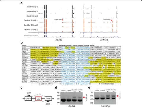
vitrostem cell culture model of Tdp-43 deletion [15], we also found cryptic exons in the brains ofCaMKIIα -Cre;-TardbpF/F knockout mice (Fig. 2a). Neuron-specific cryptic exons were still flanked by UG microsatellite repeats (Fig. 2b) and could be classified as standard cryptic exons, transcriptional start sites, exon extensions or premature polyadenylation sites (Additional file 1: Table S4, Additional file 1: Figure S1). Previously pub-lished CLIP data was also able to confirm the presence of a direct interaction with Tdp-43 (Additional file 1: Figure S2) [23]. Finally, to further validate our RNA-seq data, RT-PCR analysis was able to confirm the presence of cryptic exons in the genes Camk1g and Ap3b2. Longer PCR products, indicating cryptic exon inclusion, were detected in CaMKIIα-Cre;TardbpF/F knockout but not control mice (Fig. 2c-e).
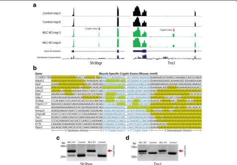
To determine whether cryptic exons of mouse myo-cytes would differ from those found in stem cells and neurons, we also performed RNA-seq analysis on quad-riceps muscle from MLC-Cre;TardbpF/F knockout mice and controls. Indeed, numerous muscle-specific cryptic
Fig. 1Generation ofCaMKIIα-Cre;TardbpF/FandMLC-Cre;TardbpF/Fknockout mice. (a) Breeding strategy to cross floxedTardbpknockout mice with
CaMKIIα-CreorMLC-Cremouse lines to conditionally delete Tdp-43 in excitatory neuron or skeletal muscle, respectively. Hippocampal protein extracts fromCaMKIIα-Cre;TardbpF/Fknockout mice were taken from p25 and 3-month old mice, as indicated. Protein extracts from various muscle groups, as
indicated, were taken from 2-month oldMLC-Cre;TardbpF/Fmice. Immunoblotting confirms deletion of Tdp-43 in the hippocampi ofCaMKIIα-Cre
;-TardbpF/Fknockout mice (b) and the quadriceps ofMLC-Cre;TardbpF/Fknockout mice (c); biological replicates of immunoblotting were performed in
excess ofn= 3 to validate knockdown. (d) Immunofluorescence staining of hippocampal sections from 3 month oldCaMKIIα-Cre;TardbpF/Fknockout
exons could be identified (Fig. 3a). Furthermore, myocyte-specific cryptic exons were also flanked by UG microsatellite repeats (Fig. 3b); the presence of cryptic exons was confirmed by RT-PCR as shown for two genes,Sh3bgrandTns1(Fig. 3c).
Unique Tdp-43 cryptic exons occur in stem cells, neurons, and myocytes
Having identified two new sets of cryptic exons belong-ing to mouse neurons and myocytes, we compared these sites with the cryptic exons previously identified in mouse stem cells [15]. Interestingly, only 66/221 (~30%) total cryptic exons showed any overlap between at least two cell types and only 32/221 (~14%) were common among all three cell types (Fig. 4a). Although the ratios varied, the majority of cryptic exons were unique to each individual cell type (155/221; ~70%). When normalized to the total number of cryptic exons in stem cells (74),
neurons (109) and myocytes (136), the number of cell type-specific cryptic exons was lower in stem cells (18; ~24%) as compared to neurons (58; ~53%) and myocytes (79; ~58%). These results indicate that a large proportion of Tdp-43’s cryptic exons are cell type-specific (Additional file 1: Table S1 and S2).
Differential levels of cryptic exon incorporation, however, increase the complexity of these cryptic exon datasets. While certain cryptic exons, such as those in Synj2bpandAdnp2, can be observed at high levels in all three cell types (Fig. 4b), it is more common to see differential usage of cryptic exons amongst stem cells, neurons, and myocytes despite abundant transcription of the associated mRNA (Fig. 4c-g). For example, the cryp-tic exon in Ube2d1is highly incorporated in stem cells, moderately incorporated in myocytes, and absent in neurons (Fig. 4c). Conversely, the cryptic exon inRrp36 is high in neurons but low in stem cells and myocytes Fig. 2Neuron-specific cryptic exons (CaMKIIα-Cre;TardbpF/Fknockout mice). (a) Visual examples of neuron-specific cryptic exons (Ap3b2,Camk1g).
(b) Neuron-specific cryptic exons are flanked by UG repeats that are present upstream, downstream or within the cryptic exon sequence itself. (ctoe) RT-PCR validation of cryptic exons (red arrows) in RNA extracted from hippocampi of 3 month oldCaMKIIα-Cre;TardbpF/Fmice. Refer to
(Fig. 4d). Thus, it appears that the activation of a cryptic exon within a specific cell type depends not only upon transcription of the associated mRNA, but also the local splicing factor environment present within the cell (Additional file 1: Figure S3).
Comparative analysis of genes affected by cryptic exon disruption
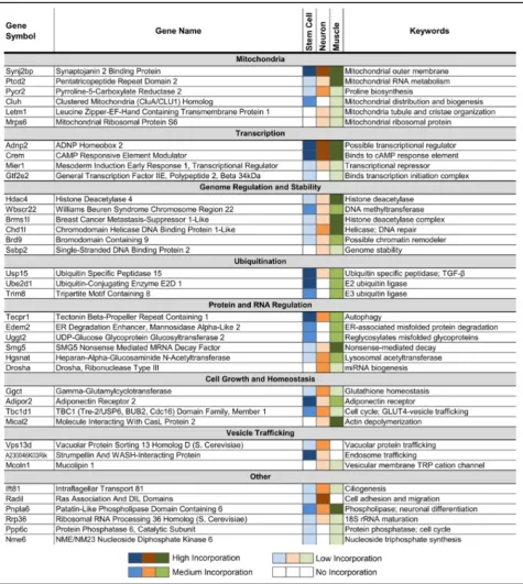
We have previously shown that Tdp-43’s nonconserved cryptic exons could disrupt gene function in cultured stem cells [15]. Similarly, while some neuron and myocyte cryptic exons reside in the 5’ or 3’ untranslated regions (~19%) with no clear effect on transcript levels, the major-ity of cryptic exons disrupt normal protein translation by introducing premature stop codons that lead to nonsense mediated decay (Additional file 1: Figure S4) or early termination of the mRNA transcript (~63%). Of these disrupted genes, numerous critical pathways are affected, ranging from mitochondrial function and protein regula-tion to transcripregula-tional control and genome stability (Table 1). These findings demonstrate that cell
type-specific pathways are altered when Tdp-43 function is lost and suggest that unique molecular pathways could differ-entially impact ALS-FTD and IBM.
Common pathways affected by Tdp-43 loss of function
Although many cryptic exons are predicted to induce nonsense mediated decay, their impact on mRNA and protein levels depends upon the frequency of cryptic exon incorporation. Across stem cells, neurons and myocytes, a broad group of genes are affected by Tdp-43 loss of function (Table 1). Many pathways are affected, from mitochondrial function and cell growth to tran-scription and genomic regulation, offering a possible explanation for the observed cell death associated with Tdp-43 deletion [24–28]; CaMKIIα-Cre;TardbpF/F ex-hibit significant cortical atrophy at 8 months of age [12] while MLC-Cre;TardbpF/F mice reach endstage by 4–5 months. Several other genes that are disrupted by cryptic exons also reflect previously reported observations: Drosha is involved in miRNA biogenesis [29],Tecpr1 is Fig. 3Muscle-specific cryptic exons (MLC-Cre;TardbpF/Fknockout mice). (a) Visual examples of muscle-specific cryptic exons (Sh3bgr,Tns1). (b) Muscle-specific cryptic exons are flanked by UG repeats that are present upstream, downstream or within the cryptic exon sequence itself. (cand
involved in autophagy [30], andTbc1d1 andAdipor2are involved in fat metabolism [20].
Interestingly, a low percentage of cryptic exons (~6%) do not induce nonsense mediated decay, but still have an impact on protein structure. These cryptic exons do not contain any stop codons and have sequence lengths that are multiples of three, thereby preventing detrimen-tal frameshifts (Additional file 1: Table S3). These inframe cryptic exons introduce short peptide insertions into the primary amino acid sequence of the protein, which may represent neoantigens.
Discussion
We have found that Tdp-43’s nonconserved cryptic exons vary widely between cell types and affect many pathways that are critical for neuronal and muscle physi-ology. This suggests that in human disease, myogenic and neurogenic TDP-43 proteinopathies exhibit cell type-specific cryptic exons that could influence disease progression in unique ways. Although our RNA-seq data are based on a limited number of samples, future ana-lysis to increase sample sizes would strengthen our find-ings. Identifying the cryptic exons that are specific to human neurons or myocytes will also help clarify the
selective vulnerability associated with diseases such as IBM and ALS-FTD.
While it remains to be proven whether TDP-43 loss of function is a central driver of human disease, our data demonstrates that within neurons and myocytes, TDP-43 is the major splicing repressor for numerous noncon-served cryptic exons. In human disease, dysregulation of Tdp-43 function may impair other neuronal functions beyond mRNA splicing such as axonal trafficking, hyper-excitability, and liquid-liquid phase separation [31–34]. Nevertheless, mouse models of Tdp-43 have demon-strated that constitutive deletion of Tardbp results in embryonic lethality [24, 25, 35, 36]. Conditional deple-tion of Tardbp in adult mice also leads to metabolic deficits and premature death [20] and significant neuro-degeneration [26, 37, 38]. Together, these studies dem-onstrate the importance of Tdp-43 for cell survival.
contribute to cell death. Furthermore, TDP-43 belongs to a family of proteins that repress cryptic exons, suggesting that these splicing factors perform a general function in the cell to maintain splicing fidelity [42]. Thus, loss of TDP-43 splicing repression contributes to
cell death and the pathways affected by cryptic exon incorporation are likely to be relevant for disease pathogenesis.
The question then becomes, how do we prevent incorporation of nonconserved cryptic exons? Therapeutic Table 1Common pathways affected by Tdp-43 cryptic exons across mouse stem cell, muscle and neuron (cryptic exon present in at least two cell-types)
strategies that aim to directly interfere with cryptic exon splicing (e.g. anti-sense oligonucleotides) will be difficult to envision due to the sizeable number of nonconserved cryptic exons per cell. Furthermore, because noncon-served cryptic exons are different between mouse and hu-man, testing splicing modulators for human cryptic exons in animal models is essentially impossible. However, the general splicing repression function of TDP-43 is conserved. Thus, it may be possible to use mouse models of TDP-43 deletion to specifically test therapeutic strategies that rescue TDP-43 mechanism of action rather than directly targeting individual cryptic exons. One strategy would employ gene therapy to introduce designer splicing factors—chimeric proteins that would couple the UG binding domain of TDP-43 with non-aggregating splicing repressor domains [15]—into neurons or muscles. In principal, this approach would repress most of TDP-43’s nonconserved cryptic exons in a manner that would be species-independent.
If neuron loss or skeletal muscle degeneration can be attenuated, such a therapeutic strategy could be rapidly translated into the clinic. Moreover, the observation that cryptic exons can occasionally introduce inframe inser-tions into mRNA suggests that certain human TDP-43 cryptic exons could represent biomarkers for human disease. We envision the development of specific anti-bodies to detect neoantigens introduced by human inframe cryptic exons in CSF or blood from patients, serving as either diagnostic biomarkers or tools to moni-tor the efficacy of treatments in future clinical trials.
Conclusions
This study demonstrates that Tdp-43 represses a unique set of cryptic exons, depending on cellular context. Thus, the pathways impacted by Tdp-43 loss-of-function and cryptic exon incorporation are likely distinct for each cell type. These results have important implications for human disease, given that Tdp-43 proteinopathy can manifest in various tissues.
Additional files
Additional file 1:Supplemental figures and tables. (PDF 4449 kb)
Additional file 2:Cryptic Exon Data Table. (XLSX 59 kb)
Abbreviations
ALS:Amyotrophic lateral sclerosis; CaMKIIα: Calcium/calmodulin-dependent protein kinase II alpha; FTD: Frontotemporal dementia; IBM: Inclusion body myositis; MLC: Myosin light chain 1/3 locus; NMD: Nonsense-mediated decay; TDP-43: Transactive response DNA binding protein 43 kDa.
Acknowledgments
We thank V. Nehus for technical assistance.CamKIIaCreandMLC-Cremice were kindly gifted, respectively, by P. Worley (Johns Hopkins University School of Medicine) and S. Burden (New York University School of Medicine).
Funding
This work was supported in part by The Robert Packard Center for ALS Research, the Amyotrophic Lateral Sclerosis Association, Target ALS, the JHU Neuropathology Pelda fund, DoD grant W81XWH1110449, Korea Brain Research Institute basic research program Grant No. 2231–415 (to YHJ), the McKnight Memory and Cognitive Disorders Award, and NIH grant R01-NS095969.
Availability of data and materials
The datasets supporting the conclusions of this article are included within the article and its Additional files 1 and 2. RNA-seq FASTQ sequencing files have been deposited at the NCBI Sequence Read Archive under SRP061340.
Authors’contributions
All authors designed experiments and interpreted results. JPL performed cryptic exon analyses. YHJ and AND characterized neuron Tdp-43 deletion mice. SZL, KEB and TEL characterized muscle Tdp-43 deletion mice. EM. and BJT assisted with RNA-sequencing. KDL assisted with pathway analysis. JPL and PCW wrote the paper and all authors approved the manuscript.
Authors’information
Not applicable.
Competing interests
J.P.L. and P.C.W. have filed a patent application in the United States that refers to the use of cryptic exon incorporation in RNA transcripts identified in human diseases that exhibit TDP-43 proteinopathy as the basis for biomarkers and therapeutic targets/strategies.
Consent for publication
Not applicable.
Ethical approval and consent to participate
Not applicable.
Author details
1Departments of Pathology, Johns Hopkins University School of Medicine, Baltimore, MD 21205, USA.2Departments of Neuroscience, Johns Hopkins University School of Medicine, Baltimore, MD 21205, USA.3Departments of Neurology, Johns Hopkins University School of Medicine, Baltimore, MD 21205, USA.4Laboratory of Neurogenetics, NIA, NIH, Bethesda, MD 20892, USA.5Neural Development and Disease Department, Korea Brain Research Institute, Daegu 701-300, South Korea.6Present address: Institute of Psychological Medicine and Clinical Neurosciences, Cardiff University School of Medicine, Cardiff CF24 4HQ, UK.
Received: 25 May 2016 Accepted: 20 December 2016
References
1. Renton AE, et al. A hexanucleotide repeat expansion in C9ORF72 is the cause of chromosome 9p21-linked ALS-FTD. Neuron. 2011;72(2):257–68. 2. DeJesus-Hernandez M, et al. Expanded GGGGCC hexanucleotide repeat in
noncoding region of C9ORF72 causes chromosome 9p-linked FTD and ALS. Neuron. 2011;72(2):245–56.
3. Freischmidt A, et al. Haploinsufficiency of TBK1 causes familial ALS and fronto-temporal dementia. Nat Neurosci. 2015;18(5):631–6.
4. Cirulli ET, et al. Exome sequencing in amyotrophic lateral sclerosis identifies risk genes and pathways. Science. 2015;347(6229):1436–41.
5. Ling S-CC, Polymenidou M, Cleveland DW. Converging mechanisms in ALS and FTD: disrupted RNA and protein homeostasis. Neuron. 2013;79(3):416–38. 6. Neumann M, et al. Ubiquitinated TDP-43 in frontotemporal lobar degeneration
and amyotrophic lateral sclerosis. Science. 2006;314(5796):130–3.
7. Josephs KA, et al. Staging TDP-43 pathology in Alzheimer’s disease. Acta Neuropathol. 2014;127:441–50.
8. Josephs KA, et al. Updated TDP-43 in Alzheimer’s disease staging scheme. Acta Neuropathol. 2016;131(4):571–85.
10. Weihl CC, et al. TDP-43 accumulation in inclusion body myopathy muscle suggests a common pathogenic mechanism with frontotemporal dementia. J Neurol Neurosurg Psychiatry. 2008;79:1186–9.
11. Hiniker A, Daniels BH, Margeta M. T-Cell-Mediated Inflammatory Myopathies in HIV-Positive Individuals: A Histologic Study of 19 Cases. J Neuropathol Exp Neurol. 2016;75(3):239–45.
12. LaClair KD, et al. Depletion of TDP-43 decreases fibril and plaqueβ-amyloid and exacerbates neurodegeneration in an Alzheimer’s mouse model. Acta Neuropathol. 2016;132(6):859–73.
13. Lloyd TE. Novel therapeutic approaches for inclusion body myositis. Curr Opin Rheumatol. 2010;22(6):658–64.
14. Lloyd TE, et al. Evaluation and construction of diagnostic criteria for inclusion body myositis. Neurology. 2014;83(5):426–33.
15. Ling JP, Pletnikova O, Troncoso JC, Wong PC. TDP-43 repression of nonconserved cryptic exons is compromised in ALS-FTD. Science. 2015; 349(6248):650–5.
16. Kim D, Langmead B, Salzberg SL. HISAT: a fast spliced aligner with low memory requirements. Nat Methods. 2015;12(4):357–60.
17. Trapnell C, et al. Differential gene and transcript expression analysis of RNA-seq experiments with TopHat and Cufflinks. Nat Protoc. 2012;7:562–78. 18. James Kent W, et al. The human genome browser at UCSC. Genome Res.
2002;12:996–1006.
19. Dennis G, et al. DAVID: Database for Annotation, Visualization, and Integrated Discovery. Genome Biol. 2003;4:P3.
20. Chiang P-M, et al. Deletion of TDP-43 down-regulates Tbc1d1, a gene linked to obesity, and alters body fat metabolism. Proc Natl Acad Sci U S A. 2010;107(37):16320–4.
21. Casanova E, et al. A CamKIIalpha iCre BAC allows brain-specific gene inactivation. Genesis. 2001;31(1):37–42.
22. Mourkioti F, Slonimsky E, Huth M, Berno V, Rosenthal N. Analysis of CRE-mediated recombination driven by myosin light chain 1/3 regulatory elements in embryonic and adult skeletal muscle: a tool to study fiber specification. Genesis. 2008;46(8):424–30.
23. Polymenidou M, et al. Long pre-mRNA depletion and RNA missplicing contribute to neuronal vulnerability from loss of TDP-43. Nat Neurosci. 2011;14(4):459–68. 24. Sephton CF, et al. TDP-43 is a developmentally regulated protein essential
for early embryonic development. J Biol Chem. 2010;285(9):6826–34. 25. Kraemer BC, et al. Loss of Murine TDP-43 disrupts motor function and plays
an essential role in embryogenesis. Acta Neuropathol. 2010;119(4):409–19. 26. Yang C, et al. Partial loss of TDP-43 function causes phenotypes of
amyotrophic lateral sclerosis. Proc Natl Acad Sci U S A. 2014;111(12):E1121–9. 27. Feiguin F, et al. Depletion of TDP-43 affects Drosophila motoneurons
terminal synapsis and locomotive behavior. FEBS Lett. 2009;583(10):1586–92. 28. Schmid B, et al. Loss of ALS-associated TDP-43 in zebrafish causes muscle
degeneration, vascular dysfunction, and reduced motor neuron axon outgrowth. Proc Natl Acad Sci U S A. 2013;110:4986–91.
29. Kawahara Y, Mieda-Sato A. TDP-43 promotes microRNA biogenesis as a component of the Drosha and Dicer complexes. Proc Natl Acad Sci U S A. 2012;109(9):3347–52.
30. Bose JK, Huang C-C, Shen C-KJ. Regulation of autophagy by neuropathological protein TDP-43. J Biol Chem. 2011;286(52):44441–8. 31. Alami NH, et al. Axonal transport of TDP-43 mRNA granules is impaired by
ALS-causing mutations. Neuron. 2014;81(3):536–43.
32. Zhang W, et al. Hyperactive somatostatin interneurons contribute to excitotoxicity in neurodegenerative disorders. Nat Neurosci. 2016;19(4):2–6. 33. Wegorzewska I, Bell S, Cairns NJ, Miller TM, Baloh RH. TDP-43 mutant
transgenic mice develop features of ALS and frontotemporal lobar degeneration. Proc Natl Acad Sci U S A. 2009;106(44):18809–14. 34. Taylor JP, Brown RH, Cleveland DW. Decoding ALS: from genes to
mechanism. Nature. 2016;539(7628):197–206.
35. Wu L-S, et al. TDP-43, a neuro-pathosignature factor, is essential for early mouse embryogenesis. Genesis. 2010;48(1):56–62.
36. Tsao W, et al. Rodent models of TDP-43: recent advances. Brain Res. 2012;1462:26–39.
37. Schwenk BM, et al. TDP-43 loss of function inhibits endosomal trafficking and alters trophic signaling in neurons. EMBO J. 2016;35(21):2350–70. 38. Walker AK, et al. Functional recovery in new mouse models of ALS/FTLD
after clearance of pathological cytoplasmic TDP-43. Acta Neuropathol. 2015;130(5):643–60.
39. Tan Q, et al. Extensive cryptic splicing upon loss of RBM17 and TDP43 in neurodegeneration models. Hum Mol Genet. 2016. doi:10.1093/hmg/ddw337.
40. Humphrey J, Emmett W, Fratta Pi, Isaacs AM, Plagnol V. Quantitative analysis of cryptic splicing associated with TDP-43 depletion. bioRxiv: 2016;1–21. https://doi.org/10.1101/076117.
41. Li Z, Vuong JK, Zhang M, Stork C, Zheng S. Inhibition of nonsense-mediated RNA decay by ER stress. RNA. 2016. doi:10.1261/rna.058040.116.
42. Ling JP, et al. PTBP1 and PTBP2 Repress Nonconserved Cryptic Exons. Cell Rep. 2016;17(1):104–13.
• We accept pre-submission inquiries
• Our selector tool helps you to find the most relevant journal
• We provide round the clock customer support
• Convenient online submission
• Thorough peer review
• Inclusion in PubMed and all major indexing services
• Maximum visibility for your research
Submit your manuscript at www.biomedcentral.com/submit

![Fig. 4 Tdp-43 cryptic exons are highly variable between cell types. (a) While some cryptic exons are common between cell types, many crypticexons are unique to neurons (58), muscle (79) and stem cell [22]](https://thumb-us.123doks.com/thumbv2/123dok_us/276775.1520512/6.595.56.540.87.380/cryptic-highly-variable-cryptic-common-crypticexons-neurons-muscle.webp)