Abnormal Central Complex Is a Marker of Severity in the Presence of
Partial Ciliary Defect
Aline Tamalet, MD*; Annick Clement, MD*; Francoise Roudot-Thoraval, MD‡; Pascale Desmarquest, MD*; Gilles Roger, MD*; Miche`le Boule´, MD*; Marie Claude Millepied, TA§; Armelle Baculard, MD*; and
Estelle Escudier, MD储
ABSTRACT. Background. Ciliary ultrastructural de-fects with total lack of dynein arms (DA) cause abnormal mucociliary function leading to the chronic infections observed in primary ciliary dyskinesia. The role of par-tial ciliary ultrastructural defects, especially those in-volving the central complex, and their relationship with respiratory symptoms have been less thoroughly inves-tigated.
Objective. In a pediatric population with partial cili-ary defects, we determined the relationship(s) between ultrastructural findings, ciliary motility, and clinical and functional features, and evaluated the outcome of this population.
Design. We analyzed the clinical presentation and pulmonary function of 43 children with chronic bronchi-tis and partial ultrastructural defects (from 15% to 90%) of their respiratory cilia demonstrated on bronchial bi-opsies. The study population was divided into 3 groups according to ciliary ultrastructure: the main ultrastruc-tural defect concerned the central complex in 23 patients (CC group), peripheral microtubules in 8 patients (PMT group), and DA in 12 patients (DA group).
Results. The percentage of ciliary defects was lower in the PMT group than in the CC and DA groups. Pa-tients in the PMT group had less severe disease with frequent normal ciliary motility. Patients in the CC group had initially a higher incidence of respiratory tract infec-tions, extensive bronchiectasis frequently requiring sur-gery, and arguments in favor of a congenital origin (high proportion of sibling form). Partial absence of DA, al-though of congenital origin, was associated with a good prognosis. In all groups, follow-up showed that the func-tional prognosis remained good with appropriate treat-ment.
Conclusions. In children with chronic respiratory in-fections, presence of situs inversus, sibling form, ob-structive pulmonary syndrome, or bronchiectasis re-quired ultrastructural analysis, regardless of ciliary motility. Detection of CC abnormalities is a marker of severity and required intensive therapy and close follow-up. Pediatrics 2001;108(5). URL: http://www.
pediatrics.org/cgi/content/full/108/5/e86;;respiratory tract infection, pediatric population, ciliary beat frequency, cil-iary ultrastructure, bronchiectasis, pulmonary function.
ABBREVIATIONS. CBF, ciliary beat frequency; DA, dynein arms; PCD, primary ciliary dyskinesia; CT, computed tomography; FRC, functional residual capacity; FEV1, forced expiratory volume; CC, central complex; PMT, peripheral microtubules.
A
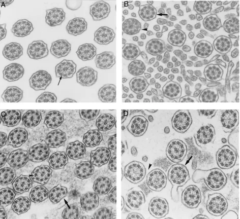
irways are lined by respiratory epithelium composed of 2 main cell types, ciliated and goblet cells, which together ensure efficient mucociliary transport.1,2Mucociliary transport is animportant defense mechanism, and ciliary beat fre-quency (CBF) is a major parameter in airway clear-ance. Each cilium beats in a coordinated fashion with its neighbors, producing unidirectional mucus flow. The structural components of the core of the cilium, known as the axoneme, is highly conserved and in-clude 9 peripheral doublet microtubules with at-tached dyneis and radial spokes, and 2 central single microtubules (Fig 1A). Inner and outer dynein arms (DA) are the transducers of mechanical force neces-sary for ciliary motion. Failure of ciln armiary struc-ture and function impairs airway clearance and can be responsible for respiratory tract infections.3–5
Cil-iary insufficiency can be caused by either an inborn error or damage inflicted on the cilia by a noxious agent.6 –10Congenital defects of ciliary ultrastructure
and function correspond to primary ciliary dyskine-sia (PCD), including Kartagener’s syndrome, which predisposes to upper and lower respiratory tract in-fections beginning in early childhood.6,11,12 In the
presence of a suggestive clinical presentation (eg, situs inversus, bronchiectasis, nasal polyposis), a 2-step protocol with CBF study followed by ultra-structural evaluation is classically proposed to con-firm ciliary abnormalities. Although the diagnosis of PCD is easy to confirm when ciliary motility is ab-normal and when all cilia share the same ultrastruc-tural defect (eg, absence of DA), it is sometimes difficult to conclude when the ultrastructural defects do not affect all cilia and when CBF seems to be normal. However, early diagnosis of PCD with ade-quate prevention and therapy of respiratory tract infections may play an important role in minimizing lung damage.
In the present study, we wanted to define, in a pediatric population with partial ciliary defects, the From the *Pediatric Pulmonology and ENT Departments, Armand
Trous-seau Hospital (AP-HP), Paris, France; the ‡Evaluation and Biostatistics Department, H. Mondor Hospital (AP-HP), Cre´teil, France; the §Depart-ment of Pathology (Electron Microscopy Laboratory), Intercommunal Hos-pital, Cre´teil, France; the㛳Departments of Genetics, Cytogenetics and Em-bryology (Biology of Reproduction Unit), Pitie´ Salpetrie`re Hospital (AP-HP), Paris, France; and INSERM U492, Cre´teil, France.
Received for publication Jan 16, 2001; accepted Jun 27, 2001.
Reprint requests to (A.T.) De´partement de Pneumologie Pe´diatrique, Ho-pital Armand-Trousseau, 26 av Dr Netter, 75012 Paris, France. E-mail: [email protected]
relationships between ultrastructural findings, cili-ary motility, and clinical and functional features. We also evaluated the clinical and functional outcome of this specific population.
PATIENTS AND METHODS Patients
Since 1985, it has been our policy to include examination of ciliary structure and function when investigating children with chronic respiratory tract bronchitis of unknown cause. Examina-tion of ciliary structure and funcExamina-tion was performed, in combina-tion with other investigacombina-tions, in children with chronic or recur-rent lower respiratory tract infections, who were seen in the pediatric pneumology department of our institution. All these children were investigated because of the increased incidence and severity of their respiratory tract infections, occurring more than once a month for at least 6 consecutive months. Respiratory tract infections were defined by a persistent cough with bronchial ronchi, associated with or without fever. All investigations were performed in the absence of acute respiratory tract infection. Pa-tients and their parents were informed of the exact nature and the
goal any of the investigations performed and gave their informed consent. All known pathologic conditions, such as cystic fibrosis,
␣1-antitrypsin deficiency or immunodeficiency, were previously excluded in these children. The only abnormalities detected in some patients of our study population were partial ciliary defects. Between 1989 and 1999, partial ciliary defects (from 15% to 95% of abnormal cilia with various axonemal defects) possibly associ-ated with alterations of ciliary motility were detected in 50 chil-dren. Complete records for 43 of 50 patients were available for analysis in this population and these 43 children (24 boys, 19 girls, aged between 1–13 years, mean: 5.8⫾3.3 years) constituted our study population.
Initial Evaluation
radiograph and computed tomography (CT) scan in all cases. Situs in versus was noted on chest radiograph. The presence of bron-chiectasis (internal diameter of bronchus larger than that of an adjacent artery) was assessed on CT scan, and its topography was scored as absent, unilateral, or bilateral. Blood gases on arterial-ized capillary blood were determined in all patients.13Two pul-monary function tests were studied in children under 7 years old: functional residual capacity (FRC) assessed by the helium dilution method and lung dynamic compliance assessed by the esophageal catheter method.14After the age of 7, FRC study was combined with spirometry for determination of vital capacity and forced expiratory volume (FEV1). Results were expressed as a percentage of the expected value for age and considered as normal when
⬎80% of the expected value.
Follow-up
The follow-up data were collected during each consultation, scheduled every 3 months. The follow-up of our patients ranged from 1 to 7 years (median: 4 years) except for 2 children who were lost for follow-up.
We evaluated and scored the frequency of antibiotic use pre-scribed over the entire follow-up period for their lower or upper respiratory tract infections (no antibiotics, intermittent or contin-uous). The course of bronchiectasis was evaluated by CT scan performed every 2 years in 25 of 41 patients and classified as stable or progressive. Radiologic deterioration corresponded to bronchi-ectasis extension. Pulmonary function tests were performed at least twice in 35 of 41 children, at a mean interval of 6 years. Any thoracic surgery was noted (absence of surgery or lobectomy).
Ciliary Ultrastructure and Function
Bronchial biopsies were obtained with flexible forceps intro-duced through the biopsy channel (Olympus Ldt, Japan, BF type 3C30 or P20D, depending on age) during fiber-optic bronchos-copy. Two bronchial mucosa specimens were sampled from the first bronchial divisions, in the absence of acute respiratory tract infection: 1 by biopsy for ultrastructural evaluation, 1 by brushing for CBF evaluation.
Bronchial biopsies were immersed in 2.5% glutaraldehyde in 0.045 M cacodylate buffer at pH 7.4 and processed as usual for ultrastructural analysis.15After fixation, samples for transmission electron microscopy were postfixed in OsO4and routinely pro-cessed. Ultrathin sections were studied at a final magnification of 60 000. At least 50 transverse sections through the body of ciliary shafts of different cells were analyzed in each specimen to study the internal axonemal structure according to a quantitative meth-od.11 Dynein arms were considered to be absent from sections when the structure was missing from at least 6 of the 9 peripheral doublets. To facilitate the definition of axonemal abnormalities, the central structures (central microtubules and central sheath) were termed the “central complex” (CC). Abnormalities of periph-eral microtubules (PMT) included absence of doublet(s) and su-pernumerary microtubule(s). Ciliary ultrastructural results were expressed as a percentage of abnormal cilia among the total num-ber of cilia analyzed. As some cilia in control specimens can exhibit ultrastructural defects, a cut off value of 15% abnormal cilia is usually considered to be abnormal.16For each ciliary study, axonemal abnormalities (involving the CC, PMT, and dynein arm [DA]) were quantified and expressed as a percentage of each ultrastructural defect among the total number of abnormal cilia and defined the main ultrastructural defect. Ciliary orientation was systematically evaluated by comparing the position of the central pairs of adjoining cilia. Disorientation was defined as an angle greater than 25°.17
For CBF evaluation, the respiratory cells obtained by brushing were suspended in Menezo’s B2 medium and the sample was examined within 3 hours under a light microscope (Nachet NS 400, Paris, France) at magnifications of x 250 and x 400. CBF was measured at room temperature using computerized microscope photometry (Leitz/Leica, Wetzlar, Germany). Briefly, the cilia were positioned to interrupt a passage of light through a small diaphragm in the Leitz MPV compact microscope photometer. This light intensity was converted into an electric signal that was subsequently expressed as CBF in Hz using a CBF processor unit. For each patient, ciliary beat frequency was determined on at least five different areas of ciliated epithelium and a mean value was calculated. When only immotile cilia were observed, CBF was
considered to be 0 Hz. According to the literature, ciliary motility was considered to be normal when CBF was greater than 8 Hz, and abnormal in the case of immotile cilia or when CBF was less than 8 Hz.11,12
Statistical Analysis
Results are expressed as mean⫾standard deviation (SD), or as percentages. For comparison of quantitative data, Mann-Whitney and Kruskal-Wallis tests were used to compare data classified in two or more categories, respectively. When necessary, the Pearson
2test or the Fisher exact test was applied to assess the relation-ship between categorical data. P ⱕ .05 was considered to be significant.
RESULTS
The study population was divided into 3 groups according to the ciliary ultrastructural results: the main ultrastructural defect involved the CC in 23 patients (54%), PMT in 8 patients (18%), and DA in 12 patients (28%; Fig. 1).
The results of ciliary studies and the various clin-ical, radiologic, and functional parameters estab-lished at the initial assessment and during follow-up were then evaluated and compared between the 3 groups.
Analysis of Ciliary Ultrastructure and Relationship With Ciliary Motility
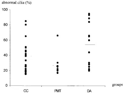
The mean percentages of ciliary abnormalities were statistically different (P⬍.03) between the CC, PMT, and DA groups (39.4 ⫾ 18.7%, 28.4⫾ 15.7%, 56⫾28%, respectively; Fig 2). The mean percentages of the main ultrastructural defect were similar in the CC, PMT, and DA groups (79.7⫾19.6%, 70⫾15.6%, 82.6⫾ 18.9%, respectively). Although the difference was not statistically significant, the percentages of patients with abnormal ciliary motility were higher in the CC and DA groups, 75% and 83%, respectively compared with 25% in the PMT group (Fig 3, Table 1). It should be stressed that no patient in the PMT group had immotile cilia and that ciliary motility was never normal when the ultrastructural defects affected⬎60% of the cilia. Ultrastructural abnormal-ities and ciliary orientation were not related to each other (data not shown).
Clinical and Functional Evaluation in the 3 Groups Comparison of the various parameters of the ini-tial and follow-up assessments between the 3 sub-groups is shown in Table 1. Mean age at diagnosis was 5.8⫾3.3 years and was similar in the CC, PMT, and DA groups (6.7⫾3.5 years, 5.4⫾3 years, 5.3⫾ 4 years, respectively). The incidence of neonatal re-spiratory distress and the age of onset was similar in the three groups. Situs in versus was only observed in the DA group (6/6), and all but 1 of the sibling forms belonged to the CC group (8/9). The incidence of consanguinity was similar in the 3 groups. Pa-tients in the CC group had more frequent lower respiratory tract infections (56.5%) than patients in the PMT or DA groups (25% and 16.7%, respectively;
P⬍.05). A similar difference was observed for bron-chiectasis: 69.6% vs 57% and 50% in the MTP and DA groups, but was not significant. It must be noted that bronchiectasis was bilateral in 47.8% of cases in the CC group versus 14% and 30% in the other groups.
Follow-Up
Serial pulmonary function tests were performed at the ages of 6, 9, and 12 years in 33 children. Mean follow-up (about 3 years) was shorter for the PMT group. The mean arterial Po2 was in the normal
range in each group (CC group: 81⫾10 mm Hg, DA group: 89 ⫾ 11 mm Hg, PMT group: 88 ⫾ 12 mm Hg). Obstruction, evaluated for the youngest pa-tients by dynamic lung compliance, was lower in the CC group (57.4% of predicted value) than in the DA group (69% of PV). After the age of 7 years, obstruc-tion evaluated by FEV1was lower in the CC group
(82⫾24% of predicted value) than in the DA group (96 ⫾ 5% of predicted value), but remained within the normal range. The results in the PMT group could not be compared with those of the other
groups, as FEV1was measured in only 2 patients in
this group.
Treatment included chest physiotherapy, and con-tinuous or intermittent antibiotics for all patients studied. Antibiotic use and radiologic deterioration were similar in the 3 groups (Table 1). The incidence of thoracic surgery was significantly higher (P⬍.05) in the CC group (52.2%) than in the other groups (12.5% in the PMT group and 14.7% in the DA group). Pulmonary function remained stable throughout the study period. FEV1showed a similar
trend and was always greater than 80% of predicted values for healthy participants (data not shown). The degree of distension, reflected by FRC, tended to increase, but remained within the normal range (ini-tially in the CC group, in the DA group and in the PMT group, 89⫾15%, 96⫾ 15% and 101⫾11%, of predictive values, respectively; after 6 years of fol-low-up in the CC group and the DA group, 99⫾15% and 93⫾10%, respectively). The results in the PMT group could not be compared with those of the other groups, as FRC was measured in only 2 patients in this group.
DISCUSSION
Comparison between ciliary studies and clinical presentation of patients with partial ultrastructural defects showed that the severity of the respiratory disease was related to the type of ultrastructural defect but not to ciliary motility. In our population, no strict relationship was demonstrated between the main ultrastructural defect and CBF in accordance with previous reports.12 Abnormal CC was
associ-ated with a higher frequency of respiratory tract infections and extensive bronchiectasis frequently re-quiring surgery, suggesting a more severe disease. On the other hand, in every case, follow-up showed that the functional prognosis remained good when the patients received appropriate therapy.
The percentage of ultrastructural defects was sig-nificantly lower in the PMT group than in the CC and DA groups. Compared with the other groups, the PMT group contained fewer patients. These pa-tients seemed to have less severe disease with fre-quently normal ciliary motility, initially fewer
infec-tions, and a lower incidence of surgery.
Abnormalities of PMT are considered to be acquired ciliary defects, usually observed in patients with chronic or recurrent infection.18 In the PMT group,
limited zones of bronchiectasis, absence of situs in-versus, and a low incidence of familial cases consti-tute arguments in favor of a non congenital origin of their respiratory disease. The cause of respiratory tract infections remains unknown in this group, even after ciliary studies and other etiologic investiga-tions. The frequently normal ciliary function with low percentage of abnormal cilia strongly suggests that ciliary abnormalities are not responsible for re-spiratory tract infections in these patients. During follow-up, these patients required more antibiotics to control their pulmonary infections, which could be related to an unknown disease (ie, as yet unidentified minor immunodeficiency, which could be at least partially controlled by antibiotics).
Absence of DA was the first ultrastructural defect described in PCD, and it remains the most common defect.11,19 However, some authors have reported
that a partial or local DA defect could be observed in some patients with acquired bronchiectasis.20
Al-though absence of DA usually occurs as a single abnormality affecting all cilia, this specific defect has also been described as a partial defect, affecting vari-able percentages of cilia in PCD patients.4,11In these
cases, it has been postulated that either dynein pro-tein synthesis or its assembly on microtubules could be partially deficient.20In our study, all cases of situs
in versus were associated with DA ultrastructural defects, as classically described.11 This is a strong
argument in favor of the congenital nature of the respiratory disease affecting patients of the DA group. The different genetically determined ultra-structural defects of cilia described in patients with PCD are also reported in flagellar mutants of Chlamy-domonas reinhardtii. Interestingly, some of the
ChlamydomonasDA mutants (eg, pf-23 and pf-13 with partial defects of inner and outer DA, respectively) are somewhat incomplete, like the comparable mu-tants observed in humans.21 Patients in the DA
group could represent sporadic cases of a congenital disease, ie, PCD with variable penetrance.
Partial CC defects were observed in 54% of the study population and were the most frequent ultra-structural defect observed. CC abnormalities are not well defined compared with the absence of DA. A consensus has not been reached concerning the sig-nificance of missing of central microtubules. This specific defect has been reported in the context of acquired and recurrent infections, but only a few cilia are affected.8,9,16On the other hand, abnormalities of
central microtubules are described in human sper-matozoa and inChlamydomonasmutants, confirming their constitutional nature.22,23 Central microtubule
defects, mainly reported in human spermatozoa, usually affect the total sperm population.24In
respi-ratory ciliated cells, CC abnormality is mostly con-sidered to be of congenital origin, although ciliary motility could be normal and ultrastructural defects
are never detected in more than half of the cilia.11,25
An association between situs in versus and abnormal CC has not been reported in the literature, but a number of arguments are in favor of the congenital origin of abnormal CC in our study. As in another study,11 a high proportion of patients presented
fa-milial CC abnormalities. An important finding is that patients with CC defects had a severe clinical pre-sentation as they tended to have a high incidence of respiratory tract infections (mainly bilateral bronchi-tis) leading to extensive bronchiectasis requiring pul-monary resection. The clinical severity observed in the CC group could not be simply explained by abnormal ciliary function, as ciliary motility abnor-malities were observed equally in both the CC and DA groups. Three hypotheses could explain partial CC defects: 1) particular instability of central micro-tubules26; 2) short central microtubules present only
in the basal part of the cilia24; and 3) quantitative
synthesis deficiency providing central microtubule structures for only some cilia.
The last interesting aspect of this pediatric study is the encouraging follow-up data showing that lung function did not deteriorate during the observation period, lasting for 3 to 6 years. The pulmonary func-tion of PCD children is initially normal in the major-ity of cases, but can deteriorate during the course of the disease.27 In our pediatric study, initial
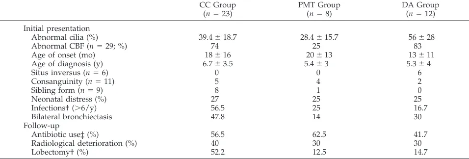
pulmo-nary function was also normal, except for dynamic lung compliance, a parameter not evaluated in pre-vious studies. In patients younger than 7 years, this functional test demonstrated that lung obstruction was more pronounced in the CC group than in the other groups. In older children, in whom spirometry was performed, patients in the CC group still exhib-ited a certain degree of obstruction compared with the other patients. These findings are in agreement with the few previous reports concerning the fol-low-up of pulmonary function in PCD patients. Air-way obstruction and air trapping with distension are frequently observed in adult PCD patients. How-ever, in the literature, as in our study, pulmonary function stabilizes for several years with appropriate TABLE 1. Clinical and Familial Characteristics in the 3 Groups Defined by the Main Ultrastructural Defect*
CC Group (n⫽23)
PMT Group (n⫽8)
DA Group (n⫽12)
Initial presentation
Abnormal cilia (%) 39.4⫾18.7 28.4⫾15.7 56⫾28
Abnormal CBF (n⫽29; %) 74 25 83
Age of onset (mo) 18⫾16 20⫾13 13⫾11
Age of diagnosis (y) 6.7⫾3.5 5.4⫾3 5.3⫾4
Situs inversus (n⫽6) 0 0 6
Consanguinity (n⫽11) 5 4 2
Sibling form (n⫽9) 8 1 0
Neonatal distress (%) 27 25 25
Infections† (⬎6/y) 56.5 25 16.7
Bilateral bronchiectasis 47.8 14 30
Follow-up
Antibiotic use‡ (%) 56.5 62.5 41.7
Radiological deterioration (%) 40 30 30
Lobectomy† (%) 52.2 12.5 14.7
* Results are expressed as the percentage of patients in each group, except for situs inversus, sibling form, and consanguinity, which are expressed as the number of patients in each group.
†P⬍.05.
treatment.28Similarly, extension of bronchiectasis was
observed in⬍30% of our patients and was not related to the type of ultrastructural defect, as already suggest-ed.29In fact, the slow progression of radiologic lesions
seems to be exclusively related to age.30These findings
emphasize the importance of early diagnosis of ciliary dyskinesia, and propose a close follow-up, with appro-priate antibiotic treatment and physiotherapy and to prevent lung damage.
CONCLUSION
In the presence of a suggestive clinical pattern, the diagnosis of ciliary dyskinesia is difficult to confirm when some cilia are normal or when all cilia do not share the same ultrastructural defect. In the present study, we defined the relationships between ultra-structural findings, ciliary function and clinical fea-tures. Significant differences were found when comparing clinical features and ultrastructural phe-notype showing that the type of the main ultrastruc-tural defect was more closely correlated with the clinical presentation than the percentage of abnormal cilia.
Analysis of the main ultrastructural defect demon-strated significant differences between patients. We identified that in children with chronic respiratory tract infections and partial ciliary defects, CC abnor-malities was a marker of severity. This specific ax-onemal defect, already reported in PCD, never con-cerns more than half of the cilia, but is probably congenital. For these patients, it is first necessary to confirm the ultrastructural defect on another biopsy, preferably performed at an other level of the respi-ratory tract, eg, nasal biopsy. Secondly, intensive antibiotic therapy and close follow-up are especially required for these patients. Finally, it will be inter-esting to proceed to genetic analysis, when it will be possible. Conversely, partial absence of DA is asso-ciated with a good prognosis, but only identification of a specific genetic abnormality will be able to con-firm that this specific axonemal defect represents a partial form of PCD. Finally, the origin of respiratory tract infections is not explained by ciliary studies in patients in the PMT group and requires further in-vestigation. It should be noted that some CC and DA defects won’t be detected when ciliary ultrastruc-tural analysis is only performed in cases of abnormal ciliary beat frequency. Some clinical features, such as situs inversus, sibling form, obstructive pulmonary function parameters, and extensive bronchiectasis are more suggestive of PCD than age of onset of respiratory tract infections, neonatal respiratory dis-tress or consanguinity. In the presence of such a suggestive clinical pattern, quantitative and qualita-tive ultrastructural analysis should be recom-mended, regardless of CBF results. Early diagnosis is particularly important, as the prognosis remains good even after lung surgery with physiotherapy and appropriate antibiotic treatment.
ACKNOWLEDGMENT
We thank Professor Andre´ Coste (Hoˆpital H. Mondor, Cre´teil) for his pertinent advice in preparation of this manuscript.
REFERENCES
1. Breeze R, Wheeldon EB. The cells of the pulmonary airways.Am Rev Respir Dis. 1977;116:705–777
2. Sleigh M. Ciliary function in transport of mucus. Eur J Respir Dis. 1983;64(suppl 128):287–292
3. Afzelius B. A human syndrome caused by immobile cilia. Science. 1976;193:317–319
4. Rutland J, Cox T, Dewar A, Cole P. Screening for ciliary dyskinesia. A spectrum of defects of motility and structure.Eur J Respir Dis. 1983; 64(suppl 127):71–77
5. Rossman C, Newhouse M. Primary ciliary dyskinesia: evaluation and Management.Pediatr Pulmonol. 1988;5:36 –50
6. Mygind N, Pedersen M, Nielsen M. Primary and secondary ciliary dyskinesia.Acta Otolaryngol. 1983;95:688 – 694
7. Afzelius B, Cammer P, Mossberg B. Acquired ciliary defects compared to those seen in the immobile-cilia syndrome.Eur J Respir Dis. 1983; 64(suppl 127):5–10
8. Lungarella G, Fonzi L, Ermini G. Abnormalities of bronchial cilia in patients with chronic bronchitis. An ultrastructural and quantitative analysis.Lung. 1983;161:147–156
9. Carson J, Collier A, Hu S. Acquired ciliary defects in nasal epithelium of children with acute viral upper respiratory infections.N Engl J Med. 1985;312:463– 468
10. Ballenger J. Acquired ultrastructural alterations of respiratory cilia and clinical disease. A review.Ann Otol Rhinol Laryngol. 1988;97:253–258 11. Escalier D, Jouannet P, David G. Abnormalities of the ciliary axonemal
complex in children: an ultrastructural and kinetic study in a series of 34 cases.Biol Cell. 1982;44:271–282
12. Rossman C, Lee R, Forrest J, Newhouse M. Nasal ciliary ultrastructure and function in patients with primary ciliary dyskinesia compared with that in normal subjects and in subjects with various respiratory diseases. Am Rev Respir Dis. 1984;129:161–167
13. Gaultier C, Boule´ M, Allaire Y, Clement A, Buvry A, Girard F. Deter-mination of capillary oxygen tension in infants and children.Bull Eur Physiopath Resp. 1979;14:287–297
14. Gaultier C, Allaire Y, Pappo A, Girard F, Gerbeaux J. Re´sistance pul-monaire totale, compliance pulpul-monaire dynamique chez l’enfant de 3 a` 15 ans.Sem Hop Paris. 1974;50:1045–1052
15. Verra F, Fleury-Feith J, Boucherat M, Pinchon M, Bignon J, Escudier E. Do nasal ciliary changes reflect bronchial changes? An ultrastructural study.Am Rev Respir Dis. 1993;147:908 –913
16. de Iongh R, Rutland J. Ciliary defects in healthy subjects, bronchiectasis, and primary ciliary dyskinesia.Am J Respir Crit Care Med. 1995;151: 1559 –1567
17. Rautiainen M, Collan Y, Nutien J. A method for measuring the orien-tation (beat direction) of respiratory cilia.Arch Otorhinolaryngol. 1986; 243:265–268
18. Torkkeli T, Nuutinen J, Rautiainen M. Clinical relevance of tubulus anomalies and compound cilia.Acta Otolaryngol. 1997;529:140 –143 19. Min YG, Shin JS, Cho SH, Chi JG, Yoon CJ. Primary ciliary dyskinesia:
ultrastructural defects and clinical features.Rhinology. 1995;33:189 –193 20. Cornillie F, Lauweryns J. Atypical bronchial cilia in children with recurrent respiratory tract infections: a comparative ultrastructural study.Path Res Pract. 1984;178:595– 604
21. Huang B, Piperno G, Luck D. Paralysed flagella mutants of Chlamydo-monas reinhardtiidefective for axonemal doublet microtubule arms. J Biol Chem. 1979;254:3091–3099
22. Eliasson R, Mossberg B, Cammer P, Afzelius BA. The immotile-cilia sndrome. A congenital ciliary abnormality as an etiologic factor in chronic airway infections and male sterility. N Engl J Med. 1977;297:1– 6 23. Afzelius B. The immobile-cilia syndrome and other ciliary disease.Int
Rev Exp Path. 1979;19:1– 43
24. Afzelius B, Eliasson R. Flagellar mutants in man: on heterogeneity of the immobile-cilia syndrome.J Ultrastruct Res. 1979;69:43–52
25. Afzelius B. The immobile-cilia syndrome: a microtubule-associated de-fect.Rev Biochem. 1985;19:63– 87
26. Dentler W. Microtubule-membrane interactions in cilia. Isolation and characterization of ciliary membranes from Tetrahymena pyriforis. J Cell Biol. 1980;84:364 –380
27. Ellerman A, Bisgaard H. Longitudinal study of lung function in a cohort of primary ciliary dyskinesia.Eur Respir J. 1997;10:2376 –2379 28. Corkey C, Levison H, Turner J. The immobile cilia syndrome, a
longi-tudinal survey.Am Rev Respir Dis. 1981;124:544 –548
29. Nadel H, Stringer D, Levison H, Turner J, Sturgess J. The immobile cilia syndrome: radiological manifestations.Radiology. 1985;154:651– 655 30. Faure´ C, Verderi D, Schmit P, Sirinelli D, Salomon J, Escalier D. The chest
DOI: 10.1542/peds.108.5.e86
2001;108;e86
Pediatrics
Estelle Escudier
Gilles Roger, Michèle Boulé, Marie Claude Millepied, TA§; Armelle Baculard and
Aline Tamalet, Annick Clement, Francoise Roudot-Thoraval, Pascale Desmarquest,
Ciliary Defect
Abnormal Central Complex Is a Marker of Severity in the Presence of Partial
Services
Updated Information &
http://pediatrics.aappublications.org/content/108/5/e86
including high resolution figures, can be found at:
References
http://pediatrics.aappublications.org/content/108/5/e86#BIBL
This article cites 30 articles, 4 of which you can access for free at:
Subspecialty Collections
b
http://www.aappublications.org/cgi/collection/infectious_diseases_su
Infectious Disease
following collection(s):
This article, along with others on similar topics, appears in the
Permissions & Licensing
http://www.aappublications.org/site/misc/Permissions.xhtml
in its entirety can be found online at:
Information about reproducing this article in parts (figures, tables) or
Reprints
http://www.aappublications.org/site/misc/reprints.xhtml
DOI: 10.1542/peds.108.5.e86
2001;108;e86
Pediatrics
Estelle Escudier
Gilles Roger, Michèle Boulé, Marie Claude Millepied, TA§; Armelle Baculard and
Aline Tamalet, Annick Clement, Francoise Roudot-Thoraval, Pascale Desmarquest,
Ciliary Defect
Abnormal Central Complex Is a Marker of Severity in the Presence of Partial
http://pediatrics.aappublications.org/content/108/5/e86
located on the World Wide Web at:
The online version of this article, along with updated information and services, is
by the American Academy of Pediatrics. All rights reserved. Print ISSN: 1073-0397.