INTERNATIONAL RESEARCH JOURNAL OF PHARMACY
www.irjponline.com
ISSN 2230 – 8407
Research Article
IDENTIFICATION OF NOVEL PPARγ MODULATORS / PARTIAL AGONISTS THROUGH
VIRTUAL SCREENING WORKFLOW
Nagashree K.S.
1, Praveen T.K.*
2, Rajini Kolure
11
Department of Pharmacology, JSS College of Pharmacy, JSS Academy of Higher Education & Research, Mysuru,
Karnataka, India
2
Department of Pharmacology, JSS College of Pharmacy, JSS Academy of Higher Education & Research, Ooty,
Nilgiris, Tamil Nadu, India
*Corresponding Author Email: [email protected]
Article Received on: 05/08/19 Approved for publication: 02/11/19
DOI: 10.7897/2230-8407.1011321
ABSTRACT
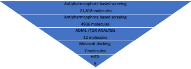
Thiazolidinedione’s (TZDs) being insulin sensitizer’s act as agonists of PPARγ used in the treatment of type 2 diabetes but suffered with serious side effects. After understanding the trans-activation mechanism of PPAR receptors and in order to overcome these side effects a new path has been led to new approaches like, PPAR-α/γ dual agonists, PPAR-δ/γ dual agonists, PPAR-pan agonists, selective PPAR-γ modulators (SPPARγMs) / partial agonists. Among them SPPARγMs) / partial agonists attracted due to their selectivity and expression in the selective tissue. The present study aims at identifying novel SPPARγMs) / partial agonists by using VS workflow and molecular docking.Virtual screening workflow is fallowed which consists of several steps like (a) Ligand based anti-pharmacophore screening(b) Ligand based Pharmacophore screening (c) ADME /Toxicity analysis and (d) Molecular Docking.Out of 21,818 molecules subjected to anti pharmacophore model, 4936 molecules qualify for the next step i.e., pharmacophore model screening. Out of these molecules only 12 molecules showed Qfit > 70. Therefore, these molecules were further subjected to ADME /TOX filter step in which 7 molecules passed the step. Further these molecules subjected to docking studies. In the docking studies based on the typical binding modes of the standard partial agonist (INT131) 5 molecules were found to have good binding mode required for a typical partial agonist. The Virtual screening workflow used in the study identifies 5 molecules as partial agonists.
Keywords: Insulin sensitizers, PPARγ partial agonists, Pharmacophore model, Virtual screening
INTRODUCTION
Peroxisome Proliferator Activated Receptors (PPARs) are transcription factor1, which act by coordinating the activities of
multiple pathways involved in metabolism instead of acting through one major target like one enzyme or one pathwa2. This
unique property of PPARs has created lot of interest for their possible use in a complex metabolic disorder such as type 2 diabetes mellitus (T2DM)3. Thiazolidinedione’s (TZDs) or
glitazones are one such class of anti-diabetic drugs which act by increasing the trans-activation activity of Peroxisome Proliferator Activated Receptors (PPARs)4. Further, TZDs reverse insulin
resistance without causing hypoglycaemic effect which is major side effect of most widely used anti diabetic drugs such as sulfonylureas. They reduce hepatic output of glucose and increase peripheral uptake, leading to reducing both pre-load and after load on the beta cell. These actions enhance the effectiveness of endogenous insulin, and reduce the amount of exogenous insulin needed to maintain a given level of blood glucose5; thus,
providing an excellent rationale for the use of glitazonesin T2DM.
Unfortunately, these glitazones used in the clinic today suffer with some serious side effects such as, hepatotoxicity6, increase
in body weight, fluid retention, weight gain, risk of bone fracture7,
bladder cancer8 and many more. These side effects reported to be
due to their non-selective activation of PPARγ in the off-target
the use of Rosiglitazone and changed the label claim of Pioglitazone for the risk of bladder cancer7. One of the reasons
for the failure of these clinically used glitazones is, their time of development. These drugs were developed when there was very little scientific data available on structure and the transcriptional mechanisms of the target peroxisome proliferator activated receptors (PPARs). After understanding the trans-activation mechanism of PPAR receptors has led to the newer approaches to the discovery of development PPAR-α/γ dual agonists, PPAR-δ/γ dual agonists, PPAR-pan agonists, selective PPAR-γ modulators or partial agonists. Among them SPPARγMs attracted many researchers due to their selectivity and expression in the selective tissue.
SPPARγMs hypothesis is based on recruitment of certain
differential receptor binding and co-factor
recruitment/displacement with specificity to the selective tissue and their expression in favourable target cells. This concept is similar to selective estrogen receptor modulators (SERMs)9,10.
SPPARγMs provide a target oriented therapeutic profile by maintaining the desired therapeutic benefits and at the same time have minimal adverse effects due to their inability to fully activate the receptor as that of a full agonist11. SPPARMs are reported to
In this study virtual screening (VS) work flow is efficiently used in order to identify novel partial agonist. We followed different strategy to screen the compounds instead of screening only from ADMET analysis. Ligand based Pharmacophore based screening was initially implemented followed by usual VS screening. Alternatively this method of screening is defined as Pharmacophore based screening of library of compounds with prescribed generated Pharmacophore from known biological value.
Unfortunately, the glitazones used in the clinic today suffer with some serious side effects such as, increase in body weight, fluid retention, weight gain, risk of bone fracture, bladder cancer, etc.5, 13-15. Dose responsive curve of the therapeutic effects and
side effect of TZDs coincide each other such a way that increase in dose increases efficacy and also degree of side effect16 hence
multiple activities are appears to be linked17, as a result they
received black box safety warning. One of the reasons for the failure of these clinically used glitazones is, their time of development. These drugs were developed when there was very little scientific data available on structure and the transcriptional mechanisms of the target peroxisome proliferator activated receptors (PPARs). After understanding the trans-activation mechanism of PPAR receptors has led to the newer approaches to the discovery of development PPAR-α/γ dual agonists, PPAR-δ/γ dual agonists, PPAR-pan agonists, selective PPAR-γ modulators or partial agonists. Among them SPPARγ Ms attracted many researchers due to their selectivity and expression in the selective tissue.
SPPARγ Ms hypothesis is based on recruitment of certain
differential receptor binding and co-factor
recruitment/displacement with specificity to the selective tissue and their expression in favourable target cells. This concept is similar to selective estrogen receptor modulators (SERMs)9,10.
SPPARγ Ms provide a target oriented therapeutic profile by maintaining the desired therapeutic benefits and at the same time have minimal adverse effects due to their inability to fully activate the receptor as that of a full agonist11. SPPARMs are reported to
achieve these effects by selectively recruiting the co-activators to PPAR receptors and thus selectively activating the genes responsible for insulin sensitization, adipogenesis, fluid retention and bone remodelling12.
In this study virtual screening (VS) work flow is efficiently used in order to identify novel partial agonist. We followed different strategy to screen the compounds instead of screening only from ADMET analysis. Ligand based Pharmacophore based screening was initially implemented followed by usual VS screening. Alternatively, this method of screening is defined as pharmacophore based screening of library of compounds with prescribed generated pharmacophore from known biological value. After this hits were subjected to molecular docking studies.
MATERIALS AND METHODS
Virtual screening workflow consists of several steps18 and was
carried out by using Discovery studio software 4.0v (Discovery studio 4.0v., San Diego, CA, USA; http://www.accelrys.com).
Ligand based pharmacopore model: Pharmacophore generation and screening
• ADME/TOX analysis
• Molecular docking
The compounds for initial screening were retrieved from various online databases like ZINC database pub chem in 3:1 ratio and more preferably 75 percent compounds are taken form Zinc database because it contains commercially available natural products and natural-product derivatives. Thus, we could purchase and test in vitro the bioactivity of the selected compounds.
Totally, initial dataset contains 21, 818 compounds were processed in this study. The 3D structures of this initial dataset compounds were processed with prepare ligands program
(Discovery studio 4.0v., San Diego, CA, USA;
http://www.accelrys.com) to remove duplicates, enumerating isomers and tautomers, and generating 3D conformations. This process was carried out with the following parameters (a) clean geometry and the force field used was CHARMm (b) Ionization-pH based, (c) Generate isomers to remove Unknown Stereo atoms and Unknown Stereo bonds, (d) Duplicates removal using SMIRKS Acid Templates, SMIRKS Base Templates and SMARTS Charge Templates. Conformations were built with the cat Conf is the Catalyst conformer generation tool generating in vacuo a maximum number of 255 conformers per structure with energy threshold of 20 kcal/mol.
Anti-Pharmacophore model generation
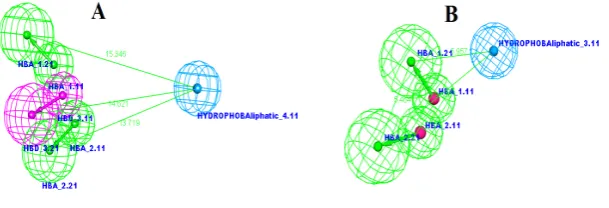
Pharmacophore generation (Discovery studio 4.0v., San Diego, CA, USA; http://www.accelrys.com) was used for the analysis of the PPAR-gamma structures are Rosiglitazone-2PRG, Pioglitazone-2XKW, Troglitazone-2VN0. Pharmacophore of the three combined structures were generated using biological activity with uncertainty. This pharmacophore (Figure 1A) is formed by 4 sites (two hydrogen-bond acceptors, one hydrogen bond donor and hydrophobic aliphatic sites) that are present in most of the complexes of full agonists analyzed and are therefore assumed to be responsible for the intermolecular interactions that are essential for the activity of PPAR-gamma full agonists. This pharmacophore is named as anti-pharmacophore since we used to exclude these most fit structures.
Pharmacophore model generation
Pharmacophore model generation: Common features of 3 existing PPARγ partial agonist (INT131-3FUR, nTZDpa-2Q5S, SF147-2Q6R) were used to generate pharmacophore model.
Figure 1: Pharmacophores used for the identification of (A) PPAR-gamma full agonists and (B) PPARγ partial agonists. Hydrophobic and acceptor/donor sites are colored in Cyan, green and pink, respectively. (C) Compared model of anti-pharmacophore and pharmacophore
features
Pharmacophore based and ADMET screening
Screening the compounds on the basis of pharmacophore is crucial process. It is projected to carry the Structure Based Pharmacophore tools within Discovery Studio. We screened initial datasets of compounds in anti-pharmacophore model with multiple hypotheses to retrieve the potent compound for biological activity. Further the screening was extended for pharmacophore model with more than one hypothesis. The compounds form second level screening was subjected to further virtual screening using ADMET property. The objective of this screening is to retrieve the biological active compound for further analysis.
Molecular docking
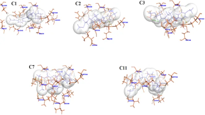
Docking studies of the PPARγ partial agonists C1, C2, C3, C7, C9, C11 and C12 were performed with the Simulation and annealing based docking was carried out using C-docker protocol of Discovery studio software 4.0v (Discovery studio 4.0v., San Diego, CA, USA ;http://www.accelrys.com) on the PPARγ crystal structure 2PRG. The binding site was defined using the define and edit binding tool the sphere is placed 3D coordinate of
49.72 X -36.98Y 19.29 Z with radius of 8.415Å. The docking system was set up with 1000 steps of dynamics with refine orientation to remove bad clashes along with simulation of heating and cooling of 2000 and 5000 steps respectively; whereas target temperature was defined as 300k for both heating and cooling along with default energy threshold for refine docking process. Additionally, CHARMm force filed was applied to each docked pose with momany-rone Ligand Partial charge. The results are screened based on the docking poses along with interaction investigation of docking poses. Discovery studio Visualizer 4.5 (Bio via Discovery studio, Tokyo, Japan; http://www.Dassault Systemes Biovia.com) was used for analyzing and visually investigating the ligand-protein interactions of the best docking poses.
RESULTS AND DISCUSSION Virtual screening of datasets
The aim of this research work is to identify the novel PPAR-gamma partial agonists. Virtual screening workflow implemented in this study is summarized in Figure 2.
Figure 2: Schematic overview of the VS workflow and the procedure used for selecting the VS hits
The number of compounds that passed each step and the programs used are shown. From an initial set of 21,818 compounds, 12 compounds were identified as putative PPAR-gamma partial agonists by the VS workflow. Five of these compounds after
passing ADMET show best docking poses with PPAR-gamma partial agonists.
PPAR-three known PPAR-gamma full agonists and PPAR-gamma partial agonists. We followed different strategy to screen the compounds instead of screening from ADMET, Pharmacophore based screening was initial implemented followed by usual VS screening method alternatively this method of screening is defined as pharmacophore-based screening of library of compounds with prescribed generated pharmacophore from known biological value. The fitting between the molecules and the pharmacophore was analyzed with the Catalyst program. The
compounds are mapped to the pharmacophore and evaluated on the basis of fit value. Anti-pharmacophore and pharmacophore model mapping to features is displayed on Figure 3, initial dataset compounds are first and foremost screened with anti-pharmacophore model now compounds which fits more than fifty percent were drop down, the subset of molecules that did not match the anti-pharmacophore was then used identify the PPAR-gamma partial agonists.
Figure 3: Mapped Pharmacophores (A) PPAR-gamma full agonists and (B) PPARγ partial agonists
Hydrophobic and acceptor / donor sites are colored in Cyan, green and pink, respectively. Subsequently to identify the novel partial agonist pharmacophores obtained using IC50 of known PPAR-gamma partial agonists was used. The subset of molecules that did match and fits more than 70% to the pharmacophore model reveals that identification of novel PPAR-gamma partial agonists.
Only 12 compounds satisfied pharmacophore model were screened for ADMET level study using discovery study (ADMET/Topkat tools). The ADMET analysis results of novel 12 PPAR-gamma partial agonists are tabulated in Table 1 and Figure 4.
Table 1: ADMET screening of compounds
Compound No ZINC ID Solubility BBB CYPD26 Hepatotoxic Absorption
C1 13259979 3 3 FALSE FALSE 0
C2 38861161 3 2 FALSE FALSE 0
C3 13586428 3 2 FALSE FALSE 0
C4 31932883 3 3 FALSE TRUE 0
C5 38974587 3 3 FALSE TRUE 0
C6 68478286 3 2 FALSE TRUE 0
C7 13849555 2 2 FALSE FALSE 0
C8 36592901 3 3 FALSE TRUE 0
C9 31367097 4 3 TRUE FALSE 0
C10 63059572 3 3 FALSE TRUE 0
C11 51425645 3 2 FALSE FALSE 0
C12 59433934 4 3 FALSE FALSE 0
Solubility levels (2-Low, 3-Good, 4-Optimal), BBB = Blood brain barrier (2-Medium, 3-Low), Absorption (0-Good), Hepatotoxic (TRUE-Toxic, FALSE-Non-toxic), CYPD26 (TRUE-Inhibitor, FALSE-Non-Inhibitor)
Compounds like C1, C2, C3, C7, C9, C11 and C12 are non -toxic, non-inhibitor with good absorption an optimal to low solubility has a competence to across the blood brain barrier from medium to low.
Our post docking analysis results elicits that, no hydrogen bond interaction and neither makes a hydrogen bond network as extensive as full agonist rosiglitazone with Tyr473 from arm I in AF2 branch I portion of the LBD of PPAR-gamma19, instead all
the 8 compounds formed hydrogen bond interaction with residue Tyr37220 lie between H3 and the 𝛽-sheet, extending from branch
II to branch III of the ligand binding pocket.
Figure 5: Hydrogen bond interactions of the test compounds with LBD of PPARγ (PDB ID: 3FUR)
DISCUSSION
SPPARγ Ms similar to selective estrogen receptor modulators (SERMs), SPPARMs are reported to achieve effects by selectively activating on the genes responsible for insulin sensitization, adipogenesis, fluid retention and bone remodeling12,21. Partial agonists act by partially agonizing PPARγ
receptors and also exhibit low trans-activation activity when compared to full agonists22.
Therefore, in this study such novel SPPARγMs / Partial agonists were identified by using VS workflow where each step it consists was able to identify putative PPARγ partial agonists. Pharmacophore-based virtual screening helped us to enrich active molecules in the hit list compared to a random selection of test compounds. VS workflow is able to predict 8 Hits.
In molecular docking confirmation analysis of existing full agonists extend from the TZD head group to intermolecular hydrogen bonds side chains of PPAR𝛾 residues like H323 (2.9˚A), H449 (2.7˚A) and Y473 (2.6˚A) allowing for stabilization of the AF2 surface23. Among these the hydrogen
bonding with the Tyr473 residue is reported to play a vital role in the stabilization of AF-2 helix through H12, allowing less of an entropic penalty for co-activator binding and thus full transcriptional output which is essential for the recruitment of co-activators necessary for transcriptional activation and these interactions are more prominent for PPAR𝛾 full agonists24,25.
PPAR𝛾 partial agonists operate through different structural and mechanistic methods than full agonists rather than simply exhibiting lowered transcriptional output due to suboptimal potency and/or affinity26. Partial agonists stabilize the LBD in a
distinct manner in comparison to full agonists. Partial agonists were shown to preferentially stabilize other regions of the ligand binding domain, especially the 𝛽-sheet region and H3 helix especially interacting with Tyr327 amino acid residue27-30.
CONCLUSION
In conclusion, among 21,818 compounds, we were able to identify 5 compounds as putative partial agonists with help of virtual screening workflow i.e. both pharmacophore model and molecular docking. Post docking analysis show all 8 compounds interacted with Tyr327 residues as that of existing partial agonists like INT131.
REFERENCES
1. Tyagi S, Gupta P, Saini AS, Kaushal C, Sharma SJ Joapt, research. The peroxisome proliferator-activated receptor: A family of nuclear receptors role in various diseases 2011; 2(4): 236.
2. Rosen CJ. Revisiting the rosiglitazone story—lessons learned. New England Journal of Medicine 2010; 363(9): 803-6.
3. Blanquart C, Barbier O, Fruchart JC, Staels B, Glineur CJT Josb, biology m. Peroxisome proliferator-activated receptors: regulation of transcriptional activities and roles in inflammation 2003; 85(2-5): 267-73.
4. Lehmann JM, Moore LB, Smith Oliver TA, Wilkison WO,
Willson TM, Kliewer SAJ JoBC. An anti diabetic thiazolidinedione is a high affinity ligand for peroxisome proliferator-activated receptor γ (PPARγ) 1995; 270(22): 12953-6.
5. Mankovsky B, Kurashvili RB. Glitazones: Beyond glucose lowering! Diabetes and Metabolic Syndrome: Clinical Research and Reviews 2007; 1(3): 197-207.
6. Watkins PB, Whitcomb RWJNE JoM. Hepatic dysfunction
associated with troglitazone 1998; 338(13): 916-7. 7. Idris I, Gray S, Donnelly RJD. Rosiglitazone and
pulmonary edema: an acute dose-dependent effect on human endothelial cell permeability 2003; 46(2): 288-90. 8. Shah P, Mudaliar SJ Eoods. Pioglitazone: side effect and
safety profile 2010; 9(2): 347-54.
10. Smith CL, O’malley BW. Co-regulator function: a key to understanding tissue specificity of selective receptor modulators. Endocrine reviews 2004; 25(1): 45-71. 11. Horita S, Nakamura M, Satoh N, Suzuki M, Seki G.
Thiazolidinediones and edema: recent advances in the pathogenesis of thiazolidinediones-induced renal sodium retention. PPAR research 2015; 2015.
12. Kubota N, Terauchi Y, Miki H, Tamemoto H, Yamauchi T, Komeda K, et al. PPARγ mediates high-fat diet–induced adipocyte hypertrophy and insulin resistance. Molecular cell 1999; 4(4): 597-609.
13. Lalloyer F, Staels B. Fibrates, glitazones, and peroxisome proliferator–activated receptors. Arteriosclerosis, thrombosis and vascular biology 2010; 30(5): 894-9. 14. Gale EA. Troglitazone: the lesson that nobody learned?
Diabetologia 2006; 49(1): 1-6.
15. Shukla R, Kalra S. Pioglitazone: Indian perspective. Indian Journal of Endocrinology and Metabolism 2011; 15(4): 294.
16. Lago RM, Singh PP, Nesto RW. Congestive heart failure and cardiovascular death in patients with pre diabetes and type 2 diabetes given thiazolidinediones: a meta-analysis of randomized clinical trials. The Lancet 2007; 370(9593): 1129-36.
17. Higgins LS, DePaoli AM. Selective peroxisome
proliferator-activated receptor γ (PPARγ) modulation as a strategy for safer therapeutic PPARγ activation. The American Journal of Clinical Nutrition 2010; 91(1): 267S-72S.
18. Guasch L, Sala E, Castell-Auví A, Cedó L, Liedl KR, Wolber G, et al. Identification of PPAR gamma partial agonists of natural origin (I): development of a virtual screening procedure and in vitro validation 2012; 7(11): e50816.
19. Vidović D, Busby SA, Griffin PR, Schürer SCJC. A Combined Ligand-and Structure-Based Virtual Screening Protocol Identifies Sub micromolar PPARγ Partial Agonists 2011; 6(1): 94-103.
20. Motani A, Wang Z, Weiszmann J, McGee LR, Lee G, Liu
Q, et al. INT131: a selective modulator of PPARγ. 2009; 386(5): 1301-11.
21. Pinaire JA, Miller AR, Gregoire FM. Development of Synthetic Modulators of PPARs: Current Challenges and Future Opportunities. PPAR research 2008; 2008: 7. 22. Hughes TS, Chalmers MJ, Novick S, Kuruvilla DS, Chang
MR, Kamenecka TM, et al. Ligand and receptor dynamics contribute to the mechanism of graded PPARγ agonism 2012; 20(1): 139-50.
23. Einstein M, Akiyama TE, Castriota GA, Wang CF, McKeever B, Mosley RT, et al. The differential interaction of peroxisome proliferator-activated receptor γ ligands with Tyr473 is a physical basis for their unique biological activities 2008; 73(1): 62-74.
24. Nolte RT, Wisely GB, Westin S, Cobb JE, Lambert MH,
Kurokawa R, et al. Ligand binding and co-activator assembly of the peroxisome proliferator-activated receptor-γ. Nature 1998; 395(6698): 137.
25. Zoete V, Grosdidier A, Michielin O. Peroxisome proliferator-activated receptor structures: ligand specificity, molecular switch and interactions with regulators. Biochimica et Biophysica Acta (BBA)-Molecular and Cell Biology of Lipids 2007; 1771(8): 915-25.
26. Burgermeister E, Schnoebelen A, Flament A, Benz Jr, Stihle M, Gsell B, et al. A novel partial agonist of peroxisome proliferator-activated receptor-γ (PPARγ) recruits PPARγ-coactivator-1α, prevents triglyceride accumulation, and potentiates insulin signaling in vitro. Molecular Endocrinology 2006; 20(4): 809-30.
27. Bruning JB, Chalmers MJ, Prasad S, Busby SA,
Kamenecka TM, He Y, et al. Partial agonists activate PPARγ using a helix 12 independent mechanism. Structure 2007; 15(10): 1258-71.
28. Oberfield JL, Collins JL, Holmes CP, Goreham DM, Cooper JP, Cobb JE, et al. A peroxisome proliferator-activated receptor γ ligand inhibits adipocyte differentiation. Proceedings of the National Academy of Sciences 1999; 96(11): 6102-6.
29. Bruning JB, Chalmers MJ, Prasad S, Busby SA,
Kamenecka TM, He Y, et al. Partial agonists activate PPARγ using a helix 12 independent mechanism 2007; 15(10): 1258-71.
30. Pochetti G, Godio C, Mitro N, Caruso D, Galmozzi A, Scurati S, et al. Insights into the mechanism of partial agonism crystal structures of the peroxisome proliferator-activated receptor γ ligand-binding domain in the complex with two enantiomeric ligands 2007; 282(23): 17314-24.
Cite this article as:
Nagashree K. S. et al.Identification of Novel PPARγ modulators / partial agonists through Virtual Screening Workflow. Int. Res. J. Pharm. 2019;10(11):69-74 http://dx.doi.org/10.7897/2230-8407.1011321
Source of support: Nil, Conflict of interest: None Declared