R E S E A R C H A R T I C L E
Open Access
Neuroprotective effects of vitamin D on
high fat diet- and palmitic acid-induced
enteric neuronal loss in mice
Sara Larsson
1and Ulrikke Voss
2*Abstract
Background:The role of vitamin D in obesity and diabetes is debated. Obese and/or diabetic patients have elevated levels of free fatty acids, increased susceptibility to gastrointestinal symptoms and are suggested to have altered vitamin D balance. The enteric nervous system is pivotal in regulating gastrointestinal activity and high fat diet (HFD) has been shown to cause loss of enteric neurons in ileum and colon. This study investigates the effect of vitamin D on HFD- and palmitic acid-induced enteric neuronal loss in vivo and in vitro.
Methods:Mice were fed either a normal diet (ND) or HFD supplemented with varying levels of vitamin D (from 0x
to 20x normal vitamin D level) for 19 weeks. Ileum and colon were analyzed for neuronal numbers and remodeling. Primary cultures of myenteric neurons from mouse small intestine were treated with palmitic acid (4x10⁻⁴M) and/or 1α,25-hydroxy-vitamin D3 (VD, 10-11- 10-7M) with or without modulators of lipid metabolism and VD pathways. Cultures were analyzed by immunocyto- and histochemical methods.
Results:Vitamin D supplementation had no effect on enteric neuronal survival in the ND group. HFD caused
substantial loss of myenteric neurons in ileum and colon. Vitamin D supplementation between 0-2x normal had no effect on HFD-induced neuronal loss. Supplementation with 20x normal, prevented the HFD-induced neuronal loss. In vitro supplementation of VD prevented the palmitic acid-induced neuronal loss. The VD receptor (VDR) was not identified in enteric neurons. Enteric glia expressed the alternative VD receptor, protein disulphide isomerase family A member 3 (PDIA3), but PDIA3 was not found to mediate the VD response in vitro. Inhibition of peroxisome proliferator-activated receptor gamma (PPARγ) and immune neutralization of isocitrate lyase prevented the VD mediated neuroprotection to palmitic acid exposure.
Conclusions:Results show that VD protect enteric neurons against HFD and palmitic acid induced neuronal loss. The mechanism behind is suggested to be through activation of PPARγleading to improved neuronal peroxisome function and metabolism of neuronal lipid intermediates.
Keywords:Enteric nervous system, Vitamin D, High fat diet, Neuronal survival, Gastrointestinal tract, Peroxisome proliferator-activated receptor gamma
* Correspondence:[email protected]
2Unit of Neurogastroenterology, Department of Experimental Medical
Science, Lund University, Sölvegatan 19, BMC B11, 22184 Lund, Sweden Full list of author information is available at the end of the article
Background
The role of vitamin D has expanded beyond its classical role in calcium homeostasis. Since animal studies have shown that vitamin D supplementation protects against high fat diet (HFD) induced metabolic parameters [1–3], several efforts have been made to elucidate the role of vitamin D in type-2 diabetes and in obesity, as well as in their complications [4–6]. Recently animal models have likewise shown that various dietary vitamin D supplemen-tations are able to protect against neurodegeneration found in i.e. models of Alzheimer’s disease [7, 8]. While the clinical data on vitamin D supplementation are incon-clusive [9,10], the need for exploring the metabolic poten-tial and cellular mechanisms of vitamin D remains.
Whether produced in the skin through UV radiation or ingested in the diet, vitamin D (25-hydroxy vitamin D3) is in the body converted to its active form 1α,25-hydroxy-vitamin D3 (VD), by the enzyme 1α-hydroxylase also known as cytochrome P450. This enzyme is found mainly in the kidneys but other cells types including immune cells and neurons have been shown to express this enzyme [11,12]. VD mediates the majority of its effects by binding to, and thereby activat-ing the nuclear VD receptor (VDR). Upon activation, VDR dimerizes with the retinoid-X receptor (RXR) forming a heterodimer, that in turn binds to specific VD response elements (VDREs). [13,14] However, it is sug-gested that initiation of VDR mediated responses cannot account for the fast responses seen upon VD stimulation in certain cell types e.g. osteoblasts and intestinal epithe-lial cells. [15–17] Protein disulphide isomerase family A member 3 (PDIA3), also known as 1,25MAARS, has been suggested as an alternative VD receptor, capable of mediating the fast VD responses and acting as a modula-tor of cellular function independent of VDR [15,18]. Be-sides the receptor mediated VD responses VD, per se, has been suggested to act as an antioxidant preventing lipid peroxidation and stabilizing cell membranes [19]. The anti-oxidative potential of VD, independent of ei-ther VDR or PDIA3, still needs to be furei-ther assessed. In addition, VD is reported to decrease reactive oxygen spe-cies (ROS) by increasing levels of the key anti-oxidative enzymes superoxide dismutase (SOD), catalase and glutathione (GSH) [20–22].
Peripheral neuropathy is a common complication of diabetes suggested to be caused by the combination of abnormal blood glucose and lipid levels, oxidative and inflammatory stress and accumulation of advanced gly-cation products (AGE) [23, 24]. VD deficiency is sug-gested to be involved in the development of diabetic neuropathy [25]. Further, it is increasingly recognized that obesity and HFD models also are associated with a neurotoxic phenotype. In animals HFD induces anxiety-like behavior and neuronal loss in hippocampus
[26, 27], as well as loss of neurons in all parts of the gastrointestinal (GI) tract [28–30]. Clinical data indir-ectly support these findings as both depression and GI dysfunction are common comorbidities of obesity and the metabolic syndrome [31–33]. The enteric nervous system (ENS), innervates the entire GI tract and is piv-otal in the regulation of all GI functions, including mo-tility, secretion and blood flow [34]. The HFD-induced neuronal loss has been suggested to be mediated mainly by the saturated fatty acid palmitic acid (PA) and involve increased oxidative stress and derangement of metabolic pathways [28]. The aim of this study was to investigate effects of VD on HFD-induced neuronal loss and to as-sess possible pathways by which VD mediates it effects on PA-induced enteric neuronal loss in vitro.
Methods
In vivo experiments
C57BL/6J mice (n=42, Taconic Bioscience) aged 4 weeks were randomized divided into seven groups (n=6 each group), animals were housed in groups. Three groups re-ceived normal diet (ND) with different doses of Vitamin D (no vitamin D, ND 0x; normal vitamin D 1.3IU/g [35], ND 1x; twenty times vitamin D, ND 20x). Four groups re-ceived high fat diet (HFD) with different doses of vitamin D (no vitamin D, HFD 0x; normal vitamin D, HFD 1x; double vitamin D HFD 2x; twenty times the normal vita-min D, HFD 20x). All diets were purified diets (Research Diets Inc., USA) Table 1 gives nutritional overview and diet details. After 19 weeks mice were sacrificed by cer-vical dislocation. The GI tract from cardia to rectum was collected, opened along the mesenteric border and emp-tied. Segments of ileum and transverse colon were fixed and processed for cryo-sectioning [36].
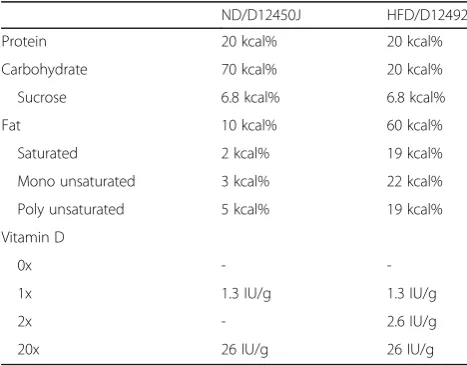
Table 1Nutritional content and vitamin D concentration in the normal diet (ND) and high fat diet (HFD)
ND/D12450J HFD/D12492
Protein 20 kcal% 20 kcal%
Carbohydrate 70 kcal% 20 kcal%
Sucrose 6.8 kcal% 6.8 kcal%
Fat 10 kcal% 60 kcal%
Saturated 2 kcal% 19 kcal%
Mono unsaturated 3 kcal% 22 kcal%
Poly unsaturated 5 kcal% 19 kcal%
Vitamin D
0x -
-1x 1.3 IU/g 1.3 IU/g
2x - 2.6 IU/g
In vitro experiments
Primary enteric cultures
C57Bl/6J mice (n=46, 20-23g, Janvier Labs, FR) on standard chow diet (R36 Lactamin, SE) were used for in vitro experimentations, animals were housed in groups. Primary cultures of myenteric neurons were prepared from the small intestine. Isolation was done in the morning. Neurons were dissociated using a modification of a previously described method [28,37]. In brief, anes-thetized mice (i.p. injection with Ketalar/Rompun) had their small intestine exposed via midline incisions. The longitudinal muscle layer with attached myenteric gan-glia was stripped from approximately 15 cm of the distal small intestine. Tissues were placed in Ca2+ and Mg2+ free Hank’s balanced salt solution (HBSS) containing 1.9 collagen digestion units (CDU)/mL collagenase 1-A (1.5 mg/mL, Merck, SE) and 4.7μU/mL protease IX (1.5mg/ mL, Sigma-Aldrich, SE) and enzymatically and mechan-ically separated. Trypsin (0.4 mg/mL, BioChrom AG) and EDTA (0.003%; Merck) were added. Trypsin was inactivated by addition of 50% v/v fetal bovine serum (FBS, Biowest, FR). Cell suspension was centrifuged at 15.6 g for 10 min. Supernatant was removed and pellet washed three times in HBSS, centrifuged at 15.6 g for 10 min and diluted to 0.8 mL in Neurobasal A (NBA) cell culture medium (ThermoFisher Scientific, SE) contain-ing 10% FBS (Biowest, FR), 0.5 mM L-glutamine (K0282, BioChrom AG), 50 U/mL penicillin and 50 μg/mL streptomycin (A2213, BioChrom AG). Cell cultures were prepared by adding 50 μL of the constantly mixed cell suspension into 8-well chambers (30108, SPL, Saveen Werner) prefilled with 450 μL of the NBA cell culture medium. From each animal two 8-well chambers (69mm2/well) were prepared. Suspensions from multiple animals were never pooled. Cultures were incubated 4 days prior to experimental treatments.
Experimental treatment agents
Stock solutions of palmitic acid (PA, P9767, Merck), VD (D1530, Merck), 7-dihydrocholesterol (7DHC), 16F16 (SML0021 Merck, PDIA3 inhibitor [38]), GW69662 (M6191, Merck, peroxisome proliferator-activated receptor gamma (PPARγ) inhibitor [28]), were prepared, aliquoted and stored at -20 °C. Isocitrate lyase (ICL) antibody (AS10713, Agrisera, SE) and pre-immunization sera (AS03 027, Agrisera, SE) were aliquoted and stored at 4 °C.
Experimental set ups
Various sets of experiments were performed. 1. Cultures were exposed to medium containing either PA(4x10⁻⁴M), VD (10-11-10-7M), 7DHC (10-11-10-7M), 16F16 (7x10⁻⁸-2x10⁻⁵M), GW69662 (10-6M), ICL antibody (1:250) or pre-immunization sera (1:250). 2 Cultures were exposed to either VD (10-11-10-7M) + PA
(4x10⁻⁴M) or 7DHC (10-9-10-7M) + PA (4x10⁻⁴M). 3. Cultures were exposed to either VD (10-7M) + experi-mental treatment agent, PA(4x10⁻⁴M) + experimental treatment agent or VD (10-7M) + PA (4x10⁻⁴M) + experi-mental treatment agent. Cultures were incubated 4 days in treatment media with controls run in parallel, fixated in Stefaninins and processed for immunocytochemistry.
Histology and immunocytochemistry
For details on primary and secondary antibodies see Table 2. All antibodies were diluted in phosphate buff-ered saline (PBS) containing 0.25% Triton X-100 and 0.25% BSA (PBS-T-B). For visualization of submucous and myenteric neurons, cryo-sections from ND and HFD mice were immunolabeled with HuC/HuD-biotin and vectastain ABC kit (Vector laboratories Inc., USA), according to manufacturer’s protocol [28, 39]. Morpho-metric analyses of intestines were on Toluidine blue (0.01% in 60% ethanol for 1.5min) stained cryo-sections.
Double immunolabeling of cultures were by overnight incubation in moist chamber at 4°C with a mixture of primary antibodies. Secondary antibodies were mixed and incubated 1h at RT. Hoechst (ThermoFisher Scien-tific, SE) cell nuclei counter staining was performed ac-cording to manufacturer’s protocol. Mounting was in PBS:glycerol 1:1 followed by fluorescence microscopy (Olympus BX43, LRI, SE) with appropriate filter setting.
Analyses
Mucosa and muscularis propria thicknesses were esti-mated for each mouse using the mean from ten mea-surements (Aperio ScanScope CS/GL SS5082 and ImageScope). The numbers of HuC/HuD-immunoreac-tive submucous and myenteric neuronal cell bodies were counted per mm section length. From each mouse, cryo-sections (10μm) cut longitudinally at 3-4 different depths comprising a total length of 30-50mm, were used. Analyses were done blinded to treatment.
Neuronal survival was estimated according to previously described protocol [28, 37]. In brief, neuronal survival after exposure to the various treatments was calculated by counting the total number of HuC/HuD-immunoreactive (IR) neurons in the entire culture well (69 mm2) and expressed as percentage of the number of total neurons in the control well run in parallel (% neuronal survival of control). With a minimum of 6 observations in 3 bio-logical repeats, in each treatment group.
Results
In vivo
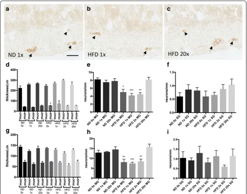
All animals were included in the study. Morphometric analyses of muscularis propria and mucosa in ileum (Fig.
1d) and colon (Fig.1g) revealed no effect of dietary vita-min D supplementation on intestinal morphology. A slight thickening (p<0.05) of ileum muscularis propria in the HFD 0x group was observed compared to the ND 1x group (Fig. 1d). None of the dietary vitamin D supple-mentations affected neuronal survival in either ileum (Fig. 1e, f) or colon (Fig. 1h, i) in animals fed the ND. HFD is known to induce loss of myenteric, but not sub-mucosal, enteric neurons in mice [28,30,40]. Vitamin D supplementation in the 0-2x range did not attenuate the HFD induced enteric neuronal loss, in ileum (Fig. 1e, f) or colon (Fig.1h, i). However, animals receiving 20x the normal vitamin D concentration were protected against the HFD-induced neuronal loss in both ileum (Fig.1e, f) and colon (Fig.1h, i).
In vitro, VD effects on primary enteric cultures
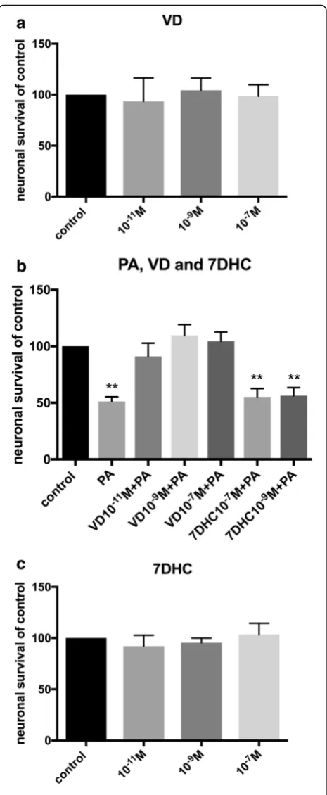
Primary cultures isolated from the longitudinal muscle layer of mouse small intestine contains myenteric gan-glia, glia and smooth muscle cells, effects of PA exposure on these has previously been described [28]. Control wells displayed 2.2±0.1 neurons per mm2. Exposure to VD (10-11 – 10-7M) per se did not affect neuronal sur-vival in cultures (Fig. 2a). VD serum concentration in mice are suggested to be in the 10-10M range [41]. Pal-mitic acid exposure alone induced a significant loss of cultured neurons (p<0.01, Fig. 2b), the loss was similar in magnitude to the previous described PA induced loss [28]. The combined exposure of PA (4x10⁻⁴M) and VD (10-11–10-7M) did not affect neuronal survival (Fig.2b). Thus, the presence of VD (10-11 –10-7M) abolished the previous described PA-induced neuronal loss. To test if the effect was due to the activated form of vitamin D (1α,25-hydroxy-vitamin D3, VD) cultures were exposed to the vitamin D precursor 7-dihydrocholesterol (7DHC). Exposure of 7DHC (10-11 – 10-7M) per se
didn’t affect neuronal survival (Fig. 2c). Addition of 7DHC (10-9–10-7M) was unable to protect enteric neu-rons against the PA-induced neuronal loss (Fig.2b).
Localization of VDR and PDIA3in vivoandin vitro
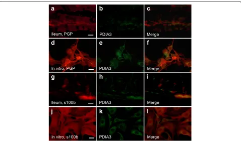
Immunocytochemistry revealed VDR expression in muco-sal epithelial cells of both small and large intestine while no VDR expression was detected in enteric neurons in vivo regardless of diet. In primary cultures of enteric neu-rons, no VDR expression was found in neurons. The alter-native vitamin D receptor PDIA3 has been suggested to mediate the rapid effects of vitamin D in intestinal epithe-lial cells [15]. Immunocytochemistry revealed PDIA3 not to be expressed on enteric neurons (Fig.3a-f) in vivo or in vitro, but to be expressed in intestinal glia in close connec-tion to enteric neurons (Fig.3g-i). This was evident in en-teric neuronal cultures as well (Fig.3j-l).
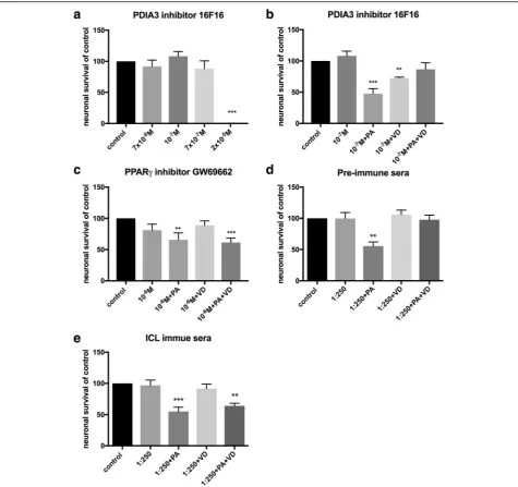
In vitro inhibition of PDIA3 in primary enteric cultures To evaluate if the VD effect was acting indirect by way of PDIA3 activation in enteric glia, primary enteric cul-tures were exposed to 16F16, known to inhibit PDI’s in-cluding PDIA3 [38]. The inhibitor (7x10⁻⁸M - 2x10⁻⁵M) induced neuronal loss at concentrations above 7x10⁻⁷, with all cells in culture lost at 2x10⁻⁵M (Fig. 4a). Sup-plementation with the PDIA3 inhibitor (10-7M) did not affect PA-induced neuronal loss in vitro nor did supple-mentation prevent the VD-mediated protection of the PA-induced neuronal loss. However, supplementation with the PDIA3 inhibitor (10-7M) significantly reduced neuronal survival in VD treated cultures (Fig.4b).
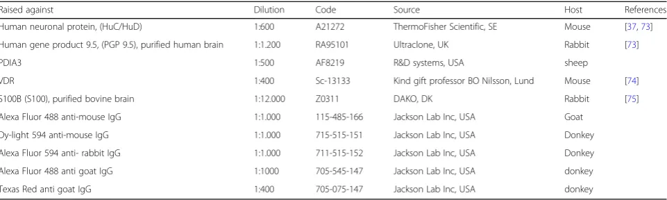
In vitro inhibition of PPARγin primary enteric cultures VDR is, like peroxisome proliferator-activated receptor (PPAR), a member of the nuclear hormone receptor family that form heterodimers with RXR. VDR and PPAR have interacting signaling pathways [42]. Primary enteric cultures were supplemented with the PPARγ in-hibitor GW9662 (10-6M). The effect of GW9662 on cul-tured enteric neurons and glia, has been assessed in a Table 2Overview of primary and secondary antibodies used in immunocytochemistry
Raised against Dilution Code Source Host References
Human neuronal protein, (HuC/HuD) 1:600 A21272 ThermoFisher Scientific, SE Mouse [37,73]
Human gene product 9.5, (PGP 9.5), purified human brain 1:1.200 RA95101 Ultraclone, UK Rabbit [73]
PDIA3 1:500 AF8219 R&D systems, USA sheep
VDR 1:400 Sc-13133 Kind gift professor BO Nilsson, Lund Mouse [74]
S100B (S100), purified bovine brain 1:12.000 Z0311 DAKO, DK Rabbit [75]
Alexa Fluor 488 anti-mouse IgG 1:1.000 115-485-166 Jackson Lab Inc, USA Goat
Dy-light 594 anti-mouse IgG 1:1.000 715-515-151 Jackson Lab Inc, USA Donkey
Alexa Fluor 594 anti- rabbit IgG 1:1.000 711-515-152 Jackson Lab Inc, USA Donkey
Alexa Fluor 488 anti goat IgG 1:1000 705-545-147 Jackson Lab Inc, USA donkey
previous study [28]. Supplementation of the PPARγ in-hibitor did not prevent the PA induced neuronal loss, but did prevent the VD-mediated protection of the PA-induced neuronal loss (Fig.4c).
In vitro ICL antibody neutralization
Bacteria, fungi and plants use the glyoxylate cycle to by-pass the CO2producing step in the Krebs cycle, utilizing two enzymes, ICL and malate synthase. Exposing enteric neuronal cultures to pre-immune serum (1:250) did not affect neuronal survival per se or VD’s effect on PA-induced neuronal loss (Fig. 4d). Exposing cultures to
ICL immune sera (1:250) did not affect neuronal survival
per se nor the PA-induced neuronal loss. However, ICL immune sera supplementation prevented the protective effect of vitamin D on PA-induced neuronal loss (Fig.4e).
Discussion
Current study investigated the neuroprotective potential of vitamin D and VD on HFD- and PA-induced enteric neuronal loss. Vitamin D supplementation with 1.3 IU/g to 2.6 IU/g had no effect on either ND or HFD enteric neuronal survival. However, supplementation of vitamin D at 26 IU/g (20x) prevented the HFD-induced neuronal
Fig. 1Effects of dietary vitamin D supplementation on high fat diet induced enteric neuronal loss and intestinal morphometric. Representative micrographs (a-c), morphometrics (d,g) and numbers of neurons in myenteric (MG) and submucosal (SG) ganglia (e,f,h,i) in ileum and colon from mice fed either normal diet (ND) or high fat diet (HFD) supplemented with different concentrations of vitamin D (0 to 20x normal concentration).a-ccryosections from ileum of ND 1x vitamin D concentration (a), HFD 1x vitamin D concentration (b) and HFD 20x vitamin D concentration (c) immunostained for HuC/HuD-biotin. Myenteric neurons are indicated with arrows and submucosal neurons with arrowheads.d,
gNo effect on intestinal morphology is observed in either ileum (d) or colon (g) in ND or HFD regardless of vitamin D concentration.e,f,h,i
loss, without affecting ND neuronal survival. In vitro studies showed VD (10-11M-10-7M) to protect against PA-induced neuronal loss, this effect was due to the presence of VD as the biological inactive precursor 7HDC was unable to mimic this neuroprotective effect. Serum concentration of VD were not assessed in current study. However, it has been demonstrated that supple-mentation of 10x the normal vitamin D dose in mice does not lead to elevated serum VD levels (10-10M) nor induce hypercalcemia [41]. It is therefore not believed that supplementing with 20 times the normal dose would lead to toxicity. Further it has been shown that obesity is associated with lower vitamin D serum con-centration, and patients should increase daily vitamin D intake [43].
HFD- and PA-induced neurotoxicity
VD is a lipid soluble vitamin and adipocytes function as an important VD reservoir [44]. With the obesity epi-demic and subsequent increase in type 2-diabetes [45], together with data suggesting that VD deficiency is a global health problem among all age groups [46], it is not surprising that the ability of VD to interfere with metabolic pathways have especially been studied in adi-pocytes and liver. In adiadi-pocytes, VD is involved in the regulation of differentiation, apoptosis, lipogenesis as well as acting as an anti-inflammatory agent [2, 47]. In liver, VD has experimentally been shown to be protective in HFD-induced steatosis. This is suggested to be through modulation of lipid metabolism, by increasing expression of enzymes and transcription factors involved in fatty acid oxidation i.e. carnitine-palmitoyl transferase 1a and 1b (CPT1a, b), pyruvate dehydrogenase kinase 4 (PDK4) and PPAR 1α. [1–3, 48] While studies have shown neurons capable of both mitochondrial and per-oxisomal fatty acid oxidation [49], they are not believed to rely on lipids for energy production [50], this is seen by e.g. their low expression of CPT1a and CPT1b in mitochondrial membranes and their expression of CPT1c on endoplasmic reticulum (ER) membranes [51,
52]. HFD- and PA-induced enteric neuronal losses have been investigated previously [28, 29]. Using pharmaco-logical agents interfering with intracellular lipid handling
pathways it was suggested that PA-induced neuronal loss in vitro was through the culmination of multiple meta-bolic disturbances. This Includes excessive palmitoylcar-nitine formation, exhausted carpalmitoylcar-nitine stores, increased acetate-coenzyme A (-CoA) levels and energy depletion as well as increased levels of oxidative stress and mem-brane destabilization [28]. The ability of VD to interfere with these metabolic pathways was, in current study, tested in vitro.
Receptor mediated VD effects
VD classically mediates its responses by binding to the nu-clear VDR, leading to altered gene transcription, VDRs have also been found in plasma membranes, co-localized with caveolin 1, where they are suggested to mediate a fast response [53]. Upon activation VDR forms homodimers with RXR and these complexes modulate gene expression through binding to VDREs [13, 14]. In man more than 2700 genes have been identified to be regulated by VDR [14]. By immunocytochemistry VDR was not found in en-teric neurons either in vivo or in vitro.It should, in this re-gard be noted that muscle cells (cardiac, skeletal and smooth) display positive VD mediated effects without clear evidence of the VDR being present [54]. It has been
suggested that the non-genomic rapid effect of VD is through activation of the alternative VD receptor, PDIA3 [15]. PDIA3 has been ascribed pleiotropic effects, includ-ing reduction of cellular stress in neurons [55,56], protec-tion against autophagy induced apoptosis in beta cells [57], and protection against free fatty acid induced hepa-tosteatosis [58]. Immunocytochemistry showed PDIA3 to be found in enteric glia in vivo and in vitro. Enteric glia play important roles in the gut including acting as neur-onal support and protection [59]. Inhibiting PDIA3 in vitro did not prevent the ability of VD to protect enteric neurons against PA-induced loss. This suggests that VD does not mediate its neuroprotective effect through activa-tion of PDIA3 in enteric glia.
Oxidative stress and membrane destabilization
VD mediates anti-inflammatory effects and has been shown to reduce HFD-induced increases in tumor ne-crosis factor α (TNFα) and pro-inflammatory cytokines such as interleukin (IL) -6 and lipid peroxidation prod-ucts [60, 61]. Besides anti-inflammatory effects VD has also been shown to have anti-oxidative effects by expres-sion of anti-oxidative enzymes such as superoxide dis-mutase (SOD), glutathione (GSH), and catalase [21,22].
a
b
c
d
e
f
g
h
i
j
k
l
However, VD has also been shown, independent of re-ceptor activation, to act as a membrane antioxidant by inhibiting lipid peroxidation. In the study, authors found the precursor 7HDC to be a more potent antioxidant than VD [19]. While current results showed 7HDC un-able to prevent PA-induced neuronal loss, a direct
anti-oxidative effect of VD in the protection against PA-induced loss cannot be entirely excluded.
Energy metabolism
HFD disrupts the energy balance and pushes the system towards a more inflammatory and reactive oxygen
Fig. 4Effects of experimental treatment agentper seand on VD and VD+PA neuronal survival and PA-induced neuronal loss in primary cultures of myenteric neurons.aSupplementation with the protein disulphide isomerase family A member 3 (PDIA3) inhibitor 16F16 in the range 7x10⁻⁸M - 7x10⁻⁷M did not affect neuronal survival, but induced loss of all cells at 2x10⁻⁵M.bSupplementation with the PDIA3 inhibitor (10-7M) had no effect on the palmitic acid (PA, 4x10⁻⁴M), the 1α,25-hydroxy-vitamin D3 (VD, 10-7M) or the PA+VD effects on neuronal survival.cPeroxisome proliferator-activated receptor gamma (PPARγ) inhibitor, GW69662 (10-6M) did not prevent the PA-induced neuronal loss but prevented the VD-induced prevention of the PA-induced loss.dSupplementation with pre-immune sera (1:250) had no effects on PA-, VD- or PA+VD-induced effects on neuronal survival.e
Supplementation of isocitrate lyase (ICL) immune sera (1:250) did not prevent the PA-induced loss but did prevent the protective effects of VD on PA-induced loss. Untreated controls were run in parallel. Data presented as mean ± SEM, controln=6-18 per treatment agent, PDIA3 inhibitor 16F16
species-rich phenotype. VD has been suggested to modulate energy pathways, including upregulating genes involved with fatty acid oxidation and anti-oxidation [3]. HFD has been shown to cause a reduced PPARγ expres-sion and classical PPARγtargets includingCD36in neu-rons [62], as well as in the small intestine [63]. In neurons PPARγ was suggested to act as neuronal lipid sensor, sensing and signaling to the central nervous sys-tem clues about the peripheral metabolic status [62]. In the intestinal epithelial cells, PPARγ is suggested to regulate barrier function and microbiome related inflam-mation [63]. We show that inhibition of PPARγprevents the VD induced protection against the PA-induced neur-onal loss in vitro, suggesting that high VD concentra-tions induce PPARγactivity.
Alternative VD pathway
Plants, bacteria and nematodes have maintained the abil-ity to synthesize glucose from lipids, using acetate pro-duced from peroxisomal beta oxidation. This energy bypass is called the glyoxylate cycle. It is catalyzed by two enzymes isocitrate lyase (ICL) and malate synthase (MS) and is active in situations where environmental en-ergy supply demands it [64]. In peroxisomes isocitrate is cleaved by ICL to form succinate and glyoxylate. Succin-ate enter the Krebs cycle and glyoxylSuccin-ate condenses with -CoA forming malate in a reaction catalyzed by MS. Malate can be converted into phosphoenolpyruvate by the enzyme phosphoenolpyruvate carboxykinase (PEPCK), and enter gluconeogenesis, thereby enabling lipid-derived carbon to be converted into glucose. The presence of this pathway in higher eukaryotes has been debated, including its presence and activity in peroxi-somes of liver [65, 66], cartilage [67] and brown fat [68] as well as its absence. [69–71] Those suggesting its pres-ence have shown that VD activate the glyoxylate or a glyoxylate-like cycle in liver and cartilage and stimulate the conversion of PA derived acetyl-CoA into glycogen [65,67]. It is tempting to speculate that high VD expos-ure in enteric neurons is able to stimulate this pathway. Using immunoneutralization to inhibit ICL activity we showed that supplementing cultures with ICL anti-bodies, but not pre-immune sera, prevented the VD-induced protection against the PA-induced neuronal loss. Whether the ICL immune serum contains undeter-mined mediators capable of inhibiting the VD response has not been investigated in currently study and cannot be excluded. It is however, interesting to note that PPARγis a known inducer of PEPCK, the enzyme enab-ling malate to enter gluconeogenesis. The induction of a glyoxylate or glyoxylate-like cycle has been suggested in hepatic glucose production [70, 72], where lipids in-crease hepatic gluconeogenesis. A working hypothesis in this respect could be that VD activates PAPRγ and
glyoxylate cycle-like pathways, causing PA or PA-CoA shunting towards peroxisome degradation, gluconeogen-esis and glucose production. In all creating a, for neu-rons, non-toxic metabolic intermediate that enables survival in a PA overload situation.
Conclusion
In this study, we showed neuroprotective effects of VD against HFD- and PA-induced neuronal loss. The sug-gested pathway involves VD-induced activation of PPARγ which in turn improve neuronal peroxisome function and shuttling of PA-CoA. Suggesting that sup-plementation of vitamin D in the diet could protect en-teric neurons against obesity-induced damage.
Abbreviations
7DHC:7-dihydrocholesterol; BSA: Bovine serum albumin; CDU: Collagen digestion units; -CoA: Acetyl- Coenzyme A; ENS: Enteric nervous system; ER: Endoplasmic reticulum; FBS: Fetal bovine serum; GSH: Glutathione; HBSS: Hank’s balanced salt solution; HFD: High fat diet; ICL: Isocitrate lyase; IL: Interleukin; IR: Immunoreactive; NBA: Neurobasal A; ND: Normal diet; PA: Palmitic acid; PBS: Phosphate buffered saline; PDI: Protein disulphide isomerase; PDIA3: Protein disulphide isomerase family A member 3; PDK4: Pyruvate dehydrogenase kinase 4; PEPCK: Phosphoenolpyruvate carboxykinase; PPARγ: Peroxisome proliferator-activated receptor gamma; ROS: Reactive oxygen species; RXR: Retinoid-X receptor; SOD: Superoxide dismutase; TNFα: Tumor necrosis factor alpha; VD: 1a,25-hydroxy-vitamin D3; VDR: VD receptor; VDREs: VD response elements
Acknowledgements
The authors would like to thank professor Eva Ekblad for financial and laboratory support and valuable input. The intestinal specimens used in the in vivo study were kindly donated by professor Cecilia Holm who designed, supervised and financed the animal model. The authors would also like to thank Anna Themner-Persson for excellent technical assistance.
Funding
This research was supported by grants from the Crafoord Foundation, the Royal Physiographic Foundation, the Påhlsson Foundation, the faculty of medicine Lund University, the Swedish Diabetes Foundation and the Swedish Research Council (project 11284 C. Holm).
Availability of data and materials
The datasets used and/or analysed during the current study are available from the corresponding author on reasonable request
Authors’contributions
SL designed and performed experiments, UV designed the study, analyzed the data and wrote the manuscript. All authors have read and approved the manuscript.
Competing interest
The authors declare that they have no competing interests.
Ethics approval
Experimental design, including housing, enrichment and animal welfare was approved by the animal ethics committee, Lund and Malmö, Sweden (dnr: M133-14 and dnr: M19-15).
Consent for publication
Not applicable
Publisher’s Note
Author details
1Unit of Molecular Endocrinology, Department of Experimental Medical
Science, Lund University, Sölvegatan 19, BMC C11, 22184 Lund, Sweden. 2
Unit of Neurogastroenterology, Department of Experimental Medical Science, Lund University, Sölvegatan 19, BMC B11, 22184 Lund, Sweden.
Received: 12 September 2018 Accepted: 8 November 2018
References
1. Yin Y, Yu Z, Xia M, Luo X, Lu X, Ling W. Vitamin D attenuates high fat diet-induced hepatic steatosis in rats by modulating lipid metabolism. Eur J Clin Invest. 2012;42(11):1189–96.
2. Sergeev IN, Song Q. High vitamin D and calcium intakes reduce diet-induced obesity in mice by increasing adipose tissue apoptosis. Mol Nutr Food Res. 2014;58(6):1342–8.
3. Marcotorchino J, Tourniaire F, Astier J, Karkeni E, Canault M, Amiot MJ, Bendahan D, Bernard M, Martin JC, Giannesini B, et al. Vitamin D protects against diet-induced obesity by enhancing fatty acid oxidation. J Nutr Biochem. 2014;25(10):1077–83.
4. Witham MD, Nadir MA, Struthers AD. Effect of vitamin D on blood pressure: a systematic review and meta-analysis. J Hypertens. 2009;27(10):1948–54. 5. Vimaleswaran KS, Berry DJ, Lu C, Tikkanen E, Pilz S, Hiraki LT, Cooper JD,
Dastani Z, Li R, Houston DK, et al. Causal relationship between obesity and vitamin D status: bi-directional Mendelian randomization analysis of multiple cohorts. PLoS Med. 2013;10(2):e1001383.
6. Danik JS, Manson JE. Vitamin d and cardiovascular disease. Curr Treat Options Cardiovasc Med. 2012;14(4):414–24.
7. Garcion E, Wion-Barbot N, Montero-Menei CN, Berger F, Wion D. New clues about vitamin D functions in the nervous system. Trends Endocrinol Metab. 2002;13(3):100–5.
8. Grimm MO, Mett J, Hartmann T. The Impact of Vitamin E and Other Fat-Soluble Vitamins on Alzheimer’s Disease. Int J Mol Sci. 2016;17:1785. 9. Wamberg L, Kampmann U, Stødkilde-Jørgensen H, Rejnmark L, Pedersen SB,
Richelsen B. Effects of vitamin D supplementation on body fat
accumulation, inflammation, and metabolic risk factors in obese adults with low vitamin D levels - results from a randomized trial. Eur J Intern Med. 2013;24(7):644–9.
10. Gallagher JC, Yalamanchili V, Smith LM. The effect of vitamin D supplementation on serum 25OHD in thin and obese women. J Steroid Biochem Mol Biol. 2013;136:195–200.
11. Eyles DW, Smith S, Kinobe R, Hewison M, McGrath JJ. Distribution of the vitamin D receptor and 1 alpha-hydroxylase in human brain. J Chem Neuroanat. 2005;29(1):21–30.
12. Overbergh L, Decallonne B, Valckx D, Verstuyf A, Depovere J, Laureys J, Rutgeerts O, Saint-Arnaud R, Bouillon R, Mathieu C. Identification and immune regulation of 25-hydroxyvitamin D-1-alpha-hydroxylase in murine macrophages. Clin Exp Immunol. 2000;120(1):139–46.
13. Schmidt DR, Mangelsdorf DJ. Nuclear receptors of the enteric tract: guarding the frontier. Nutr Rev. 2008;66(10 Suppl 2):S88–97.
14. Ramagopalan SV, Heger A, Berlanga AJ, Maugeri NJ, Lincoln MR, Burrell A, Handunnetthi L, Handel AE, Disanto G, Orton SM, et al. A ChIP-seq defined genome-wide map of vitamin D receptor binding: associations with disease and evolution. Genome Res. 2010;20(10):1352–60.
15. Nemere I, Hintze K. Novel hormone“receptors”. J Cell Biochem. 2008; 103(2):401–7.
16. Doroudi M, Schwartz Z, Boyan BD. Phospholipase A2 activating protein is required for 1α,25-dihydroxyvitamin D3 dependent rapid activation of protein kinase C via Pdia3. J Steroid Biochem Mol Biol. 2012;132(1-2):48–56. 17. Wang Y, Chen J, Lee CS, Nizkorodov A, Riemenschneider K, Martin D, Hyzy
S, Schwartz Z, Boyan BD. Disruption of Pdia3 gene results in bone abnormality and affects 1alpha,25-dihydroxy-vitamin D3-induced rapid activation of PKC. J Steroid Biochem Mol Biol. 2010;121(1-2):257–60. 18. Chen J, Olivares-Navarrete R, Wang Y, Herman TR, Boyan BD, Schwartz Z.
Protein-disulfide isomerase-associated 3 (Pdia3) mediates the membrane response to 1,25-dihydroxyvitamin D3 in osteoblasts. J Biol Chem. 2010; 285(47):37041–50.
19. Wiseman H. Vitamin D is a membrane antioxidant. Ability to inhibit iron-dependent lipid peroxidation in liposomes compared to cholesterol, ergosterol and tamoxifen and relevance to anticancer action. FEBS Lett. 1993;326(1-3):285–8.
20. Farhangi MA, Nameni G, Hajiluian G, Mesgari-Abbasi M. Cardiac tissue oxidative stress and inflammation after vitamin D administrations in high fat- diet induced obese rats. BMC Cardiovasc Disord. 2017;17(1):161. 21. Noyan T, Balaharoğlu R, Kömüroğlu U. The oxidant and antioxidant effects
of 25-hydroxyvitamin D3 in liver, kidney and heart tissues of diabetic rats. Clin Exp Med. 2005;5(1):31–6.
22. Bhat M, Ismail A. Vitamin D treatment protects against and reverses oxidative stress induced muscle proteolysis. J Steroid Biochem Mol Biol. 2015;152:171–9. 23. Singh VP, Bali A, Singh N, Jaggi AS. Advanced glycation end products and
diabetic complications. Korean J Physiol Pharmacol. 2014;18(1):1–14. 24. Sandireddy R, Yerra VG, Areti A, Komirishetty P, Kumar A.
Neuroinflammation and oxidative stress in diabetic neuropathy: futuristic strategies based on these targets. Int J Endocrinol. 2014;2014:674987. 25. Lv WS, Zhao WJ, Gong SL, Fang DD, Wang B, Fu ZJ, Yan SL, Wang YG.
Serum 25-hydroxyvitamin D levels and peripheral neuropathy in patients with type 2 diabetes: a systematic review and meta-analysis. J Endocrinol Invest. 2015;38(5):513–8.
26. Boitard C, Cavaroc A, Sauvant J, Aubert A, Castanon N, Layé S, Ferreira G. Impairment of hippocampal-dependent memory induced by juvenile high-fat diet intake is associated with enhanced hippocampal inflammation in rats. Brain Behav Immun. 2014;40:9–17.
27. Kaczmarczyk MM, Machaj AS, Chiu GS, Lawson MA, Gainey SJ, York JM, Meling DD, Martin SA, Kwakwa KA, Newman AF, et al. Methylphenidate prevents high-fat diet (HFD)-induced learning/memory impairment in juvenile mice. Psychoneuroendocrinology. 2013;38(9):1553–64. 28. Voss U, Sand E, Olde B, Ekblad E. Enteric neuropathy can be induced by
high fat diet in vivo and palmitic acid exposure in vitro. PLoS One. 2013; 8(12):e81413.
29. Voss U, Turesson MF, Robaye B, Boeynaems JM, Olde B, Erlinge D, Ekblad E. The enteric nervous system of P2Y13 receptor null mice is resistant against high-fat-diet- and palmitic-acid-induced neuronal loss. Purinergic Signal. 2014;10:455.
30. Stenkamp-Strahm CM, Kappmeyer AJ, Schmalz JT, Gericke M, Balemba O. High-fat diet ingestion correlates with neuropathy in the duodenum myenteric plexus of obese mice with symptoms of type 2 diabetes. Cell Tissue Res. 2013;354(2):381–94.
31. Morin JP, Rodríguez-Durán LF, Guzmán-Ramos K, Perez-Cruz C, Ferreira G, Diaz-Cintra S, Pacheco-López G. Palatable Hyper-Caloric Foods Impact on Neuronal Plasticity. Front Behav Neurosci. 2017;11:19.
32. Eslick GD, Talley NJ. Prevalence and relationship between gastrointestinal symptoms among individuals of different body mass index: A population-based study. Obes Res Clin Pract. 2016;10(2):143-50.
33. Wiltink J, Michal M, Wild PS, Zwiener I, Blettner M, Münzel T, Schulz A, Kirschner Y, Beutel ME. Associations between depression and different measures of obesity (BMI, WC, WHtR, WHR). BMC Psychiatry. 2013;13:223. 34. Furness JB, Callaghan BP, Rivera LR, Cho HJ. The enteric nervous system and
gastrointestinal innervation: integrated local and central control. Adv Exp Med Biol. 2014;817:39–71.
35. Reeves PG, Nielsen FH, Fahey GC. AIN-93 purified diets for laboratory rodents: final report of the American Institute of Nutrition ad hoc writing committee on the reformulation of the AIN-76A rodent diet. J Nutr. 1993;123(11):1939–51. 36. Sand E, Themner-Persson A, Ekblad E. Corticotropin releasing factor-Distribution
in rat intestine and role in neuroprotection. Regul Pept. 2011;166(1-3):68–75. 37. Voss U, Sand E, Hellstrom PM, Ekblad E. Glucagon-like peptides 1 and 2 and
vasoactive intestinal peptide are neuroprotective on cultured and mast cell co-cultured rat myenteric neurons. BMC Gastroenterol. 2012;12:30. 38. Hoffstrom BG, Kaplan A, Letso R, Schmid RS, Turmel GJ, Lo DC, Stockwell BR.
Inhibitors of protein disulfide isomerase suppress apoptosis induced by misfolded proteins. Nat Chem Biol. 2010;6(12):900–6.
39. Voss U, Turesson MF, Robaye B, Boeynaems J-M, Olde B, Erlinge D, Ekblad E. The enteric nervous system of P2Y13receptor null mice is resistant against high fat diet- and palmitic acid-induced neuronal loss. Purinergic Signal. 2014;10(3):455–64.
40. Nezami BG, Mwangi SM, Lee JE, Jeppsson S, Anitha M, Yarandi SS, Farris AB, Srinivasan S. MicroRNA 375 Mediates Palmitate-Induced Enteric Neuronal Damage and High-Fat Diet-Induced Delayed Intestinal Transit in Mice. Gastroenterology. 2014;146(2):473–483.e473.
42. Matsuda S, Kitagishi Y. Peroxisome proliferator-activated receptor and vitamin d receptor signaling pathways in cancer cells. Cancers (Basel). 2013;5(4):1261–70. 43. Ekwaru JP, Zwicker JD, Holick MF, Giovannucci E, Veugelers PJ. The
importance of body weight for the dose response relationship of oral vitamin D supplementation and serum 25-hydroxyvitamin D in healthy volunteers. PLoS One. 2014;9(11):e111265.
44. Landrier JF, Marcotorchino J, Tourniaire F. Lipophilic Micronutrients and Adipose Tissue Biology. Nutrients. 2012;4(11):1622–49.
45. Guariguata L, Whiting DR, Hambleton I, Beagley J, Linnenkamp U, Shaw JE. Global estimates of diabetes prevalence for 2013 and projections for 2035. Diabetes Res Clin Pract. 2014;103(2):137–49.
46. Palacios C, Gonzalez L. Is vitamin D deficiency a major global public health problem? J Steroid Biochem Mol Biol. 2014;144 Pt A:138–45.
47. Eugene C, Yangha K. Vitamin D Decreases Adipocyte Lipid Storage and increases NAD-SIRT1 Pathway in 3T3-L1 Adipocytes. Nutrition. 2016; 32(6):702–8.
48. Yin Y, Yu ZW, Xia M, Luo XQ, Lu XF, Ling WH. Vitamin D attenuates high fat diet-induced hepatic steatosis in rats by modulating lipid metabolism. Eur J Clin Invest. 2012;42(11):1189–96.
49. Berger J, Dorninger F, Forss-Petter S, Kunze M. Peroxisomes in brain development and function. Biochim Biophys Acta. 2016;1863(5):934–55. 50. Romano A, Koczwara JB, Gallelli CA, Vergara D, Micioni Di Bonaventura MV,
Gaetani S, Giudetti AM. Fats for thoughts: An update on brain fatty acid metabolism. Int J Biochem Cell Biol. 2017;84:40–5.
51. Lee J, Wolfgang MJ. Metabolomic profiling reveals a role for CPT1c in neuronal oxidative metabolism. Bmc Biochem. 2012;13.
52. Sierra AY, Gratacos E, Carrasco P, Clotet J, Urena J, Serra D, Asins G, Hegardt FG, Casals N. CPT1c is localized in endoplasmic reticulum of neurons and has carnitine palmitoyltransferase activity. J Biol Chem. 2008;283(11):6878–85.
53. Huhtakangas JA, Olivera CJ, Bishop JE, Zanello LP, Norman AW. The vitamin D receptor is present in caveolae-enriched plasma membranes and binds 1 alpha,25(OH)2-vitamin D3 in vivo and in vitro. Mol Endocrinol. 2004;18(11): 2660–71.
54. Wang Y, DeLuca HF. Is the vitamin d receptor found in muscle? BMC Biochem. 2012;13:23.
55. Castillo V, Oñate M, Woehlbier U, Rozas P, Andreu C, Medinas D, Valdés P, Osorio F, Mercado G, Vidal RL, et al. Functional Role of the Disulfide Isomerase ERp57 in Axonal Regeneration. PLoS One. 2015;10(9):e0136620. 56. Pendyala G, Ninemire C, Fox HS. Protective role for the disulfide isomerase
PDIA3 in methamphetamine neurotoxicity. PLoS One. 2012;7(6):e38909. 57. Yamamoto E, Uchida T, Abe H, Taka H, Fujimura T, Komiya K, Hara A,
Ogihara T, Fujitani Y, Ueno T, et al. Increased expression of ERp57/GRP58 is protective against pancreatic beta cell death caused by autophagic failure. Biochem Biophys Res Commun. 2014;453(1):19–24.
58. Zhang XQ, Pan Y, Yu CH, Xu CF, Xu L, Li YM, Chen WX. PDIA3 Knockdown Exacerbates Free Fatty Acid-Induced Hepatocyte Steatosis and Apoptosis. PLoS One. 2015;10(7):e0133882.
59. Neunlist M, Rolli-Derkinderen M, Latorre R, Van Landeghem L, Coron E, Derkinderen P, De Giorgio R. Enteric glial cells: recent developments and future directions. Gastroenterology. 2014;147(6):1230–7.
60. ErbaşO, Solmaz V, Aksoy D, Yavaşoğlu A, Sağcan M, Taşkıran D. Cholecalciferol (vitamin D 3) improves cognitive dysfunction and reduces inflammation in a rat fatty liver model of metabolic syndrome. Life Sci. 2014;103(2):68–72.
61. Kheder R, Hobkirk J, Saeed Z, Janus J, Carroll S, Browning MJ, Stover C. Vitamin D3 supplementation of a high fat high sugar diet ameliorates prediabetic phenotype in female LDLR(-/-) and LDLR(+/+) mice. Immun Inflamm Dis. 2017;5(2):151–62.
62. Liu C, Bookout AL, Lee S, Sun K, Jia L, Lee C, Udit S, Deng Y, Scherer PE, Mangelsdorf DJ, et al. PPARγin vagal neurons regulates high-fat diet induced thermogenesis. Cell Metab. 2014;19(4):722–30.
63. Tomas J, Mulet C, Saffarian A, Cavin JB, Ducroc R, Regnault B, Kun Tan C, Duszka K, Burcelin R, Wahli W, et al. High-fat diet modifies the PPAR-γ pathway leading to disruption of microbial and physiological ecosystem in murine small intestine. Proc Natl Acad Sci U S A. 2016;113(40):E5934–43. 64. Dunn MF, Ramírez-Trujillo JA, Hernández-Lucas I. Major roles of isocitrate
lyase and malate synthase in bacterial and fungal pathogenesis. Microbiology. 2009;155(Pt 10):3166–75.
65. Davis WL, Matthews JL, Goodman DB. Glyoxylate cycle in the rat liver: effect of vitamin D3 treatment. FASEB J. 1989;3(5):1651–5.
66. Davis WL, Goodman DB. Evidence for the glyoxylate cycle in human liver. Anat Rec. 1992;234(4):461–8.
67. Davis WL, Jones RG, Farmer GR, Cortinas E, Matthews JL, Goodman DB. The glyoxylate cycle in rat epiphyseal cartilage: the effect of vitamin-D3 on the activity of the enzymes isocitrate lyase and malate synthase. Bone. 1989;10(3):201–6. 68. Davis WL, Goodman DB, Crawford LA, Cooper OJ, Matthews JL. Hibernation
activates glyoxylate cycle and gluconeogenesis in black bear brown adipose tissue. Biochim Biophys Acta. 1990;1051(3):276–8.
69. Kondrashov FA, Koonin EV, Morgunov IG, Finogenova TV, Kondrashova MN. Evolution of glyoxylate cycle enzymes in Metazoa: evidence of multiple horizontal transfer events and pseudogene formation. Biol Direct. 2006;1:31. 70. Song S. Can the glyoxylate pathway contribute to fat-induced hepatic
insulin resistance? Med Hypotheses. 2000;54(5):739–47.
71. Popov VN, Igamberdiev AU, Schnarrenberger C, Volvenkin SV. Induction of glyoxylate cycle enzymes in rat liver upon food starvation. FEBS Lett. 1996; 390(3):258–60.
72. Song S. The role of increased liver triglyceride content: a culprit of diabetic hyperglycaemia? Diabetes Metab Res Rev. 2002;18(1):5–12.
73. Sand E, Voss U, Hammar O, Alm R, Nordin Fredrikson G, Ohlsson B, Ekblad E. Gonadotropin-releasing hormone analog buserelin causes neuronal loss in rat gastrointestinal tract. Cell Tissue Res. 2013;351(3):521–34.