A Novel Hypothetical Method to Increase the
Dimensions of the Coronary Arteries when
Required
Mark Christopher Arokiaraj
11. Cardiology, Pondicherry Institute of Medical Sciences, Pondicherry, India.
Corresponding author: Mark Christopher Arokiaraj,
Cardiology, Pondicherry Institute of Medical Sciences, Pondicherry, India. Email: [email protected]
Introduction
It is a common observation that coronary arteries appear thin (of narrow diameter) in long-standing diabetic and hyperlipidemic individuals. In patients with refractory angina, symptoms can persist even after optimal coronary artery revascularization by medical therapy, stents or coronary artery bypass grafts [1,2]. Approximately 60,000 cases are seen every year in this category in Europe or the US [1]. A method to increase the size of the coronary arteries would be of potential therapeutic interest. This would be challenging and therapeutically useful. An increase in size could result in a reduced incidence of coronary events. Especially in diabetic patients, there is a reduction in arterial sizes due to deposition of advanced glycosylated end products [3]. Hyperlipidemic conditions due to deposition of
the micro cholesterol particles also lead to a reduction in the coronary dimensions and result in increased coronary events [4]. Reduction in the caliber of the blood vessels leads to a reduced volume of blood flow as well as an increase in wall shear stress [5] and results in earlier coronary events.
The primary objective of this study was to identify a novel method which could increase the dimensions of the coronaries. Though coronary dilatation cannot prevent atherosclerosis directly, it is possible to reduce atherosclerotic processes by reducing wall shear stress, which initiates endothelial dysfunction. In this study, after a series of observations in various unexpected clinical
© 2020 Author(s). This is an Open Access article distributed under the terms of the Creative Commons Attribution CC-BY-4.0 license CC-BY-4.0 (http://creativecommons.org/ licenses/by/4.0/), which permits use, distribution and reproduction, provided the original work is properly cited. Published by Barcaray (International) Publishing.
Abstract
Background
The purpose was to develop a novel hypothetical method to increase the size of coronary arteries.
Methods
During long-term observation an increase in coronary size has been seen in three unexpected scenarios. Changes in coronary artery sizes were observed in patients with mitral stenosis (n=69) by angiogram prior to percutaneous balloon mitral valvuloplasty or valve replacement surgery for severe mitral stenosis. The coronaries of patients with patent ductus arteriosus who underwent surgical closure in the past (n=14) were examined by echocardiogram. Patients with renal failure on long-term dialysis through peripheral arterio-venous fistula without left ventricular hypertrophy (n=17) were studied by echocardiography. Normal age, weight and sex matched coronary sizes served as controls in the study. All these observations were made over a period of 12 years.
Results
The sizes of coronaries in patients with mitral stenosis, patients who underwent closure for patent ductus arteriosus, and in patients on hemodialysis through arteriovenous fistulas were higher than normal controls (p<0.05, for all). A hypothetical model to increase the coronary sizes could be developed based on the analysis of the differential equations of Poiseuille’s. A method is proposed of creating a peripheral arterio-venous fistula, which could be closed later electively by a percutaneous method/surgery. The closure time needs to be determined by experimental studies. The other methods could be a continuous exercise program or by the use of beta-blockers.
Conclusions
A novel hypothetical method of peripheral arteriovenous fistula formation is described which could potentially increase the size of the coronaries, which could be closed later.
Keywords: Coronary dimensions; Poiseuille’s Equation; Arteriovenous fistula; Coronary dilatation; Wall shear stress; Coronary blood flow; Atherosclerosis; Angiogenesis
scenarios a theoretical model has been proposed as a derivative of physical equations governing fluid dynamics.
Methods and Results
The study was developed after the observation of a series of cases in different clinical settings in which the coronary arteries appeared dilated. Three clinical conditions were identified and studied, in which the coronary arteries were widened, and the left ventricles had normal dimensions. In these three clinical conditions an increase in coronary sizes was not expected. Conditions which are known to increase or decrease the coronary and left ventricular dimensions were excluded. After observations in the initial three years, a prospective case-controlled study was planned. The primary outcome of the study was to identify dilated coronaries in patients with normal left ventricular dimensions, compared to the controls. The recruitment of the patients was on a continuous non-randomized method. This is due to a limited number of patients available for the study. The study was not blinded, and the author was aware of the subjects as well as the controls. The observations were made over a period of 12 years. Coronary diameters were measured in the left main coronary artery (LMCA), Left anterior descending artery and the right coronary artery within 3mm from the coronary ostium (Philips IE33, HP Sonos 5500 and Sonosite M-Turbo).
Rheumatic Mitral Stenosis
Observations in the coronary angiograms (n=32) (Siemens Axiom Artis) before mitral balloon valvuloplasty (figure 1) or as a preoperative workup (n=13) for a mitral valve replacement for severe calcific mitral stenosis (figure 2) showed a mild increase in the coronary sizes instead of the expected small caliber since the left ventricle is usually small in this condition compared to the controls (figure 3). During this period balloon mitral valvuloplasty was performed in 157 patients. However, a coronary angiogram was performed in only 32 patients. The mean age of the patients was 38±9 yrs. (median 37, range 18 to 57 years).
In a further subset of 24 patients out of the 157 patients who underwent balloon mitral valvuloplasty, left ventricular angiograms were performed in the initial period before the year 2011 (Figure 4). The patient profile was similar to those patients who underwent balloon mitral valvuloplasty subsequently after year 2010. In these patients, direct coronary angiogram was not performed, and it was a protocol to perform left ventricular (LV) angiogram at that time to look for the mitral valve calcification and to quantify the extent of mitral regurgitation. Diagnostic coronary angiogram as a routine was performed only in patients more than fort-five years, which is a standard guideline. These patients LV angiograms were reviewed and the mean LMCA 4.3±0.3mm and LAD 3.5±0.2mm dimensions were significantly higher than the normal controls. Also, none of these patients had coronary artery disease.
The mean age of patients who underwent surgery (mitral valve replacement) for isolated severe mitral stenosis was 43±6 yrs. (n=13). (median 45; range 28 to 58). Patients with other valvular lesions and mitral regurgitation were excluded. All these patients underwent coronary angiography before the surgical procedure (supplementary figures 6 and 7). All the patients successfully recovered after surgery. Patients with any hyperdynamic circulatory states such as anemia, pregnancy or thyrotoxicosis,
and patients with hypertension or the presence of left ventricular hypertrophy were excluded.
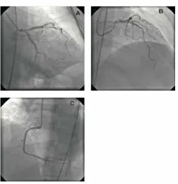
Figure 1
Shows the coronaries of a patient with rheumatic mitral stenosis (panels A, B, C and E) prior to balloon mitral valvuloplasty (panel D), magnification factor 17.
Figure 2
Creatinine levels were normal except in 1 patient where the creatinine was 1.4 mg/dl, and the coronaries were not dilated in this patient. The mean left main coronary artery (LMCA) diameter 4.2±0.2mm was, and the mean proximal right coronary artery (RCA) 3.2±0.3mm diameter and proximal LAD 3.4±0.2mm diameters and the proximal left circumflex artery (LCA) were 3.3±0.3 mm. None of these patients had coronary artery disease (figures 1 and 2, and supplementary figures 1 to 7).
The control patients (n=32) were patients with normal chamber dimensions and normal coronaries who underwent coronary angiography for evaluation of chest pain after a stress test. The control patients were age-sex-weight matched without other co-morbidities or valvular disorders, and they were selected retrospectively in a randomized non-blinded fashion. The control (figure 3 and supplementary figures 8 to 12) values of the normal coronary artery sizes in a similar age and body size-matched controls for LMCA was 3.4±0.3mm, proximal LAD was 2.9±0.3mm, the proximal RCA was 2.7±0.3mm, and the proximal LCA - 2.8±0.2mm. The values were significant by t-test (p<0.05, for each). Baseline characteristics are given in the table 1. Out of the total 69 patients in this category, 57 had dilated coronaries compared to control, six had only mildly dilated vessels, five had normal sized vessels; one patient had a smaller vessel and this patient had higher creatinine value of 1.4mg/dl.
Patent Ductus Arteriosus
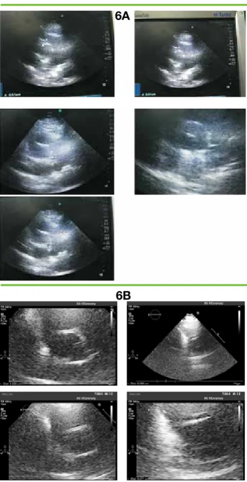
Patent ductus arteriosus is a congenital vascular connection between the aorta and pulmonary artery. Patients (n= 14) with large shunts who underwent surgery in the past were analyzed for the coronary sizes by echocardiography. Baseline characteristics of this patient group is given in table 2. It was observed that the coronary arterial sizes were larger than the usual, which was observed by echocardiography analysis. The size of the aorta in these patients was normal. In all these cases surgical/coil closure was performed at least four years before the echocardiography these observations of coronary dilatations (figure 5, upper panel, Figure 6A and 6B). The mean age of the patients was 17±7 yrs. None of the patients had hyperdynamic circulations such as with Figure 4
Shows LV angiogram in patients prior to undergoing Balloon mitral valvuloplasty (magnification factor 23).
Figure 3
Shows the coronaries of a normal patient as control (panels A to C).
Figure 5
anemia, thyrotoxicosis or peripheral arteriovenous fistula. The mean LMCA diameter was 4.28±0.4mm, LAD was 3.6±0.3, and the mean RCA diameter was 3.4±0.2mm.
The control patients (n=13) were normal age-sex-weight matched patients without other co-morbidities. They had normal chambers and coronary dimensions and there were selected by a randomized method. The control values of normal LMCA, LAD and RCA measured by the similar technique was 3.4±0.4mm, 3.0±0.3mm and 2.8±0.3mm respectively. The values were statistically significantly different (p<0.05, for each). The RCA was co-dominant or dominant in all these cases. Visual assessment (eyeballing) of the coronary arteries showed a significant increase in the size of the coronary vessels in these patients compared to the similar age and body-size matched controls (figure 7). All echocardiographic measurements of the coronary arteries were performed with the same magnifications. Also, in all the cases except one, the coronary arteries were dilated compared to the controls giving a high statistical power.
Patients on hemodialysis through the peripheral
arterio-venous (AV) fistula
Patients who undergo dialysis (n=17) with peripheral arteriovenous fistula were analyzed for their coronary sizes. Patients who already have left ventricular hypertrophy/dilatation were excluded, as a left ventricular hypertrophy/dilatation could increase the size of the coronary vessels to overcome the demand-supply mismatch in coronary blood flow. The control patients (n=17) were normal healthy age, sex and weight matched controls without other comorbidities.
The coronary dimensions of the patients with A-V fistula showed larger dimensions. These observations in the coronary arteries were made by echocardiographic evaluation (figure 5 lower panel and figure 8). The mean age of the patients was 54±9 yrs. (median 53; range 41,65). Other baseline characteristics of the patients are given in table 3. The mean LMCA diameter was 4.0±0.3mm,
Table 1. Baseline characteristics of patients undergoing balloon mitral valvuloplasty/surgery.
Parameters Values, n=69(32BMV>2010+24BMV <2011+13Surgery)
Age, yrs. 37±9 Sex, M/F 22/47 Diabetes, n 1 Hypertension, n 1 MV area Pre-procedure, cm2 0.9 ± .1
MV area Post procedure, cm2 1.8 ± 0.2
Height, cm 166 ±5 Left ventricle size, mm 46 ± 0.2 Balloon size used, size mm,
(n =52) 24, (62); 26(7) Preprocedural mitral
regurgitation (Gr 1), n 8 Preprocedural Pulmonary artery systolic pressure, mm Hg 82±10 Serum Creatinine, mg/dl 0.9±0.2 Atrial fibrillation, n 41 Periprocedural mortality, n Nil
Figure 6
(Figures 6A and 6B)Echocardiography of a patient who underwent PDA closure in the past and the coronaries were dilated.
6A
6B
Table 2. Baseline characteristics of patients underwent patent ductus arteriosus closure.
Parameters Values, n=14
Age, yrs. 18±7, median 19 (11, 43) Sex, M/F 5/9
Diabetes, n 1 Hypertension, n nil Duration after coil or surgical
LAD was 3.6±0.2mm, and the mean proximal RCA diameter was 3.3±0.3mm; in the control patients the LMCA diameter was 3.4±0.3mm, LAD 3.2±0.2mm and RCA was 2.8±0.4mm. The values were statistically significant (p<0.05 for all). The RCA was co-dominant or dominant in all these cases. Visual assessment (i.e., eyeballing) of the coronary arteries showed a distinct increase in size of the coronary vessels. All echocardiographic measurements of the coronary arteries were performed with the same magnifications. In 16 out of the 17 cases, the coronary dimensions were elevated in varying degrees, and in one patient the coronary dimensions were not increased.
Statistical power calculation
Retrospective power calculations were performed based on the observed standard deviations in each category and a minimum significant increase (minimum difference) in arterial dimensions of 0.5mm. A minimum in each group to be studied based on the standard deviation was about ten patients. Hence, the study has the minimum required power for proof of concept purposes, although this is not prospective trial proof of this effect. Based on this analysis, the study results suggest further prospective evaluation may be worthwhile [6]
Discussion
The results presented in the study are from 3 different patient subgroups, unrelated to each other, and the studies were performed over several years. They had one aspect in common, which was the varying degrees of dilatation of coronaries, without dilatation or hypertrophy of the left ventricle. In mitral stenosis, this could be due the fall in the mean arterial pressure compared to the controls, and in other two subgroups, it is possibly related to the existing or the closed arterio-ventricular fistulas. This could be due to changes in physical mechanics or cellular signaling. A hyperdynamic circulation as the only mechanism for coronary dilatation in A-V fistula patients is unlikely, as these patients had normal chamber dimensions.
Application of Poisueille’s equation
Poisueille’s law states that the velocity of the steady flow of a fluid through a narrow tube varies directly as the pressure and the fourth power of the radius of the tube and inversely as the length of the tube and the coefficient of viscosity. In these three conditions in the study, the diastolic or the coronary perfusion pressure tends to be low, and the biological response is to dilate the vessels. The study shows some interesting observations on the dilated coronary arteries in specific scenarios. The controls were normal patients, and when compared to diabetic vessels these coronary dimensions would be much higher. Analysing the Poisueille’s equation a reduction in blood pressure especially in the diastolic blood pressures would increase the radius of the blood vessels in the long term [7]. The coronary arteries are usually larger than the normal in patients with aortic regurgitation where the diastolic pressures are low. However, in this condition, the aorta is often dilated and left ventricular hypertrophy is
Table 3. Baseline characteristics of patients undergoing haemodialysis.
Parameters Values, (n=17)
Age, yrs. 54±9 Sex, M/F 14/2 Diabetes, n 3 Hypertension, n 4 Left ventricular size, mm 47±2 LV posterior wall thickness, mm 10.6 LA size, mm 29±0.3 Duration of dialysis on the fistula, yrs. 4.5±2.1 Valvular heart diseases Nil Pulmonary artery systolic pressure, mm Hg 30±4 Systolic blood pressure, mm Hg 130±6 Diastolic blood pressure, mm Hg 84±10 Nifedipine, n 4 Prazosin, n 2 Haemoglobin, gm/dl 11±0.4
Figure 7
seen. Even in other states like rheumatic mitral regurgitation the coronary arteries are dilated. However, they are often associated with left ventricular dilatation with or without hypertrophy.
The aorta and left ventricles are of standard caliber in all the three study groups. Also, in chronic renal failure on dialysis, the left ventricle is often dilated with hypertrophy. The patients chosen in this study with chronic renal failure did not have either dilatation or hypertrophy of left ventricle.
A lowering of blood pressure, could orchestrate a series of translational pathways, and result in the dilatation of the coronaries. The coronary blood flow and pressures are reduced in mitral stenosis, which improves with percutaneous balloon mitral valvuloplasty [8,9]. In patients with mitral stenosis, there is hypertrophy of the right ventricle. Hence, the right coronary or the circumflex artery would tend to be larger. However, the interesting observation is that the left anterior descending artery was also dilated in-spite of the smaller left ventricle usually seen in this condition.
Cellular mechanisms
Baroreceptors are located in the coronaries, which are similar to the baroreceptors of the aorta [10]. These baroreceptors could mediate the changes through various signaling pathways. Possibly these mediators could be vascular endothelial growth factors [11], hepatocyte growth factors [12,13]and non-canonical WNT pathways [14-17] or Mastermind proteins [18] through NOTCH signalling [19]; Hedgehog pathways [20]. Further investigations are required to identify the exact mechanism of these observations.
Growth factors such as vascular endothelial growth factors and hepatocyte growth factors are known to have a significant role in the signaling of vessel growth. There is also an interesting observation that by creating an arterio-venous fistula the progression of chronic renal failure to end-stage renal failure is delayed [21].
Hence, the formation of the arterio-venous fistula may increase the size of the coronary arteries. This could have clinical therapeutic benefits, which would be reflected by a lower incidence in coronary events. It has been observed in various studies that enhanced external counter pulsations can reduce angina in patients with refractory angina [22, 23]. The observations in this study possibly explain the variations in blood pressures induced by enhanced counter pulsations could be the mechanism for better clinical performance in these patients [24].
There is also a potential disadvantage of the formation of an arteriovenous fistula including tachycardia, which could be managed with medications. However, as observed in this study in patients with patent ductus arteriosus who underwent closure in the past the beneficial effects of an increase in the size of the coronary vessels persist for many years even after the closure of the patent ductus arteriosus. Hence, the peripheral arteriovenous fistula, which when created, could be closed later by either surgical or percutaneous methods. To reduce the complications induced by the arteriovenous fistula, the connections could be performed in distal locations, for example, in the radial/ ulnar artery instead of the proximal brachial artery.
Mass-Energy Equivalence principle in fluids
The traditional mass-energy equivalence principle is applicable not only in the atomic principles but also in the fluids and super-liquid quantum vacuum [25-27]. The presence of an arteriovenous fistula tends to increase the workload in the heart and the energy dissipated will reflect on the changes in the mass and the dimensions, which would be an increase in the coronary dimensions, LV mass and also the capillary density. However, these need to be studied in detail by prospective experiments and trials.
Limitations
The number of patients in each category is small. The incidence of normal echocardiographic findings in patients on chronic hemodialysis is rare, and also the number of patients at follow-up who remain asymptomatic several years after patent ductus arteriosus closure is also not high. Moreover, nowadays, due to early treatment by balloon mitral valvuloplasty, the number of patients treated with valve replacement for mitral stenosis is low. Hence, the principle was to recruit as large patient numbers Figure 8
as possible. Angiography during balloon mitral valvuloplasty was performed only after some initial observations of coronary dilatations. Also, in 24 cases undergoing balloon mitral valvuloplasty before 2011, the measurements of the coronaries were based on left ventricular angiogram pictures, which are not very accurate. Nevertheless, the observation was convincing, as the coronaries are dilated.
In some conditions in the study, coronary sizes were measured by echocardiography as in patients with closed patent ductus arteriosus, and in patients with renal failure on hemodialysis, which is not the gold standard to evaluate the coronary sizes. A high degree of intra and inter-observer variability was noticed for the coronary size measurements [28]. Especially when the coronary sizes are less than 3mm. However, anatomical assessment is feasible and in children with Kawasaki’s disease [29], the coronary size measurement is performed with echocardiography in many centers routinely including our institution [30]. In this study the dilated coronaries were more than 3mm. Due to ethical considerations angiography was not contemplated in these patients. Nevertheless, this is a rare and specific group of patients selected in the study, and by visual assessment the observations were outstanding. Overall, the total number of patients in each group is less, and therefore the evidence presented is not strong. Also, the study group in all the subgroups were younger than the usual age group for coronary artery disease. Hence, further studies with large numbers need to be performed, which could reveal more information.
Right heart strain
The concept of volume overload and right atrial and ventricular dilatation and RV strain and dysfunction which is progressive over time is well known [31,32]. These studies have a median follow up period of about 3.9 (IQR 2.4-6.4) years, and the incidence of RV dysfunction is about 35% [33]. The RV dysfunction is more seen in patients with brachiocephalic fistulas than in the radial venous fistulas [31]. Hence, if a peripheral arteriovenous fistula is formed for this purpose the closure time could be in the range of 1.5 to 2 years to prevent complications.
Future perspectives
The concept outlined in the study is hypothetical and further validation accurately is only possible after an animal study by creating an arteriovenous fistula, and studying the angiogram before and after the A-V fistula possibly with a long-time interval of about 6m to 1 year in large animals. Echocardiographic measurement of coronary arteries before and after the arteriovenous fistula in clinical practice can be performed, however, this method is less accurate to conclude. The effect of beta-blockers, which reduce the heart rate and blood pressure, and its impact on coronary anatomical variations need to be studied. Also, the effect of a continuous exercise program modifying the coronary sizes needs to be determined.
Conclusion
There is a possibility for a novel therapeutic method of AV fistula formation as a method to increase coronary sizes. This concept needs to be evaluated by experimental studies in animals by observing coronary dimensions before and after creating an arteriovenous fistula.
Declarations of Interest
The author declares no conflicts of interest. Source of funding: None
Ethical considerations: Informed consent for the procedures was taken, and institutional ethical review board approved the study (pims/13012011/C1/G).
Acknowledgements
The author states that he abides by the requirements for ethical publishing in biomedical journals.[34].
References
1. Cheng K, de Silva R. New advances in the management of refractory angina pectoris. Eur Cardiol. 2018;13(1):70–79. doi:10.15420/ecr.2018:1:2. 2. Manchanda A, Aggrawal A, Aggrawal N, et al. Management of refractory
angina pectoris. Cardiol J. 2011;18(4):343-51.
3. Ertan C, Ozeke O, Gul M, et al. Association of prediabetes with diffuse coronary narrowing and small-vessel disease. Journal of Cardiology. 2014; 63: 29–34. doi: 10.1016/j.jjcc.2013.06
4. Nelson RH. Hyperlipidemia as a Risk Factor for Cardiovascular Disease. Primary care. 2013;40(1):195-211. doi:10.1016/j.pop.2012.11.003. 5. Cheng C, Helderman F, Tempel D et al. Large variations in absolute wall shear
stress levels within one species and between species. Atherosclerosis. 2007;195(2):225-235. doi: 10.1016/j.atherosclerosis.2006.11.019
6. Columb M, Atkinson M. Statistical analysis: sample size and power estimations. BJA Education. 2016;16(5):159-161. doi:10.1093/bjaed/ mkv034
7. Sutera, Salvatore P.; Skalak, Richard. The History of Poiseuille’s Law. Annual Review of Fluid Mechanics. 1993; 25: 1–19.
8. Osman Bektaş, Zeki Yüksel Günaydın, Ahmet Karagöz, et al. Effects of Mitral Balloon Valvuloplasty on Coronary Blood Flow and Flow Reserve. The Journal of Heart Valve Disease 2015;24:729-735.
9. Mymin D, Sharma G. Total and effective coronary blood flow in coronary and noncoronary heart disease. Journal of Clinical Investigation. 1974;53(2):363-373. doi: 10.1172/JCI107568
10. Kincaid K, Ward M, Nair U, et al. The Coronary Baroreflex in Humans. The Journal of Extra-corporeal Technology. 2005;37(3):306-310.
11. Uruno A, Sugawara A, Kanatsuka H, et al. Hepatocyte Growth Factor Stimulates Nitric Oxide Production through Endothelial Nitric Oxide Synthase Activation by the Phosphoinositide 3-Kinase/Akt Pathway and Possibly by Mitogen-Activated Protein Kinase Kinase in Vascular Endothelial Cells. Hypertension Research. 2004;27(11):887-895. DOI:10.1291/hypres.27.887 12. Morishita R, Aoki M, Yo Y, et al. Hepatocyte Growth Factor as Cardiovascular
Hormone: Role of HGF in the Pathogenesis of Cardiovascular Disease. Endocrine Journal. 2002;49(3):273-284. doi: 10.1507/endocrj.49.273 13. Lönn J, Starkhammar Johansson C, Kälvegren H, et al. Hepatocyte
growth factor in patients with coronary artery disease and its relation to periodontal condition. Results in Immunology. 2012;2:7-12. doi: 10.1016/j. rinim.2011.12.002
14. Marinou K, Christodoulides C, Antoniades C, et al. Wnt signaling in cardiovascular physiology. Trends in Endocrinology & Metabolism. 2012;23(12):628-636. doi:10.1016/j.tem.2012.06.001
15. Reis M, Liebner S. Wnt signaling in the vasculature. Experimental Cell Research. 2013;319(9):1317-1323. doi: 10.1016/j.yexcr.2012.12.023. 16. Meyer IS, Jungmann A, Dieterich C, et al. The cardiac microenvironment
uses nonşcanonical WNT signaling to activate monocytes after myocardial infarction. EMBO Molecular Medicine. 2017;9(9):1279-1293. doi:10.15252/ emmm.201707565.
17. Mazzotta S, Neves C, Bonner R, et al. Distinctive Roles of Canonical and Noncanonical Wnt Signaling in Human Embryonic Cardiomyocyte Development. Stem Cell Reports. 2016; (7): 764–776. doi:10.1016/j. stemcr.2016.08.008
18. McElhinny A, Li J, Wu L. Mastermind-like transcriptional co-activators: emerging roles in regulating cross talk among multiple signaling pathways. Oncogene. 2008;27(38):5138-5147. doi:10.1038/onc.2008.228
19. Anderson L, Gibbons G. Notch: a mastermind of vascular morphogenesis. Journal of Clinical Investigation. 2007;117(2):299-302. doi:10.1172/ JCI31288.
20. Lee SW, Moskowitz MA, Sims JR. Sonic hedgehog inversely regulates the expression of angiopoietin-1 and angiopoietin-2 in fibroblasts. International Journal of Molecular Medicine. 19 (3); 445–51: 2007. doi.org/10.3892/ ijmm.19.3.445
21. Golper T, Hartle P, Bian A. Arteriovenous fistula creation may slow estimated glomerular filtration rate trajectory. Nephrology Dialysis Transplantation. 2015;30(12):2014-2018. doi:10.1093/ndt/gfv082
from the International EECP Patient Registry). The American Journal of Cardiology. 2006;97(1):17-20. doi:10.1016/j.amjcard.2005.07.122 23. Sharma U, Ramsey HK, Tak T. The Role of Enhanced External Counter
Pulsation Therapy in Clinical Practice. Clinical Medicine & Research. 2013;11(4):226-232. doi:10.3121/cmr.2013.1169.
24. V. Kitsou, T. Xanthos, R. Roberts, et al. Enhanced external counterpulsation: mechanisms of action and clinical applications. Acta Cardiologica. 2010;65(2):239-247. doi: 10.2143/AC.65.2.2047060
25. Einstein A. 1946, “An Elementary Derivation of the Equivalence of Mass and Energy,” in A. Einstein(1956), pp. 116–119.
26. Palacios A. The Mass-Energy Equivalence Principle in Fluid Dynamics. Journal of High Energy Physics, Gravitation and Cosmology. 2015;01(01):48-54. doi: 10.4236/jhepgc.2015.11005.
27. Šorli, A.S. Mass–Energy Equivalence Extension onto a Superfluid Quantum Vacuum. Sci Rep 9, 11737 (2019) doi:10.1038/s41598-019-48018-2. 28. Ronai C, Hamaoka-Okamoto A, Baker A, de Ferranti S, Colan S, Newburger
J et al. Coronary Artery Aneurysm Measurement and Z Score Variability in Kawasaki Disease. Journal of the American Society of Echocardiography. 2016;29(2):150-157. doi:10.1016/j.echo.2015.08.013
29. Margossian R, Lu M, Minich LL, et al. Predictors of coronary artery visualization in Kawasaki disease. JAmSocEchocardiogr.2011;24(1):53–59. doi:10.1016/j.echo.2010.10.015.
30. Alla Boshchenko, Alexander Vrublevsky and Rostislav Karpov (September 15th 2011). Transthoracic Echocardiography in the Assessment of Coronary Arteries, Coronary Angiography - Advances in Noninvasive Imaging Approach for Evaluation of Coronary Artery Disease, Branislav Baskot, IntechOpen, doi: 10.5772/21793.
31. Said K, Hassan M, Baligh E, Zayed B, Sorour K. Ventricular Function in Patients with End-Stage Renal Disease Starting Dialysis Therapy: A Tissue Doppler Imaging Study. Echocardiography. 2012;29(9):1054-1059. doi:10.1111/j.1540-8175.2012.01749.x
32. Alkhouli M, Sandhu P, Boobes K, Hatahet K, Raza F, Boobes Y. Cardiac complications of arteriovenous fistulas in patients with end-stage renal disease. Nefrología (English Edition). 2015;35(3):234-245. doi:10.1016/j. nefro.2015.03.001
33. Reddy Y, Obokata M, Dean P, Melenovsky V, Nath K, Borlaug B. Long-term cardiovascular changes following creation of arteriovenous fistula in patients with end stage renal disease. European Heart Journal. 2017;38(24):1913-1923. doi:10.1093/eurheartj/ehx045