Molecular Basis of the Dynamic Structure
of the TIM23 Complex in the Mitochondrial
Intermembrane Space
Rakhi Bajaj,
1q
ukasz Jaremko,
1,2Mariusz Jaremko,
1Stefan Becker,
1and Markus Zweckstetter
1,2,3,*
1Department of NMR-based Structural Biology, Max Planck Institute for Biophysical Chemistry, 37077 Go¨ttingen, Germany 2German Center for Neurodegenerative Diseases (DZNE), 37077 Go¨ttingen, Germany
3Center for the Molecular Physiology of the Brain, University Medicine Go¨ttingen, 37073 Go¨ttingen, Germany *Correspondence:[email protected]
http://dx.doi.org/10.1016/j.str.2014.07.015
SUMMARY
The presequence translocase TIM23 is a highly
dy-namic complex in which its subunits can adopt
multiple conformations and undergo
association-dissociation to facilitate import of proteins into
mito-chondria. Despite the importance of protein-protein
interactions in TIM23, little is known about the
mol-ecular details of these processes. Using nuclear
magnetic resonance spectroscopy, we
character-ized the dynamic interaction network of the
inter-membrane space domains of Tim23, Tim21, Tim50,
and Tom22 at single-residue level. We show that
Tim23
IMScontains multiple sites to efficiently interact
with the intermembrane space domain of Tim21
and to bind to Tim21, Tim50, and Tom22. In
addi-tion, we reveal the atomic details of the dynamic
Tim23
IMS-Tim21
IMScomplex. The combined data
support a central role of the intermembrane space
domain of Tim23 in the formation and regulation of
the presequence translocase.
INTRODUCTION
More than 99% of all mitochondrial proteins are synthesized in
the cytosol and traverse the mitochondrial membranes to reach
their final destination(
Neupert and Herrmann, 2007
). Preprotein
import is based on the coordinated action of hetero-oligomeric
translocases in the outer (TOM) and inner (TIM) mitochondrial
membrane (
Bauer et al., 2000; Pfanner, 1998; Ryan and Jensen,
1995; Schatz, 1996
). Sorting of preproteins to the mitochondrial
matrix and the inner mitochondrial membrane is achieved by
the TIM23 complex in the inner mitochondrial membrane, also
known as presequence translocase (
Chacinska et al., 2005;
Glick et al., 1992; Hutu et al., 2008; Pfanner and Geissler,
2001; van der Laan et al., 2007, 2010; Yamamoto et al., 2002
).
The presequence translocase TIM23 is a highly dynamic
com-plex in which its subunits can adopt multiple conformations and
undergo association-dissociation to facilitate preprotein import
(
Chacinska et al., 2005; Marom et al., 2011; Popov-Celeketi
c
et al., 2008; Tamura et al., 2009; van der Laan et al., 2010
).
TIM23 contains the core proteins Tim23, Tim17, and Tim50, as
well as Mgr2 and Tim21 as accessory subunits and the
motor-associated proteins Pam 17, Pam 16-18, Tim44, and mtHsp
(
Chacinska et al., 2009; Endo et al., 2011; Gebert et al., 2012;
Kutik et al., 2007; Neupert and Herrmann, 2007; Wiedemann
et al., 2004
). The functional form of TIM23 involves diverse
inter-actions between the intermembrane space (IMS) domains of its
subunits that are important for: (1) receiving the presequence (
de
la Cruz et al., 2010; Marom et al., 2011; Moczko et al., 1997;
Schulz et al., 2011
), (2) formation of the translocation contact
(
Albrecht et al., 2006; Chacinska et al., 2003; Mokranjac et al.,
2005; Shiota et al., 2011; Tamura et al., 2009
), and (3) regulation
of the pore across the inner membrane (
Martinez-Caballero
et al., 2007; Meinecke et al., 2006
). Indeed, in vivo and in vitro
crosslinking studies have provided support for a variety of IMS
interactions such as Tim23-Tim50, Tim21-Tim23,
Tom22-Tim50, Tim17-Pam18, and Pam17-Tim23 (
Chacinska et al.,
2005; Hutu et al., 2008; Lytovchenko et al., 2013; Marom
et al., 2011; Moczko et al., 1997; Shiota et al., 2011; Tamura
et al., 2009; Yamamoto et al., 2002
). In addition, the incoming
preprotein can be crosslinked to the IMS domains of many of
the aforementioned subunits (
Geissler et al., 2002; Moczko
et al., 1997
;
Schulz et al., 2011; Shiota et al., 2011; Tamura
et al., 2009
).
The IMS domain of Tim23 plays a key role for preprotein
import (
Davis et al., 2000; Donzeau et al., 2000;
Gevorkyan-Air-apetov et al., 2009; Popov-Celeketi
c et al., 2008; Tamura et al.,
2009; Truscott et al., 2001
). In
Saccharomyces cerevisiae
,
Tim23
IMSconsists of the N-terminal 96 residues of Tim23.
Tim23
IMSis intrinsically disordered in vitro and contains a
bind-ing site for presequences (
de la Cruz et al., 2010
). In intact
mitochondria, the first 20 amino acids of Tim23
IMSare sensitive
to protease cleavage and have been proposed to traverse the
outer mitochondrial membrane. In addition, residues 50–96 of
Tim23 were proposed to dimerize and regulate channel activity
(
Bauer et al., 1996
). Crosslinks and mutations in this region
affect the association with various other subunits including
Tim50 and Tim21 (
Gevorkyan-Airapetov et al., 2009; Tamura
et al., 2009
).
Despite the importance of protein-protein interactions within
the TIM23 complex, little is known about the molecular details
of these interactions. At present, only the 3D structure of a
pre-sequence in complex with the cytosolic domain of Tom20 has
been resolved (
Abe et al., 2000
), whereas no 3D structure of a
protein-protein complex within or between the translocases is
known. Here we investigated the protein interaction network of
the intermembrane space domain of Tim23, the central
compo-nent of the TIM23 complex, at the residue level and determined
the atomic details of the Tim23
IMS-Tim21
IMScomplex. The
com-bined data support a central role of Tim23
IMSin the formation
and dynamic regulation of the TIM23 complex.
RESULTS
Three Tim23 Sites Bind to Tim21
IMSThe dynamic association and dissociation of Tim21 with the
core subunits of TIM23 has been proposed to regulate the
sort-ing of the preprotein either to the inner mitochondrial
mem-brane or to the mitochondrial matrix (
Chacinska et al., 2005;
van der Laan et al., 2007, 2010
). In addition, Tim23
IMShas
been crosslinked to Tim21 in vivo (
Lytovchenko et al., 2013;
Ta-mura et al., 2009
). To obtain insight into the interaction of the
IMS domains of Tim23 and Tim21 at single-residue resolution,
we used nuclear magnetic resonance (NMR) spectroscopy.
To this end, we titrated
15N-labeled Tim23
IMSwith increasing
amounts of unlabeled Tim21
IMS. The addition of Tim21
IMScaused progressive changes in NMR signal intensity and
posi-tion in distinct regions of Tim23
IMS(
Figure 1
A). NMR signals of
residues 67–74 and 90–96 were strongly broadened and
shifted, identifying them as anchor sites for Tim21
IMS. In
addi-tion, residues 1–7 showed pronounced signal attenuation (
Fig-ure 1
A), whereas the gradual signal decrease from residues 30
to 60 is likely due to enhanced NMR relaxation times as a
consequence of binding of residues 67–74 to the globular
structure of Tim21
IMS. To validate a direct interaction of
Tim21
IMSwith Tim23
IMS, we attached the paramagnetic tag
MTSL to two sites in Tim21
IMS. The two attachment sites,
114 and 128, were in proximity to the Tim21
IMSresidues that
are involved into binding to Tim23
IMS(see below and
Figures
1
B and 1C). The MTSL-tagged Tim21
IMSvariants were then
added to Tim23
IMSin a 1:1 molar ratio. Pronounced PRE
broadening was observed in the three regions of Tim23 (
Figures
1
B and 1C), which showed strong chemical shift perturbation,
demonstrating a direct interaction of these regions with
Tim21
IMS. Residues 1–7, 68–74, and 90–96 consist of the
se-quences MSWLFGD, VEYLDLE, and SRGWTDD, respectively.
All three residue stretches contain an aromatic residue at
position i (F5, Y70, W93) and an aspartic acid at position i+2.
In addition, the stretches
68VEYLDLE
74and
90SRGWTDD
96contain at least one additional negatively charged residue,
whereas an additional aromatic residue is located at the N
ter-minus. Taken together, the data demonstrate that three distinct
regions in Tim23
IMSparticipate in complex formation with
Tim21
IMS.
Rapid Exchange of Tim23 Sites with a Single Tim21
Binding Pocket
Next, we identified the binding site of Tim23
IMSon Tim21
IMS.
Concentration-dependent changes in NMR chemical shifts
were observed for the Tim21
IMSresidues F109, V113, S114,
V116 and E117, and 138–144 upon addition of Tim23
IMS(
Fig-ure 2
A). Quantitative analysis of the binding curves determined
the K
dvalue as 153 ± 67
m
M. Notably, surface plasmon
reso-nance of immobilized Tim23
IMSpointed to a much lower K
dof
1
m
M (
Lytovchenko et al., 2013
). However, because Tim23
IMSis known to bind to hydrophobic environments such as
mem-branes (
Donzeau et al., 2000
) and the Tim23
IMS-Tim21
IMSinter-action involves three Tim23 segments that are in rapid exchange
(
Figure 1
), analysis of the interaction by surface plasmon
reso-nance is complicated. The residues identified with NMR analysis
are located in
b
strand 1 and on one side of the
a
helix 1 of
Figure 1. Single-Residue Analysis of the Binding of Tim21IMS toTim23IMS
(A) Interaction sites in Tim23IMSfor Tim21IMSas derived from 2D1H-15N-HSQC titration experiments of15
N-labeled Tim23IMS
with increasing amounts of unlabeled Tim21IMS
. Changes in NMR signal intensity and position at 16-fold excess of Tim21IMS
are shown.
(B and C) Paramagnetic relaxation enhancement induced in Tim23IMS upon addition of MTSL-tagged Tim21IMS. PRE profiles (ratio of signal intensities observed in 2D1
H-15
N HSQC spectra in the paramagnetic, Ip, and diamag-netic, Id, state) as a function of Tim23 residue number upon addition of (B) Tim21IMS
MTSL-tagged at S114C and (C) Tim21IMS
MTSL-tagged at C128. Missing data points are due to the presence of prolines or signal overlap. The error in intensity ratios were based on the signal-to-noise ratio. Cartoon re-spresentations show unlabeled Tim21IMS
(green) and15
N-labeled Tim23IMS (red) at a 1:1 molar ratio. The position of the paramagnetic tag is shown in blue.
Tim21
IMS(
Figure 2
B). Further support for the rapid exchange of
multiple binding motifs of Tim23
IMSwith a common Tim21
bind-ing site was provided by paramagnetic relaxation enhancement:
attachment of a MTSL-tag to either residue 11 of Tim23, i.e., the
N-terminal binding region of Tim23, or residue 67, which is in
proximity to the binding region 2 in Tim23, caused highly similar
paramagnetic broadening in Tim21
IMS(
Figures 2
C and 2D). The
Tim21
IMSresidues that form the shallow binding pocket for
bind-ing to Tim23
IMSare conserved (
Figure 2
E).
The striking finding of the NMR-based interaction mapping is
that three regions in Tim23
IMSare involved in binding, but on
Tim21
IMSthere is only a single binding site. To further support
Figure 2. Tim23IMSBinds to a Conserved Pocket in Tim21IMS(A) Residue specific binding curves of Tim21IMS
residues belonging to helix 1 andbstrand 1. Chemical shift changes were fit to a single site model. (B) Localization of the Tim23-binding site on the solution structure of Tim21IMS
. Residues experiencing strong NMR signal perturbation upon addition of Tim23 are shown in magenta.
(C and D) Paramagnetic relaxation enhancement induced in Tim21IMS
(green) upon addition of MTSL-tagged Tim23IMS
(red). Tim23IMS
was tagged with MTSL at position T11C (C) and G67C (D). Ip/Idare the intensity ratios obtained from 2D
15 H-15
N HSQC spectra of Tim21IMS
in the presence of MTSL-tagged Tim23IMS (Ip) and after addition of ascorbic acid to the same sample (Id). Missing data points are due to the presence of prolines or signal overlap. In (D), the location of secondary structure elements in Tim21IMS
is indicated. Thebhairpin seen in the crystal but absent in solution (seeFigure 3) is shown in gray. The most strongly attenuated residues were 138–144, defining the binding pocket in Tim21IMS
. The errors in intensity ratios were based on signal-to-noise ratio. (E) Sequence alignment for Tim21IMS
highlighting the conservation of residues in the Tim23IMS
binding site among different kingdoms with I-V asRattus norvegicus,Homo sapiens,Aspergillus flavus,Saccharomyces cerevisiae, andCandida albicans, respectively. The sequence alignment was done using ClustalW (Larkin et al., 2007) and depicted with Jalview (Clamp et al., 2004). Hydrophobic and aromatic residues are highlighted in blue; positive and negative charged as red and magenta, respectively; neutral residues in green; glycine and proline in orange and yellow, respectively. (Top) Secondary structure elements of S.cerevisiaeTim21 are shown and the dashed box highlights the Tim23IMS
binding region in Tim21IMS .
the complex nature of this interaction, we analyzed the Tim21
IMSinteraction of a peptide comprising the N-terminal 13 residues of
Tim23 (
Figure 3
A). In addition, in a separate experiment, the
binding of a peptide comprising residues 61–96 of Tim23
IMSto
Tim21
IMSwas measured (
Figure 3
B). Tim23(1–13) contains the
N-terminal Tim21-binding site, whereas Tim23(61–96) contains
the other two interacting residue stretches. Stepwise addition
of each of the Tim23 fragments caused the same chemical shift
trajectories of Tim21
IMSresidues as observed in the binding
studies with the full Tim23
IMSdomain (
Figures 3
A and 3B).
How-ever, the magnitude of chemical shift changes was significantly
smaller in case of the peptides. Quantitative analysis indicated
a K
dof 396 ± 65
m
M for Tim23(61–96), that is approximately
2-fold higher than that of Tim23(1–96) (
Figure S1
available online).
In the case of Tim23(1–13), NMR signal perturbation was even
smaller, pointing to a K
dvalue exceeding 1 mM. The observation
that the Tim23 fragments change the position of individual NMR
signals of Tim21
IMSin the same direction as the complete IMS
domain proves that the three binding motifs of Tim23
IMSbind
in a similar manner to the same site in Tim21
IMS. In addition,
due to the similarity of the chemical shift changes, we further
conclude that the Tim21-binding mode of Tim23(1–13) is highly
similar to that of residues 69–74 and 90–96, albeit at a decreased
affinity. Thus, rapid exchange of multiple Tim23 binding sites
with a single Tim21 binding pocket is essential for an effective
Tim23
IMS-Tim21
IMSassociation.
Dynamic Structure of the Tim21
IMS-Tim23
IMSComplex
To obtain an atomic resolution view of the association of
Tim23
IMSwith Tim21
IMS, we characterized the 3D structure of
Tim21
IMSin complex with the three Tim23 binding motifs. This
was achieved by determination of the solution structure of
Tim21
IMS, followed by NMR-driven docking of the Tim23
IMSbinding motifs. The 3D structure of unbound Tim21
IMSwas
determined based on nearly complete chemical shift assignment
and a large number of nuclear Overhauser effect (NOE) distance
restraints (
Figure 4
and
Table 1
). Comparison of the solution
structure of Tim21
IMSwith the one observed in the crystal
(
Albrecht et al., 2006
) showed that the core of the structures is
highly similar. However, residues 144–153, which are in spatial
proximity to the Tim23 binding site (
Figure 2
), do not form a
b
hairpin in solution but are dynamic (
Figure 4
B). Indeed, residues
144–153 are involved in crystal contacts that can stabilize the
b
hairpin. We then used the NMR chemical shift perturbation
and paramagnetic broadening data to dock the three binding
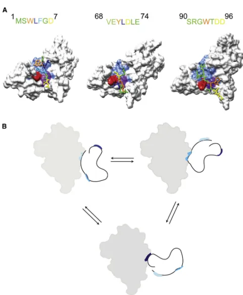
motifs of Tim23—that is,
1MSWLFGD
7,
68VEYLDLE
74, and
90
SRGWTDD
96—to the Tim21
IMSsolution structure. Because
of the low affinity of each individual motif, no large structural
re-arrangements are expected to occur in Tim21
IMSupon binding of
Tim23
IMS. For docking, the Tim23 peptide was positioned in an
extended conformation 10–12 A˚ above the binding pocket as
defined by the NMR data. Peptide docking was performed, in
which the structure of Tim21
IMSwas kept fixed but the structure
of the Tim23-motif was allowed to change.
Figures 5
A and
S2
show different docked conformations of the three Tim23 motifs
in complex with Tim21
IMS. Due to the dynamic nature of the
Tim23
IMS- Tim21
IMScomplex, it was not possible to obtain
experimental information about the structure of the three
Tim23
IMSbinding motifs when bound to Tim21
IMS. Thus, we
cannot exclude that the three Tim23
IMSmotifs can also populate
alternative conformations. Despite this uncertainty, however, the
docking models shown in
Figures 5
A and
S2
suggest that K139
and Y141 of Tim21 might be important for the interaction with
Tim23
IMS.
Notably, peptide docking did not result in a single bound
conformation, but a set of conformations with similar docking
energies. Moreover, even for a single Tim23 binding motif,
different hydrophobic residues formed contacts with Y141 of
Tim21
IMSin the docked structures (
Figures 5
A and
S2
). Part of
this structural heterogeneity might be due to limitations of the
docking algorithm. On the other hand, experimental support for
structural heterogeneity in the Tim23
IMS-Tim21
IMScomplex
comes from PRE-broadening induced in Tim21
IMSupon addition
of Tim23
IMS, which was tagged with MTSL at either residue 11
or 67 (
Figures 2
C and 2D). The MTSL attachment site (residue
11) is in one case C terminal to the binding motif (the
1
MSWLFGD
7motif), whereas in the other case (residue 67), it is
N terminal to the
68VEYLDLE
74binding motif. The PRE profiles
Figure 3. Interaction of Linear Motifs of Tim23IMSwith Tim21IMSSuperposition of selected regions of 2D1 H-15
N HSQC spectra of Tim21IMS with increasing amounts of Tim23(1–13) (A) and Tim23(61–96) (B), respectively (reference, black; 8-fold excess, blue; and 32-fold excess, red).
induced in Tim21
IMSwere, however, similar (
Figures 2
C and 2D), a
finding not expected when each motif would bind in a single
orien-tation (as in this case the MTSL tag would likely be located at
different sites with respect to Tim21
IMS). Conformational
hetero-geneity might be important for the ability of Tim21
IMSto recognize
the three different Tim23 binding motifs (
Figure 5
B). In addition, it
might allow for lower affinity and therefore efficient dissociation
despite the specificity of the Tim23-Tim21 interaction.
Interaction of Tim23 with Tim50
Within the TIM23 complex, the association of the IMS domains of
Tim23 and Tim50 plays an important role in receiving the
prese-quence carrying preprotein from the outer mitochondrial
translo-case and directing it to the inner mitochondrial pore (
Meinecke
et al., 2006; Shiota et al., 2011
). In vitro binding studies in
com-bination with in vivo chemical crosslinking revealed that Y70
and L71 of Tim23
IMSare important for binding to Tim50
IMS(
Ge-vorkyan-Airapetov et al., 2009; Mokranjac et al., 2003; Tamura
et al., 2009; Yamamoto et al., 2002
). Y70 and L71 belong to
the Tim23 residue stretch
68VEYLDLE
74that binds to Tim21
IMS(
Figure 5
). To obtain insight into the Tim23-Tim50 interaction
on a residue level, we used two different Tim50
IMSvariants.
Tim50(164–476) comprises most of the IMS domain of Tim50,
whereas for Tim50(164–361), the 3D structure is known (
Qian
et al., 2011
), but it lacks the presequence-binding domain.
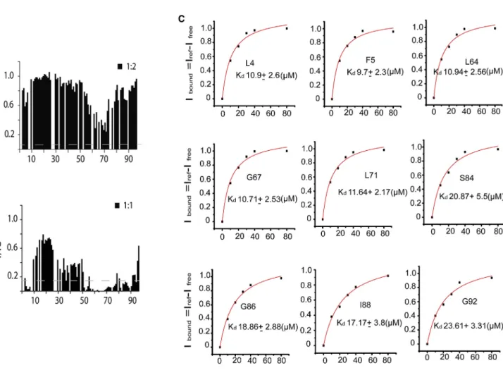
Com-plex formation between Tim23
IMSand Tim50(164–361) caused
a strong decrease in NMR signal intensity of residues 1–7 and
56–78 of Tim23
IMS(
Figure 6
A). Because no accompanying
chemical shift changes were observed, it suggests that the
bind-ing process is not fast on the NMR time scale. When usbind-ing
Tim50(164–476), more Tim23 residues participated in complex
formation and the affinity was increased (
Figure 6
B). Quantitative
analysis showed that the affinity of the Tim23
IMS-Tim50(164–
476) complex in solution is 10–20
m
M (
Figure 6
C). Taken
together, the data demonstrate that efficient formation of the
Tim23
IMS-Tim50
IMScomplex requires the C-terminal, so-called
presequence-binding domain of Tim50.
Identification of the Tim23-Tom22 Translocation
Contact Site
A direct translocation contact between the TOM40 complex
and the TIM23 complex has been established by in vivo
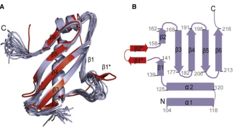
cross-Figure 4. Solution Structure of Tim21IMS
(A) Superposition of the 20 lowest energy NMR conformers (purple) of Tim21IMS
with the X-ray structure (red; Protein Data Bank code: 2CIU;
Albrecht et al., 2006).
(B) Secondary structure of Tim21IMS . Residue numbers are marked. Note that in solution, resi-dues 145–154 are flexible and do not form ab hairpin (b1*andb2*) as seen in the crystal. Resi-dues 145–154 are in immediate vicinity to the Tim23IMS
binding site.
linking of Tom22 to residue 41 of Tim23
(
Tamura et al., 2009
). Our NMR-based
binding analysis of the isolated domains
supports a translocation contact
be-tween Tim23 and Tom22. Tim23 residues V53 and L58–L61
were perturbed by addition of Tom22
IMS(
Figure 7
A). Moreover,
changes in carbon resonances of the aliphatic side chains of
Tom22
IMSwere observed upon addition of Tim23
IMS(
Figure 7
B).
Thus, both in vivo crosslinking—taking into account the length
of the crosslinker—and in vitro binding map the Tim23-Tom22
translocation contact to the central part of Tim23
IMS. In
contrast to the direct Tim23
IMS-Tom22
IMSinteraction, NMR
sig-nals of Tim23
IMSremained unperturbed in a titration with the
N-terminal tail of Tom40 (
Figure 7
C), which is predicted to be
disordered and located in the IMS. In addition, we did not
detect an interaction between Tim21
IMSand Tom22
IMS(
Fig-ure S3
), in line with the finding that Tim21 appears not to play
a primary role in linking the TOM40 and TIM23 complexes
(
Tamura et al., 2009
).
DISCUSSION
To facilitate preprotein import, the subunits of the TIM23
com-plex adopt multiple conformations and undergo
association-dissociation processes (
Popov-Celeketi
c et al., 2008; van der
Laan et al., 2007
). The Tim23 protein is the main subunit of
the presequence translocase. It forms the protein-conducting
pore in the inner mitochondrial membrane (
Truscott et al.,
2001
) and has been suggested to interact with more than 15
subunits across the translocases (
Albrecht et al., 2006;
Chacin-ska et al., 2003; Mokranjac et al., 2005; Shiota et al., 2011;
Tamura et al., 2009
). Interactions among the intermembrane
space domains of TIM23 are important in receiving and
direct-ing the preprotein toward the TIM23 channel. Usdirect-ing purified
IMS domains of Tim23 and Tim21 in combination with NMR
spectroscopy, we revealed a complex mechanism of
interac-tion between the intermembrane space domains of Tim23
and Tim21. Tim23 contains three distinct motifs that bind to a
single binding pocket in Tim21 (
Figures 1
,
2
,
3
,
4
, and
5
). The
binding pocket is formed by
b
strand 1 and
a
helix 1, a region
that is evolutionary conserved in Tim21 (
Figures 2
B and 2E).
The Tim23 binding motifs bind individually very weakly to
Tim21 (
Figure 3
). However, by being connected within one
chain, the concentration of Tim21-binding motifs effectively
increases, so that the rate of binding of Tim23 to Tim21 is
enhanced.
Binding of several short linear motifs to a single binding site
was previously observed to regulate the interaction of the
disordered cyclin-dependent kinase inhibitor Sic1 with its
re-ceptor Cdc4 (
Mittag et al., 2008
). Electrostatic interactions
be-tween multiple phosphorylated sites on Sic1 and Cdc4 resulted
in a dynamic equilibrium. In case of Tim23
IMS-Tim21
IMSa
similar dynamic complex is found (
Figure 5
). However, in
contrast to the Sic1-Cdc4 system, hydrophobic interactions
are more important for the dynamic recognition of Tim21
IMS.
In addition, our structural analysis points to the existence of
conformational heterogeneity of even a single Tim23
IMSmotif
when bound to Tim21
IMS(
Figures 5
and
S2
). This feature is
reminiscent of the dynamic binding mode of a presequence
in the binding site of the cytoplasmic domain of Tom20 (
Ko-muro et al., 2013; Saitoh et al., 2011
) and highlights the
im-portance of dynamic interactions for protein import into
mitochondria.
What is the role of the linear binding motifs of Tim23 for other
IMS interactions? Residue-specific analysis of the Tim23
IMS-Tim50
IMSinteraction showed that all three Tim21-binding motifs
of Tim23 are also involved in binding to Tim50
IMS(
Figure 6
). In
addition, the full IMS domain of Tim50 further recruits residues
29–46 of Tim23 into the complex (
Figure 6
B). The data show
that Tim23’s interaction motifs are involved in several IMS
interactions. The motifs are highly conserved (
Figure 8
A) and
can bind to multiple partners (
Figure 8
B). Multiple binding
sites are used to enhance affinity for one protein such as
Tim21 or Tim50. In addition, the presence of several distinct
interaction motifs might enable simultaneous binding of the
intermembrane space domain of Tim23 to multiple protein
components of the TIM23 complex. Such an interaction with
multiple protein partners might stabilize the TIM23 complex
and potentially enable the formation of heterooligomeric
com-plexes in the translocation contact site. The presequence
bind-ing site in Tim23 (
de la Cruz et al., 2010
) overlaps with one of
the motifs that is important for binding to both Tim21 and
Tim50 (
Figures 1
and
6
). Because the affinity of each of the
three individual motifs in Tim23 for binding to Tim21 is weak,
presequence can efficiently compete with the 68–74 motif of
Tim23 for binding to Tim21
IMS/Tim50
IMS, whereas the other
two motifs are still bound to either Tim21
IMSor Tim50
IMS. In
this way, the TIM23 complex might be regulated in a
signal-sensitive manner.
Tim23 is anchored with its C-terminal tail in the inner
mito-chondrial membrane and can contact the outer mitomito-chondrial
membrane through its IMS domain (
Donzeau et al., 2000
).
Removal of the 50 residues at the N terminus of Tim23, however,
only modulates preprotein import (
Chacinska et al., 2003
). Thus,
alternative ways to coordinate the translocases of the outer and
inner mitochondrial membrane must exist (
Chacinska et al.,
2003; de la Cruz et al., 2010; Popov-Celeketi
c et al., 2008;
Ta-mura et al., 2009
). Using NMR spectroscopy we demonstrated
that residues 53–61 of Tim23 directly bind in vitro to Tom22
IMS,
whereas no interaction with the C-terminal tail of Tom40 was
detected (
Figure 7
). In line with a direct interaction between the
central region of Tim23 and the IMS domain of Tom22, Tom22
has been crosslinked in vivo to residue 41 of Tim23 (
Tamura
et al., 2009
). The region of Tim23
IMSthat binds to Tom22
IMSis
not involved in binding to presequence (
de la Cruz et al., 2010
)
and does not participate in the complex with Tim21
IMS(
Figure 8
).
Formation of the Tim23-Tom22 translocation contact is
there-fore possible even when Tim21 is associated with the TIM23
complex.
In summary, we provided residue-level insight into the
interac-tion of the intermembrane space domains of Tim23, Tim21, and
Tim50, key components of the TIM23 complex, and determined
the dynamic structure of the Tim23
IMS-Tim21
IMScomplex. In
addition, we identified the translocation contact site between
Tim23 and Tom22, a major component of the TOM40
translo-case in the outer mitochondrial membrane. We showed that
Tim23
IMScontains multiple sites to efficiently interact with the
intermembrane space domain of Tim21 and to bind to multiple
partners. Our data support a central role of the intermembrane
Table 1. NMR Constraints and Structural Statistics for the Ensemble of 20 Lowest Energy Conformers of Tim21IMS
Calculated in Xplor-NIH 2.2.1
NOE Distance Constraintsa 3,134
Intraresidual and sequential (jijjR1) 1,654 Medium range (1 <jijj< 5) 373
Long range (jijjR5) 1,107
Restraints per residue 24.7
Torsion angle constraints
Backbone (4/c) 95/95
Mean rmsd from experimental restraints (±SD)
NOE (A˚) 0.0054 ± 0.0002
Dihedral angles () 0.2502 ± 0.0003
Rmsd from idealized covalent geometry (region 1..127) (±SD)
Bonds (A˚) 0.0015 ± 0.0002
Angles () 0.3859 ± 0.0019
Impropers () 0.2200 ± 0.0071
Ramachandran plot (1..127)b
Residues in most favored regions (%) 74.4 ± 1.4 Residues in additional allowed regions (%) 19.4 ± 1.4 Residues in generously allowed regions (%) 5.1 ± 1.1 Residues in disallowed regions 1.1 ± 1.0 Ramachandran plot (5..46,57..120)
Residues in most favored regions (%) 83.0 ± 1.0 Residues in additional allowed regions (%) 15.8 ± 1.0 Residues in generously allowed regions (%) 1.2 ± 0.7 Residues in disallowed regions 0.0 ± 0.7 Rmsd to the mean structurec
Ordered backbone atoms (1..127) (A˚) 2.10 ± 0.47 Ordered heavy atoms (1..127) (A˚) 2.38 ± 0.41 Rmsd to the mean structure
Ordered backbone atoms (5..46,57..120) (A˚) 0.52 ± 0.09 Ordered heavy atoms (5..46,57..120) (A˚) 0.92 ± 0.08 FromSchwieters et al., 2003.
aNone of the 20 structures had a distance violation more than 0.2 A˚ and dihedral angle violations more than 5.
bThe quality of the 20 Tim21IMS conformers was evaluated using PROCHECK-NMR (v.3.4) (Laskowski et al., 1996).
c
space domain of Tim23 in the formation and dynamic regulation
of the presequence translocase.
EXPERIMENTAL PROCEDURES Protein Preparation
Constructs corresponding to the IMS domains of Tim23(1–96), Tim50(164– 361), Tim50(164–476), Tim21(103–225), and Tom22(120–153) were amplified from c-DNA templates obtained from the Harvard Plasmid Repository and were confirmed by DNA sequencing. Tim23(1–96) was expressed and purified as described earlier (de la Cruz et al., 2010), whereas all other constructs were fused with an N-terminal Z2domain using a modified pET28a vector (
Bogomo-lovas et al., 2009). The growth medium was selected based on the type of sample needed, such as M9 medium supplemented by 4 g13C glucose, 1 g 15
N NH4Cl for13C15N samples, and 1 g15N NH4Cl for15N-labeled samples. For all constructs, protein expression inEscherichia coliBL21 (DE3) cells was induced with 1 mM isopropylb-D-1-thiogalactopyranoside at an optical density 600 of 0.6. Tom22IMS
was expressed at 37C for 5 hr, Tim21IMS at 25C for 10 hr, and Tim50IMS
at 16C for 16 hr. The fusion proteins were puri-fied using IMAC (Ni-NTA) followed by TEV cleavage at room temperature. The cleaved proteins were reloaded onto Ni-NTA beads to remove the Z2domain and TEV. Gel filtration on a Superdex 75 HiLoad column (GE Healthcare) was used to further purify Tim21IMS
and Tim50IMS
. Tom22IMS
was purified by reverse-phase high-performance liquid chromatography (HPLC). The N-terminal residues 361–387 of Tom40 were predicted to be disordered using
Figure 5. Dynamic Structure of the Tim21IMS-Tim23IMSComplex
(A) Structures of the interaction motifs of Tim23IMS (represented as sticks) in complex with Tim21IMS obtained by NMR-driven flexible peptide docking. On top, the primary sequence of the interaction motifs of Tim23IMS
is shown (color coding as in complex structure). K139 (red) and Y141 (purple) of Tim21 act as the main hydrophobic anchor. (B) Schematic representation of the complex nature of the Tim23IMS
- Tim21IMS
interaction. Three interaction motifs of Tim23IMS
(colored blue, red, and purple) bind to a single binding pocket in Tim21IMS(filled gray surface). Three representa-tions are shown, highlighting that three interaction motifs of Tim23IMS
rapidly exchange with the Tim21IMS
binding pocket. By connection of three interaction motifs in one chain, the overall affinity toward Tim21 is increased. At the same time, the presence of multiple interaction motifs enables simultaneous binding to multiple partners and therefore dynamic regulation of complex formation.
the I-TASSER server (Zhang, 2008). The mutants Tim23IMS
(T11C), Tim23IMS
(G67C), and Tim21IMS (S114C, C128A) were generated using the Quick Change Mutagenesis kit from Stratagene and were verified by DNA sequencing. All protein sam-ples were dialyzed against NMR buffer (20 mM HEPES, 50 mM NaCl, pH = 7.2) prior to NMR studies.
Tom40(361–387), Tim23(1–13), and Tim23(61– 96) were prepared by solid-phase synthesis. Tom40(361–387) as well as Tim23(61–96) were pepared as acetylated peptides at the N terminus, to avoid an additional charge at the N terminus (for example when compared to the corresponding motif in Tim23[1–96]). All three peptides were puri-fied with reverse-phase HPLC.
For paramagnetic relaxation enhancement studies, a 5-fold molar excess of MTSL—(S-(2, 2, 5, 5-tetramethyl-2, 5-dihydro-1H-pyrrol-3-yl) methyl metha-nesulfonothioate—; purchased from Toronto Research Chemicals—was added to the protein and allowed to react for 2 hr at 4C. Excess MTSL was removed using a PD-10 desalting column (GE Healthcare). After loading with MTSL, protein samples were dialyzed against NMR buffer (20 mM HEPES, 50 mM NaCl, pH = 7. 2). The covalent attachment of MTSL to the protein of interest was confirmed with electrospray mass spectrometry.
NMR Spectroscopy
NMR spectra were recorded on 600 and 700 MHz Bruker spectrometers equipped with cryogenic probes (Bruker Biospin). NMR spectra of Tim23IMS and Tim21IMS
were measured at 288 K and 298 K, respectively. NMR data were processed using NMR Pipe (Delaglio et al., 1995) and analyzed using SPARKY (T.D. Goddard and D.G. Kneller, University of California, San Francisco).
Sequence-specific resonance assignment of Tim21IMS
was accomplished using conventional 3D NMR experiments (HNCA, HNCACB, CBCACONH, HNCO, HCCH-TOCSY, 13
C-edited NOESY-heteronuclear single-quantum correlation [HSQC],15
N edited-NOESY-HSQC;Sattler et al., 1999). The struc-ture of Tim21IMSwas calculated using the distance restraints derived from 13
C-edited NOESY-HSQC (aliphatic and aromatic) and15
N edited-NOESY-HSQC spectra. The 20 lowest-energy structures were further refined in explicit solvent using X-PLOR NIH (Schwieters et al., 2003).
15 N-1
H HSQC spectra were recorded in the presence and absence of the ligand at different molar ratios using identical NMR acquisition parameters.
The normalized average chemical shift perturbation (CSP),DHN, was calcu-lated asDHN ={[(dN/5)2+ (dH)2] /2}1/2.
To determine the binding affinity, Kd, changes in CSP as a function of con-centration of the ligand were fitted to a single-site binding model according to
DHN=Ddmax
½LT+½PT+Kd
n
½LT+½PT+Kd
2
4½LT ½PT
o1=2
=2½PT
;
whereDdmaxis the maximal chemical shift perturbation value at saturation, and [P]Tand [L]Tare the total concentration of protein and ligand.
Intermolecular paramagnetic relaxation enhancement was determined based on1
H-15
N-HSQC spectra using samples that contained a 1:1 ratio of labeled protein and unlabeled binding partner. The diamagnetic state was measured with identical acquisition parameters after addition of ascorbic acid (10 molar equivalents to protein) to the same sample. The intensity error in each spectrum was obtained from the signal-to-noise ratio.
NMR-Based Docking of the Tim23IMS-Tim21IMSComplex
Peptides corresponding to the Tim21-interaction motifs of Tim23 (1
MSWLFGD7 ,68
VEYLDLE74 , and90
SRGWTDD96
) were manually placed 10– 12 A˚ away from the binding site in Tim21IMS(lowest energy conformer of the
NMR structure) according to the experimentally observed chemical shift perturbation and intermolecular paramagnetic relaxation enhancement data. Subsequently, the protein-peptide complex models were docked and refined using the FlexPepDock refinement protocol in Rosetta3.2 (Raveh et al., 2010, 2011). For each of the three interaction motifs, 25,000 decoys were created and ranked according to the reweighted Rosetta energy score. The top 500 models were then clustered using a backbone root-mean-square deviation (rmsd) of 2 A˚. The top scoring five clusters were in agreement with PREs and CSP and were analyzed manually.
ACCESSION NUMBERS
The Protein Data Bank accession number for the structure coordinates of Tim21IMS
is 2MF7, and the BioMagRes Bank accession number for its chem-ical shifts is 19538.
SUPPLEMENTAL INFORMATION
Supplemental Information includes three figures and can be found with this article online athttp://dx.doi.org/10.1016/j.str.2014.07.015.
Figure 6. Recognition of Tim50 by Tim23
(A and B) Normalized NMR signal intensity changes (I/Io) as a function of the primary sequence of Tim23IMS
at a 2-fold excess of Tim50(164–361) (A) and at an equimolar concentration of Tim50(164–476) (B).
(C) Residue-specific binding curves for Tim23IMS
upon binding to Tim50(164–476). Iboundis the fraction of Tim23 IMS
bound to Tim50(164–476) as obtained from NMR signal intensity changes in 2D1H-15N HSQC spectra of Tim23IMS(Ifree) upon addition of Tim50(164–476) (Iref). The solid curve shows the fit to a single site binding model.
AUTHOR CONTRIBUTIONS
R.B. prepared proteins and performed NMR experiments and analyzed data; q.J. and M.J. determined the Tim21IMS
structure; S.B. supervised the protein preparation; R.B. and M.Z. wrote the manuscript; and S.B. and M.Z. super-vised the project.
ACKNOWLEDGMENTS
We thank Kerstin Overkamp for peptide synthesis and Peter Rehling for help in the initial phase of the project. This work was supported by the Foundation for Polish Science (Fundacja na rzecz Nauki Polskiej, FNP) START, the Ventures Programme (to M.J. and L.J.), and cofinanced by the EU European Regional Development Fund and the DFG Collaborative Research Center 860, project B2 (to M.Z.).
Received: November 21, 2013 Revised: July 4, 2014 Accepted: July 19, 2014 Published: September 25, 2014 REFERENCES
Abe, Y., Shodai, T., Muto, T., Mihara, K., Torii, H., Nishikawa, S., Endo, T., and Kohda, D. (2000). Structural basis of presequence recognition by the mito-chondrial protein import receptor Tom20. Cell100, 551–560.
Albrecht, R., Rehling, P., Chacinska, A., Brix, J., Cadamuro, S.A., Volkmer, R., Guiard, B., Pfanner, N., and Zeth, K. (2006). The Tim21 binding domain con-nects the preprotein translocases of both mitochondrial membranes. EMBO Rep.7, 1233–1238.
Bauer, M.F., Sirrenberg, C., Neupert, W., and Brunner, M. (1996). Role of Tim23 as voltage sensor and presequence receptor in protein import into mitochondria. Cell87, 33–41.
Bauer, M.F., Hofmann, S., Neupert, W., and Brunner, M. (2000). Protein trans-location into mitochondria: the role of TIM complexes. Trends Cell Biol.10, 25–31.
Bogomolovas, J., Simon, B., Sattler, M., and Stier, G. (2009). Screening of fusion partners for high yield expression and purification of bioactive viscotox-ins. Protein Expr. Purif.64, 16–23.
Chacinska, A., Rehling, P., Guiard, B., Frazier, A.E., Schulze-Specking, A., Pfanner, N., Voos, W., and Meisinger, C. (2003). Mitochondrial translocation contact sites: separation of dynamic and stabilizing elements in formation of a TOM-TIM-preprotein supercomplex. EMBO J.22, 5370–5381.
Chacinska, A., Lind, M., Frazier, A.E., Dudek, J., Meisinger, C., Geissler, A., Sickmann, A., Meyer, H.E., Truscott, K.N., Guiard, B., et al. (2005). Mitochondrial presequence translocase: switching between TOM tethering and motor recruitment involves Tim21 and Tim17. Cell120, 817–829. Chacinska, A., Koehler, C.M., Milenkovic, D., Lithgow, T., and Pfanner, N. (2009). Importing mitochondrial proteins: machineries and mechanisms. Cell 138, 628–644.
Clamp, M., Cuff, J., Searle, S.M., and Barton, G.J. (2004). The Jalview Java alignment editor. Bioinformatics20, 426–427.
Davis, A.J., Sepuri, N.B., Holder, J., Johnson, A.E., and Jensen, R.E. (2000). Two intermembrane space TIM complexes interact with different domains of Tim23p during its import into mitochondria. J. Cell Biol.150, 1271–1282. de la Cruz, L., Bajaj, R., Becker, S., and Zweckstetter, M. (2010). The inter-membrane space domain of Tim23 is intrinsically disordered with a distinct binding region for presequences. Protein Sci.19, 2045–2054.
Figure 7. The Tim23-Tom22 Translocation Contact
(A) Tom22IMS
binds to a distinct site in the central region of Tim23IMS as evi-denced by1
H-15
N NMR chemical shift perturbation of Tim23IMS
in the pres-ence of a 16-fold excess of Tom22IMS
. The gray dashed line indicates the estimated error in the chemical shift perturbation analysis.
(B) Superposition of selected regions from 2D 13 C-1
H HSQC spectra of Tom22IMS
in the presence (green) and absence (red) of Tim23IMS . (C) Influence of addition of Tom40(361-387) on 1
H-15
N HSQC spectra of Tim23IMS
. Chemical shifts remained below the estimated uncertainty (gray dashed line) indicating that Tom40(361–387) does not bind to Tim23IMSin vitro.
Delaglio, F., Grzesiek, S., Vuister, G.W., Zhu, G., Pfeifer, J., and Bax, A. (1995). NMRPipe: a multidimensional spectral processing system based on UNIX pipes. J. Biomol. NMR6, 277–293.
Donzeau, M., Ka´ldi, K., Adam, A., Paschen, S., Wanner, G., Guiard, B., Bauer, M.F., Neupert, W., and Brunner, M. (2000). Tim23 links the inner and outer mitochondrial membranes. Cell101, 401–412.
Endo, T., Yamano, K., and Kawano, S. (2011). Structural insight into the mito-chondrial protein import system. Biochim. Biophys. Acta1808, 955–970. Gebert, M., Schrempp, S.G., Mehnert, C.S., Heißwolf, A.K., Oeljeklaus, S., Ieva, R., Bohnert, M., von der Malsburg, K., Wiese, S., Kleinschroth, T., et al. (2012). Mgr2 promotes coupling of the mitochondrial presequence translocase to partner complexes. J. Cell Biol.197, 595–604.
Geissler, A., Chacinska, A., Truscott, K.N., Wiedemann, N., Brandner, K., Sickmann, A., Meyer, H.E., Meisinger, C., Pfanner, N., and Rehling, P. (2002). The mitochondrial presequence translocase: an essential role of Tim50 in directing preproteins to the import channel. Cell111, 507–518. Gevorkyan-Airapetov, L., Zohary, K., Popov-Celeketic, D., Mapa, K., Hell, K., Neupert, W., Azem, A., and Mokranjac, D. (2009). Interaction of Tim23 with Tim50 Is essential for protein translocation by the mitochondrial TIM23 com-plex. J. Biol. Chem.284, 4865–4872.
Glick, B.S., Brandt, A., Cunningham, K., Mu¨ller, S., Hallberg, R.L., and Schatz, G. (1992). Cytochromes c1 and b2 are sorted to the intermembrane space of yeast mitochondria by a stop-transfer mechanism. Cell69, 809–822. Hutu, D.P., Guiard, B., Chacinska, A., Becker, D., Pfanner, N., Rehling, P., and van der Laan, M. (2008). Mitochondrial protein import motor: differential role of Tim44 in the recruitment of Pam17 and J-complex to the presequence trans-locase. Mol. Biol. Cell19, 2642–2649.
Komuro, Y., Miyashita, N., Mori, T., Muneyuki, E., Saitoh, T., Kohda, D., and Sugita, Y. (2013). Energetics of the presequence-binding poses in mitochon-drial protein import through Tom20. J. Phys. Chem. B117, 2864–2871. Koradi, R., Billeter, M., and Wu¨thrich, K. (1996). MOLMOL: a program for display and analysis of macromolecular structures. J. Mol. Graph. 14, 51–55, 29–32.
Kutik, S., Guiard, B., Meyer, H.E., Wiedemann, N., and Pfanner, N. (2007). Cooperation of translocase complexes in mitochondrial protein import. J. Cell Biol.179, 585–591.
Larkin, M.A., Blackshields, G., Brown, N.P., Chenna, R., McGettigan, P.A., McWilliam, H., Valentin, F., Wallace, I.M., Wilm, A., Lopez, R., et al. (2007). Clustal W and Clustal X version 2.0. Bioinformatics23, 2947–2948. Laskowski, R.A., Rullmannn, J.A., MacArthur, M.W., Kaptein, R., and Thornton, J.M. (1996). AQUA and PROCHECK-NMR: programs for checking the quality of protein structures solved by NMR. J. Biomol. NMR8, 477–486. Lytovchenko, O., Melin, J., Schulz, C., Kilisch, M., Hutu, D.P., and Rehling, P. (2013). Signal recognition initiates reorganization of the presequence translo-case during protein import. EMBO J.32, 886–898.
Marom, M., Dayan, D., Demishtein-Zohary, K., Mokranjac, D., Neupert, W., and Azem, A. (2011). Direct interaction of mitochondrial targeting presequen-ces with purified components of the TIM23 protein complex. J. Biol. Chem. 286, 43809–43815.
Martinez-Caballero, S., Grigoriev, S.M., Herrmann, J.M., Campo, M.L., and Kinnally, K.W. (2007). Tim17p regulates the twin pore structure and voltage gating of the mitochondrial protein import complex TIM23. J. Biol. Chem. 282, 3584–3593.
Meinecke, M., Wagner, R., Kovermann, P., Guiard, B., Mick, D.U., Hutu, D.P., Voos, W., Truscott, K.N., Chacinska, A., Pfanner, N., and Rehling, P. (2006). Tim50 maintains the permeability barrier of the mitochondrial inner membrane. Science312, 1523–1526.
Mittag, T., Orlicky, S., Choy, W.Y., Tang, X., Lin, H., Sicheri, F., Kay, L.E., Tyers, M., and Forman-Kay, J.D. (2008). Dynamic equilibrium engagement of a poly-valent ligand with a single-site receptor. Proc. Natl. Acad. Sci. USA 105, 17772–17777.
Moczko, M., Bo¨mer, U., Ku¨brich, M., Zufall, N., Ho¨nlinger, A., and Pfanner, N. (1997). The intermembrane space domain of mitochondrial Tom22 functions as a trans binding site for preproteins with N-terminal targeting sequences. Mol. Cell. Biol.17, 6574–6584.
Figure 8. Protein Interaction Network of Tim23IMS
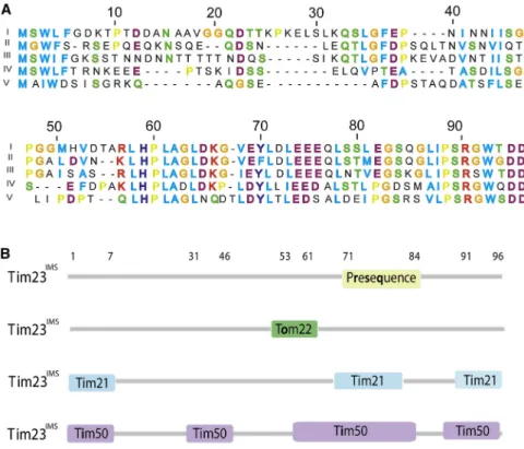
(A) Sequence alignment for Tim23IMS
highlighting the conservation of residues among different Fungi kingdoms with 1-5 as S. cerevisaiae, ZygoSaccharomyces rouxii, Candida albicans, Schizosaccharomyces pombe, and Aspergillus fumigatus, respectively. Hydrophobic and aro-matic residues are highlighted in blue; positive and negative charged residues are shown in red and magenta, respectively; neutral residues in green; and glycine and proline in orange and yellow, respectively.
(B) Summary of interaction motifs in Tim23IMS used for binding to Tim21IMS
, Tom22IMS , prese-quence (de la Cruz et al., 2010), and Tim50IMS
. Each interaction motif is used for binding to mul-tiple protein partners.
Mokranjac, D., Paschen, S.A., Kozany, C., Prokisch, H., Hoppins, S.C., Nargang, F.E., Neupert, W., and Hell, K. (2003). Tim50, a novel component of the TIM23 preprotein translocase of mitochondria. EMBO J.22, 816–825. Mokranjac, D., Popov-Celeketic, D., Hell, K., and Neupert, W. (2005). Role of Tim21 in mitochondrial translocation contact sites. J. Biol. Chem. 280, 23437–23440.
Neupert, W., and Herrmann, J.M. (2007). Translocation of proteins into mito-chondria. Annu. Rev. Biochem.76, 723–749.
Pfanner, N. (1998). Mitochondrial import: crossing the aqueous intermem-brane space. Curr. Biol.8, R262–R265.
Pfanner, N., and Geissler, A. (2001). Versatility of the mitochondrial protein import machinery. Nat. Rev. Mol. Cell Biol.2, 339–349.
Popov-Celeketic, D., Mapa, K., Neupert, W., and Mokranjac, D. (2008). Active remodelling of the TIM23 complex during translocation of preproteins into mitochondria. EMBO J.27, 1469–1480.
Qian, X., Gebert, M., Ho¨pker, J., Yan, M., Li, J., Wiedemann, N., van der Laan, M., Pfanner, N., and Sha, B. (2011). Structural basis for the function of Tim50 in the mitochondrial presequence translocase. J. Mol. Biol.411, 513–519. Raveh, B., London, N., and Schueler-Furman, O. (2010). Sub-angstrom modeling of complexes between flexible peptides and globular proteins. Proteins78, 2029–2040.
Raveh, B., London, N., Zimmerman, L., and Schueler-Furman, O. (2011). Rosetta FlexPepDock ab-initio: simultaneous folding, docking and refinement of peptides onto their receptors. PLoS ONE6, e18934.
Ryan, K.R., and Jensen, R.E. (1995). Protein translocation across mitochon-drial membranes: what a long, strange trip it is. Cell83, 517–519.
Saitoh, T., Igura, M., Miyazaki, Y., Ose, T., Maita, N., and Kohda, D. (2011). Crystallographic snapshots of Tom20-mitochondrial presequence interac-tions with disulfide-stabilized peptides. Biochemistry50, 5487–5496. Sattler, M., Schleucher, J., and Griesinger, C. (1999). Heteronuclear multidi-mensional NMR experiments for the structure determination of proteins in solution employing pulsed field gradients. Prog. Nucl. Magn. Reson. Spectrosc.34, 93–158.
Schatz, G. (1996). The protein import system of mitochondria. J. Biol. Chem. 271, 31763–31766.
Schulz, C., Lytovchenko, O., Melin, J., Chacinska, A., Guiard, B., Neumann, P., Ficner, R., Jahn, O., Schmidt, B., and Rehling, P. (2011). Tim50’s presequence receptor domain is essential for signal driven transport across the TIM23 complex. J. Cell Biol.195, 643–656.
Schwieters, C.D., Kuszewski, J.J., Tjandra, N., and Clore, G.M. (2003). The Xplor-NIH NMR molecular structure determination package. J. Magn. Reson.160, 65–73.
Shiota, T., Mabuchi, H., Tanaka-Yamano, S., Yamano, K., and Endo, T. (2011). In vivo protein-interaction mapping of a mitochondrial translocator protein Tom22 at work. Proc. Natl. Acad. Sci. USA108, 15179–15183.
Tamura, Y., Harada, Y., Shiota, T., Yamano, K., Watanabe, K., Yokota, M., Yamamoto, H., Sesaki, H., and Endo, T. (2009). Tim23-Tim50 pair coordinates functions of translocators and motor proteins in mitochondrial protein import. J. Cell Biol.184, 129–141.
Truscott, K.N., Kovermann, P., Geissler, A., Merlin, A., Meijer, M., Driessen, A.J., Rassow, J., Pfanner, N., and Wagner, R. (2001). A presequence- and voltage-sensitive channel of the mitochondrial preprotein translocase formed by Tim23. Nat. Struct. Biol.8, 1074–1082.
van der Laan, M., Meinecke, M., Dudek, J., Hutu, D.P., Lind, M., Perschil, I., Guiard, B., Wagner, R., Pfanner, N., and Rehling, P. (2007). Motor-free mito-chondrial presequence translocase drives membrane integration of prepro-teins. Nat. Cell Biol.9, 1152–1159.
van der Laan, M., Hutu, D.P., and Rehling, P. (2010). On the mechanism of preprotein import by the mitochondrial presequence translocase. Biochimica et Biophysica Acta (BBA) -. Mol. Cell Res.1803, 732–739. Wiedemann, N., Frazier, A.E., and Pfanner, N. (2004). The protein import machinery of mitochondria. J. Biol. Chem.279, 14473–14476.
Yamamoto, H., Esaki, M., Kanamori, T., Tamura, Y., Nishikawa, Si., and Endo, T. (2002). Tim50 is a subunit of the TIM23 complex that links protein transloca-tion across the outer and inner mitochondrial membranes. Cell111, 519–528. Zhang, Y. (2008). I-TASSER server for protein 3D structure prediction. BMC Bioinformatics9, 40.