Received 29 Jul 2015
|
Accepted 8 Oct 2015
|
Published 7 Dec 2015
Identification of an allosteric binding site
for ROR
g
t inhibition
Marcel Scheepstra
1,
*, Seppe Leysen
1,
*, Geert C. van Almen
1
, J. Richard Miller
2
, Jennifer Piesvaux
2
,
Victoria Kutilek
2
, Hans van Eenennaam
3,
w
, Hongjun Zhang
2
, Kenneth Barr
2,
w
, Sunil Nagpal
2,
w
,
Stephen M. Soisson
4
, Maria Kornienko
5
, Kristen Wiley
5
, Nathaniel Elsen
5,
w
, Sujata Sharma
5
, Craig C. Correll
2
,
B. Wesley Trotter
2
, Mario van der Stelt
3,
w
, Arthur Oubrie
3,
w
, Christian Ottmann
1
, Gopal Parthasarathy
4
& Luc Brunsveld
1
ROR
g
t is critical for the differentiation and proliferation of Th17 cells associated with several
chronic autoimmune diseases. We report the discovery of a novel allosteric binding site on
the nuclear receptor ROR
g
t. Co-crystallization of the ligand binding domain (LBD) of ROR
g
t
with a series of small-molecule antagonists demonstrates occupancy of a previously
unreported allosteric binding pocket. Binding at this non-canonical site induces an
unprecedented conformational reorientation of helix 12 in the ROR
g
t LBD, which blocks
cofactor binding. The functional consequence of this allosteric ligand-mediated conformation
is inhibition of function as evidenced by both biochemical and cellular studies. ROR
g
t function
is thus antagonized in a manner molecularly distinct from that of previously described
orthosteric ROR
g
t ligands. This brings forward an approach to target ROR
g
t for the treatment
of Th17-mediated autoimmune diseases. The elucidation of an unprecedented modality of
pharmacological antagonism establishes a mechanism for modulation of nuclear receptors.
DOI: 10.1038/ncomms9833
OPEN
1Laboratory of Chemical Biology, Department of Biomedical Engineering and Institute of Complex Molecular Systems, Eindhoven University of Technology,
PO Box 513, Eindhoven 5600MB, The Netherlands.2Merck Research Laboratories, 33 Avenue Louis Pasteur, Boston, Massachusetts 02115, USA.
3Merck Research Laboratories, Molenstraat 110, Oss 5342 CC, The Netherlands.4Merck Research Laboratories, 770 Sumneytown Pike, West Point,
N
uclear
receptors
(NRs)
modulate
transcription
of
particular sets of genes on binding of small lipophilic
ligands and thereby regulate physiological parameters of
cellular function
1. NRs are also important pathological regulators
in diseases such as cancer, diabetes and autoimmune disorders.
This combination of characteristics of NRs has given rise to some
of the most notable pharmaceutical agents of the past century
2.
The retinoic-acid-receptor-related orphan receptor (ROR) is a
NR subclass that demonstrates great therapeutic potential
3.
In particular, ROR
g
t, whose activity is required for the
proliferation and functionality of immune Th17 cells, is the
subject of intense investigation to modulate its activity to achieve
clinical benefit
4–6. Th17 cells exert an inflammatory, pathological
role in autoimmune diseases
7,8and on stimulation produce
pro-inflammatory cytokines
9. Antibodies directed against the cytokine
IL17 have been clinically successful, proving the potential of
targeting the Th17/IL17 axis
10. Active ROR
g
t is a prerequisite for
the differentiation of T cells into Th17 cells
11,12. Small-molecule
inhibition of ROR
g
t has therefore been brought forward as a
novel strategy for the treatment of autoimmune diseases
13,14.
NRs are characterized by the ability to bind small ligands at a
highly conserved hydrophobic orthosteric-binding pocket located
within the protein’s ligand-binding domain (LBD)
1. A typical NR
LBD exhibits a three-layered fold of
B
12 alpha helices and 2–3
b
-strands. Ligand binding in this pocket can activate or inhibit
the receptor to various degrees
15. Helix 12 (H12, also called
activation function-2, AF-2) can adopt distinct conformations in
response to ligand binding, regulating the interaction of the
LBD with cofactor proteins with resulting changes in gene
transcription at a particular locus. Typically, on the binding of an
agonist, H12 is stabilized in a conformation that facilitates the
binding of a coactivator
16,17. Conversely, antagonist binding
induces a different H12 conformation unsuitable for coactivator
binding. NR drugs thus bind to this orthosteric-binding pocket
and act as molecular ‘switches’ that control NR transcriptional
activity due to the positioning of H12 (ref. 18). This canonical
ligand binding is associated with selectivity issues and
mutation-induced antagonist/agonist switches for different NRs and
therefore molecules that occupy allosteric-binding sites on NRs
are highly sought after
19–21. Such allosteric modulation might be
expected to induce conformational effects that are not dependent
on competition with endogenous ligands and could provide
enhanced potency/efficacy or greater specificity over canonical
ligands.
We previously identified a novel series of ROR
g
t inhibitors
22.
Here we characterize the mode of action of these inhibitors to
guide an optimization program and surprisingly find a novel
binding mode, thereby identifying the first allosteric-binding
pocket for a highly potent, cellular active small NR ligand.
Structural, biochemical and cellular data reveal that the
unprecedented allosteric-binding modality confers both high
potency and selectivity to ROR
g
t for these novel antagonists.
Results
Helix 12 repositions to generate a novel binding pocket
.
Literature suggests that the RORs feature ligand-independent
transcription, with their LBDs partially in a conformation
promoting coactivator binding
23. Biological data and the
co-crystal structures of ROR
g
t LBD bound to hydroxycholesterols
24,
and synthetic inverse agonistic ligands such as T0901317 (Fig. 1a)
have shown that the ROR
g
t LBD is still structurally responsive to
ligands. For ROR
g
t, reports suggest that multiple small molecules
affect antagonism via binding to the canonical orthosteric site
13.
A high-throughput screen for molecules that disrupt the
interaction
of
the
ROR
g
t-LBD
with
steroid
receptor
coactivator-1 (SRC-1) cofactor peptide, followed by a hit
optimization program, led to the identification of indazoles as a
novel class of ROR
g
t inhibitors, typified by
MRL-871
(Fig. 1a)
22.
To elucidate the molecular basis of ROR
g
t modulation by
MRL-871
, we performed co-crystallization studies with an
equimolar complex of ROR
g
t-LBD and
MRL-871
. Co-crystals
grew in two different space groups (Table 1). Crystals in space
group R32:H had unit cell dimensions of
a
¼
b
¼
173.8,
c
¼
67.2 Å, diffracted to 2.3 Å resolution and contain one
molecule per asymmetric unit. Crystals in space group P6122
had unit cell dimensions of
a
¼
b
¼
108.5,
c
¼
104.7 Å, diffracted
to 2.2 Å resolution and also contain one molecule per asymmetric
unit. The experimental electron density maps from both
conditions showed clear density for all features of the protein,
including
MRL-871
. The overall structures from both sets were
identical in all the features and the ligand conformation. The
ROR
g
t LBD crystallized with the typical NR arrangement of
helices 1–11, but with the H12 positioned in a conformation
unprecedented for all NR LBD complexes reported to date
(Fig. 1c, green). On top of that, the crystal structure revealed the
binding site of
MRL-871
to be different from the canonical
orthosteric NR ligand-binding site.
MRL-871
binds instead to a
previously unidentified allosteric pocket in the ROR
g
t LBD,
located distal to the classical binding site (Fig. 1c, orange). Crystal
structure analysis shows that this allosteric pocket, absent in the
classical NR-folding motif, is formed by helices 4, 5, 11 and the
reoriented flexible H12 (Fig. 1b). The ortho-substituted
trifluoromethyl and chloro moieties are spatially clearly
positioned and impart a specific rotation to the phenyl group of
MRL-871
and address hydrophobic sites in the allosteric pocket
(Fig. 1b,d). Hydrogen-bonding interactions exist between the
molecule’s carboxylic acid group and the side chain of ROR
g
t
residue Q329 as well as the main-chain amide hydrogen atoms of
residues A497 and F498. Apart from these distinct polar
interactions,
the
newly
generated
allosteric
is
predominantly hydrophobic, because of the amino-acid side
chains of residues on helices 4, 5, 11 and, notably, H12 and the
activation function loop between helices 11 and 12 (AF-2
domain). The arrangement of the ROR
g
t LBD in the presence
of
MRL-871
generates a druggable molecular-binding site
critically regulated by favourable interactions of
MRL-871
with
the AF-2 domain. These interactions reposition the highly flexible
H12 in a conformation unique among reported NR ligand
structures, of either the agonist or antagonist type, and distinct
from the previously reported ROR
g
t agonist-bound state
1,6,13,14.
The unique conformation induced by the binding of
MRL-871
prevents interaction with cofactor peptides, which typically bind
ROR
g
t through a conserved LXXLL motif, at the AF-2.
Functional ROR
c
t inhibition via allosteric inverse agonism
.
To determine the functional effect of the binding of these novel
modulators and the resulting ROR
g
t conformational changes,
two variants of
MRL-871
(
MRL-058
and
MRL-003
) were also
prepared and tested alongside an agonist and canonical inverse
agonist in an AlphaScreen cofactor peptide recruitment assay.
Indazoles
MRL-871
and its derivatives
MRL-058
and
MRL-003
all inhibited coactivator binding in a dose-dependent manner
(Fig. 2a) with half-maximum inhibitory concentration (IC50)
values of 7
±
1 nM (
MRL-871
), 98
±
23 nM (
MRL-058
) and
280
±
117 nM (
MRL-003
), showing the antagonistic profile of the
series. Cholesterol, binding as agonist to the LBD, functioned
as a weak activator, further enhancing the interaction of the
ROR
g
t–LBD with a cofactor peptide
25. The inverse agonist
T0901319 demonstrated an IC50
value of 24
±
13 nM in this assay,
less potent than
MRL-871
(refs 26,27). The data thus show that
the allosteric-binding mode of the novel modulators as observed
in the crystal structures translates into potent inhibition of
coactivator peptide binding to ROR
g
t–LBD. Furthermore, these
data also indicate that both ortho-substituents on the benzamide
moiety are required to enhance ROR
g
t-binding affinity, which
suggest that the observed binding mode in the crystal structure is
most optimal.
To corroborate our structural findings and gain more insight in
the mode of action for these compounds, competitive binding
assays for both
MRL-871
(Fig. 2c) and T0901317 (Fig. 2d) were
performed against fixed concentrations of cholesterol. On the
basis of the structural data, we hypothesized that
MRL-871
should not compete with cholesterol for the orthosteric
ligand-binding pocket, because they bind into different pockets. As such,
the IC50
value of
MRL-871
for inhibition of the ROR
g
t
coactivator interaction should be independent of cholesterol
concentration. Increasing concentrations of cholesterol increase
the ROR
g
t coactivator interaction, in line with the agonistic
properties of cholesterol, as evidenced by an increased
fluores-cence ratiometric level with cofactor peptide (Fig. 2c,d).
MRL-871
, however, effectively antagonizes the ROR
g
t coactivator
peptide recruitment interaction, independent of cholesterol
concentration. The competitive cofactor-binding assay for
MRL-871
showed no significant change in IC50
value when
performed in the presence of different concentrations of
cholesterol (Fig. 2c). In contrast, the inverse agonist T0901317
competes for the same binding site as cholesterol, resulting in a
competitive displacement and a cholesterol
concentration-dependent increase of the IC50
value for T0901317 (Fig. 2d);
that is, ROR
g
t binding and inhibition by T0901317 becomes less
potent in the presence of agonistic ligands. Together, these data
demonstrate that the allosteric inhibition of coactivator binding
to ROR
g
t by
MRL-871
is both potent and independent of
orthosteric site occupancy.
ROR
g
t mediates IL17a gene expression in EL4 cells
4. The EL4
murine lymphoblast cell line constitutively expresses ROR
g
t,
which drives production of IL17a (ref. 28). To confirm that
allosteric ROR
g
t modulation has also functional relevance at the
cellular level, EL4 cells were treated with 10
m
M of modulators
MRL-871
,
MRL-058
and
MRL-003
for 24 h and IL17a messenger
RNA (mRNA) levels were measured by quantitative reverse
transcriptase PCR (RT–PCR) (Fig. 2b). Treatment of EL4 cells
with the more potent
MRL-871
and
MRL-058
significantly
reduced the IL17a mRNA levels, while the weaker
MRL-003
did
not reduce the mRNA levels, consistent with the lower
Helix 5 T325 Helix 4 Helix 11 Helix 12 A321 W317 L483 Q329 I328 A497
F498 L353A496
Y502 L505 F506 T0901317 Allosteric site (MRL-871) Orthosteric site (T0901317)
MRL-871. R1=Cl, R2=CF 3
MRL-058. R1=H, R2=CF3
MRL-003. R1=H, R2=H
Orthosteric inverse agonist
OH OH CF3 CF3 CF3 N S O O O N N R1 R2 O
Allosteric inverse agonist
a
b
c
d
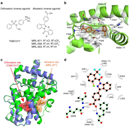
A321 W317 T325 A497 2.96 Å 2.61 Å 2.83 Å A496 (Helix 12) F498 L505 L353 I328 K354 F506 (Helix 11) L483 F2 C15 F1 F O2 C9 C8 N1 C16 C21 C7 C20 C19 C18 C17 N C1 C10 C11 C14 C12 C13 C5 C6 C3 C2 C1C4 CE1 O N CG CD1 CD2 CE2 CZ CB CA C O C O1 NE2 N CA CB O C OE1 CD CG CB CA N C O Q329 Y502 (Helix 12)Figure 1 | Co-crystal structure of RORct with MRL-871.(a) Chemical structures of a representative orthosteric ligand T0901317 (ref. 27), binding to
the canonical site of NRs, and indazolesMRL-871,MRL-058andMRL-003. (b) Zoomed-in view ofMRL-871in the novel allosteric-binding pocket of
RORgt formed by helices 4, 5, 11 and 12.MRL-871shown as orange sticks in the electron density. The RORgt residues involved in hydrophobic interactions
and hydrogen bonding are shown as white and green sticks, respectively. (c) Superposition of RORgt (blue) in complex with orthosteric ligand T0901317
shown in red (PDB ID 4NB6)26and RORgt (green) in complex withMRL-871, shown in orange.MRL-871(orange) sits in a new allosteric pocket in
direct contact with H12 inducing its repositioning. Orthosteric ligand T0901317 destabilizes H12, which is therefore not visible in the blue structure.
biochemical activity observed for this compound. This result thus
demonstrates that functional modulation of the allosteric pocket
by small molecules results in cellular responses (that is, reduced
gene transcription).
Structural basis for ligand potency
. The cofactor recruitment
assay described above provides an indirect assessment of
modulator potency since the site of cofactor interaction does not
directly overlap with the binding site of the indazoles. Therefore,
an orthogonal assay that directly and selectively probes the
novel allosteric ROR
g
t-binding site is desirable for screening,
characterization and optimization purposes. We used the
co-crystal structure of ROR
g
t and
MRL-871
(Fig. 1c) to rationally
design a synthetic ligand analogue containing a time-resolved
fluorescence resonance energy transfer (TR-FRET) acceptor.
AlexaFluor 647 was connected to the six position of
MRL-871
via
a short molecular spacer (Supplementary Fig. 1); this site of the
molecule protrudes into an open channel in the crystal structure.
This probe molecule proved to have a robust TR-FRET signal at
low concentrations in the presence of His-tagged ROR
g
t–LBD
and an anti-His Europium chelate antibody. Titration studies
in a TR-FRET assay revealed a
K
dof 100
±
24 nM with the
ROR
g
t–LBD (Supplementary Fig. 1). When the parent
MRL-871
was tested for the ability to compete with the allosteric probe, the
resulting IC50
value was similar to that observed in cofactor
recruitment assays (Table 2).
We explored the synthetic optimization of
MRL-871
to establish
structure–activity relationships around the indazole series. These
compounds were tested in biochemical assays, including the
competition assay using the fluorescent-labelled allosteric probe,
cellular chimeric receptor reporter assays in HEK-293 cells and
Th17 differentiation/IL17a production assays in primary human
peripheral blood mononuclear cells (PBMCs), as well as structural
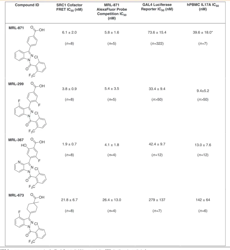
studies (Fig. 3). Table 2 describes several informative examples.
MRL-299
features two additional fluoro groups, one at the
indazole ring and one at the phenyl substituent.
MRL-367
contains
additional polar functionality—the phenyl substituent contains a
hydroxyl substituent ortho to the carboxylic acid, and the C-4 of
the indazole is replaced by nitrogen. In the case of
MRL-673
, the
benzoic acid functionality is saturated, and the carbon atom
adjacent to the carboxylate is alkylated, resulting in a
tetrasub-stituted carbon centre. This modification induces a displacement of
the carboxyl group and highlights the importance of the polar
interactions in the binding site.
The additional fluorine groups (
MRL-299
) and the polar
functionalities (
MRL-367
) were both important for ROR
g
t-binding potency and functional inhibition (Table 2). In particular,
MRL-367
exhibited enhanced potency in the cofactor
displace-ment and direct binding assay, as well as increased inhibitory
activity in a luciferase reporter assay and in functional inhibition
of IL-17 production. To insure that the functional activity of the
indazole series is attributable to inhibition of ROR
g
t,
MRL-299
was tested against a commercially available panel of cell-based
NR reporter assays (Supplementary Table 1). Across this panel of
NR assays,
MRL-299
was
4
100-fold selective for ROR
g
t. The
only significant off-target activity was against PPAR
g
(PPAR
g
activity was also recently reported for a structurally similar series
of molecules
29), which was de-risked (Supplementary Note 1).
Structural elucidation of these compounds reveals that all
antagonists occupy the same allosteric-binding pocket as
MRL-871
(Fig. 3). Detailed comparison of, for example,
MRL-871
and
MRL-367
(Fig. 3b) reveals that both polar
molecular additions to the scaffold are fully tolerated and lead
only to very minor changes in the surrounding amino-acid
orientations.
MRL-673
, in contrast, exhibits 10-fold weaker
Table 1 | Data collection and refinement statistics (molecular replacement).
PDB ID 4YPQ 5C4O 5C4U 5C4S 5C4T
Ligand name MRL-871 MRL-871 MRL-367 MRL-299 MRL-673
Data collection
Space group R32:H P 61 2 2 P 61 2 2 P 61 2 2 P 61 2 2
Cell dimensions
a,b,c(Å) 173.8, 173.8, 67.2 108.5, 108.5, 104.7 108.1, 108.1, 106.5 108.4, 108.4, 106.3 107.3, 107.3, 100.4
a,b,g(°) 90, 90, 120 90, 90, 120 90, 90, 120 90, 90, 120 90, 90, 120
Resolution (Å) 35.47–2.32 (2.40–2.32)* 69.91–2.24 (2.32–2.24) 93.6–2.08 (2.154–2.08) 93.92–2.23 (2.31–2.23) 92.9–1.77 (1.836–1.77)
Rsym 0.121 (0.886) 0.047 (1.45) 0.0420 (1.236) 0.056 (1.402) 0.0565 (1.239)
I/sI 12.1 (2.7) 35.91 (2.57) 38.87 (2.62) 30.03 (2.51) 29.46 (2.61)
Completeness (%) 100.0 (100.0) 99.94 (99.89) 99.78 (99.36) 99.99 (100.0) 99.99 (100.00)
Redundancy 11.7 (11.6) 16.0 (15.8) 19.3 (19.9) 18.8 (19.2) 19.1 (19.7)
Refinement
Resolution (Å) 35.47–2.32 69.91–2.24 93.6–2.08 93.9–2.23 92.9–1.77
No. reflections 16882 17932 22579 18547 33582
Rwork/Rfree 0.174/0.227 0.227/0.266 0.228/0.273 0.218/0.261 0.192/0.223
No. atoms
Protein 1987 1996 1994 1987 1985
Ligand/ion 32 60 51 45 118
Water 130 14 23 18 101
B-factors
Protein 45.70 67.90 59.80 67.9 39.6
Ligand/ion 41.50 102.5 57.70 64.1 50.50
Water 47.30 64.20 57.80 63.9 45.30
R.m.s. deviations
Bond lengths (Å) 0.008 0.014 0.014 0.014 0.015
Bond angles (°) 1.04 1.78 1.81 1.75 1.64
R.m.s., root mean squared.
potency in both the biochemical and cellular assays relative to
MRL-367
. Structural comparison of, for example,
MRL-367
and
MRL-673
(Fig. 3c) reveals that, while these compounds bind the
same allosteric site, the position of the carboxylate ring of
MRL-673
distorts the binding pocket. The modification induces a
displacement of the carboxyl group and highlights the importance
of the polar interactions in the binding site. Although the
carboxylate of
MRL-673
makes the same number of polar
contacts with ROR
g
t as
MRL-367
, the methylation induces the
reorientation of the side chains of Phe498 and Tyr502. This shift
in the side chains and the accompanying shifts in the backbone of
the C-terminal residues of Ala497 and Phe498, explains the lower
receptor affinity and functional inhibitory potency.
Discussion
NR drug development has been based primarily on the ability of
NRs to bind ligands at the highly conserved hydrophobic
orthosteric pocket of the LBD, which is also the binding site of
endogenous ligands
1,18. Compared with unbound NRs, the
binding of an agonist induces a conformational change that
stabilizes the positioning of H12 and AF-2 domain, resulting in
coactivator recruitment. The binding of antagonist or inverse
agonist ligands typically destabilizes H12 and the AF-2 domain
(possibly unfolding it) resulting in a lack of coactivator or
enhanced corepressor recruitment, ultimately resulting in
transcriptional inhibition at specific loci. Numerous successful
drugs targeting NRs have been developed, all working via this
mechanism, but increasing resistance against certain cancer
therapy oriented NR antagonists, as well as the challenge of
targeting orphan receptors has increased the need for alternative
site modulators of NRs
19–21. Similar to, for example, the efforts
to target allosteric sites in protein kinases
30, allosteric NR
modulators may differentiate favourably versus orthosteric
ligands. Potential advantages of allosteric NR inhibitors are
enhanced selectivity, no competition with increasing endogenous
ligands during pathological conditions, and no sensitivity to
agonist/antagonist switching due to mutations.
The recently reported crystal structure of ROR
g
t with a potent
tertiary amine orthosteric agonist revealed the
coactivator-binding site on the LBD, in a classical NR H12 switch agonist
mode and bound to a cofactor peptide motif (Fig. 4a)
31. The new
crystal structures of ROR
g
t reported here with
MRL-871
and its
analogues (Fig. 3) reveal that these indazole modulators bind to a
receptor position normally occupied by H12 in the non-liganded
or agonist ligand-bound conformation. The final orientation of
H12 in the presence of the novel indazole modulators is such that
the classical binding surface for the cofactor LXXLL motif is not
only modified, but actively blocked (Fig. 4e). This overall
orientation effectively antagonizes cofactor binding to ROR
g
t.
The consequence of this allosteric modulator-mediated refolding
of ROR
g
t is functional inhibition as evidenced by biochemical
and cellular studies. The unique mode of allosteric binding
reported thus provides a structural rationale for targeting NRs
with small molecules in an orthogonal manner that does not
require competition with canonical ligands.
Cholesterol (μM) IC50 (nM) 0
1 10 25
4.7 ± 1.4 2.5 ± 0.9 1.8 ± 1.5 2.3 ± 1.8
Cholesterol (μM) IC50 (nM) 0
1 10 25
11 ± 3.4 18 ± 4.9 195 ± 59 548 ± 152
T0901317 Cholesterol MRL-871 MRL-058 MRL-003
DMSO
MRL-058 MRL-871 MRL-003
2,000 1,800 1,600 1,400 1,200 1,000 800
1,200 1,100
1.5
1.0
0.5
0.0
**
Relativ
e mRNA e
xpression
**
1,000 900 800 700 600 500 1,000
800 600 400
Relativ
e units (665 nm/620 nm)
Relativ
e units (665 nm/620 nm)
Relativ
e units (665 nm/620 nm)
–12 –10 –8 –6 –4 Concentration ligand (log(M))
Concentration MRL-871 (log(M)) –12 –10 –8 –6 –4
Concentration T0901317 (log(M)) –12 –10 –8 –6 –4
a
b
c
d
Figure 2 | Allosteric inhibition.(a) TR-FRET assay showing the effect of the agonist cholesterol, the inverse agonist T0901317 and indazoles
MRL-871,MRL-058andMRL-003on cofactor recruitment to the RORgt LBD in a dose-dependent manner.MRL-871,MRL-058,MRL-003and T0901317 function as inhibitors, while cholesterol promotes cofactor binding to RORgt. Error bars are defined as s.d. (n¼3). (b) IL17a mRNA expression in EL4 cells
treated withMRL-871,MRL-058,MRL-003(10mM, 24 h) or DMSO. The level of IL17a mRNA expression was normalized to that of GAPDH expression. All
data are expressed as the mean±s.e.m. (n¼6). Statistical analysis was performed using an one-way analysis of variance comparing against the DMSO
The X-ray structures demonstrate that these indazoles all
occupy the novel allosteric binding pocket. Reported crystal
structures with orthosteric inverse agonists, binding to the
canonical site of ROR
g
t, lack clear structural data for the
position of the H12 and the resulting cofactor-binding site
(for example, Fig. 1c with T0901317 and Fig. 4b). Synthetic
ROR
g
t inverse agonists T0901317 (ref. 27) and tertiary
sulfonamides
26bind at the canonical orthosteric NR
ligand-binding pocket. Their ligand-binding distorts the structure of the
LBD, leading to helices 11
0and 12 being unstructured in the
crystallized protein (Fig. 4b). A superposition of the ROR
g
t
structures with an inverse agonist in the orthosteric site and an
indazole modulator in the allosteric site reveals the additional
benefit of the novel site (Fig. 1c; Supplementary Fig. 4).
Whereas both compounds induce the partial unfolding of
helix 11
0(ref. 32),
MRL-871
subsequently also stabilizes the
folding of H12 via direct interactions. The unfolded helix 11
0spans the distance to the displaced H12 N terminus (Fig. 4d).
Table 2 | Potency (mean
±
s.d.) of selected ROR
g
t allosteric inverse agonists in biochemical and cellular assays.
Compound ID
MRL-871
MRL-299
MRL-367
MRL-673
SRC1 Cofactor FRET IC50 (nM)
6.1 ± 2.0 O
3.8 ± 0.9
1.9 ± 0.7
21.8 ± 6.7 26.4 ± 13.0 279 ± 137 142 ± 64 5.8 ± 1.6
5.4 ± 3.5
4.1 ± 1.8
73.6 ± 15.4
33.4 ± 9.4
42.4 ± 9.7
39.6 ± 18.0*
9.4±5.2
13.0 ± 7.6
MRL-871 AlexaFluor Probe Competition IC50
(nM)
GAL4 Luciferase Reporter IC50 (nM)
hPBMC IL17A IC50
(nM)
(n=8) (n=5) (n=322) (n=7)
(n=8) (n=4) (n=12) (n=12)
(n=8) (n=4) (n=7) (n=6) (n=8) (n=5) (n>50) (n>50) OH
N N CI
O
F3C
O OH
N F F
N CI
O
F3C
O OH
N F
N CI
O
F3C
O OH HO
N
N F
N CI
O
F3C
FRET, fluorescence resonance energy transfer; IC50, half-maximal inhibitory concentration; SRC1, steroid receptor coactivator 1.
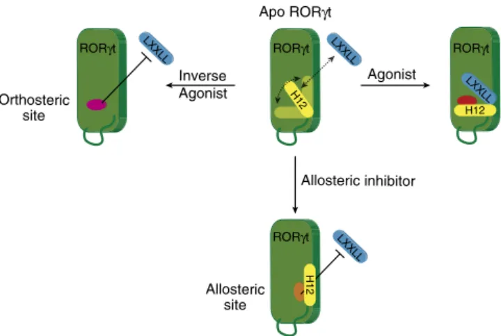
This results in ROR
g
t taking on a stably folded antagonistic
state and directly blocking cofactor peptide binding (Fig. 4e).
These antagonists thus induce a conformational change in
the ROR
g
t LBD, which blocks cofactor binding through the
stabilization of the H12 subdomain in an unprecedented folded
state (Fig. 5).
In summary, we demonstrate the structural and functional
elucidation of an unprecedented NR allosteric inhibitory
mechanism, based on highly potent small-molecule modulators
for ROR
g
t. This is the first allosteric binding pocket with
highly potent small drug-like molecules, functionally active in
Th17 differentiation/IL17a production inhibition. This allosteric
inhibitory mechanism offers great advantages for NR drug
development, including selectivity, independence of endogenous
ligands and less responsive to point mutations within the
orthosteric ligand binding site. ROR
g
t regulates a variety of
physiological processes and has emerged as a highly promising
drug target for autoimmune diseases. The discovery of this novel
allosteric NR pocket and concomitant selective molecules that
target this site offers a new strategic opportunity to develop
agonist-independent therapeutics for Th17-mediated
autoim-mune disorders.
A497 F498
Y502
c
b
a
Figure 3 | Co-crystal structure of RORct with MRL-871, MRL-299, MRL-367 and MRL-673.(a) Overlay of RORgt (green) co-crystallized withMRL-871,
MRL-299,MRL-367andMRL-673. For an overall structural comparison of RORgt in complex with MRL-299 and MRL-367 see Supplementary Fig. 3.
(b) Magnified view of the overlay ofMRL-871(orange) andMRL-367(cyan) in the novel allosteric binding pocket of RORgt (overall root mean squared
deviation (r.m.s.d) 0.219). (c) Magnified view of overlay ofMRL-367(cyan) andMRL-673(yellow) in the novel allosteric binding pocket of RORgt (overall
r.m.s.d 0.38).MRL-673is a racemic mixture and the two isomers are depicted. Arrows highlight repositioning of the side chains of residues Tyr502 and
Phe498 and the accompanying shift in the backbone (for example, Ala497) on bindingMRL-673.
H12
a
b
c
d
e
H11′
H11′
H12
H12 H11
H11
Agonist
Orthosteric inverse agonist Allosteric inhibitor
Figure 4 | Molecular mechanism of allosteric RORct modulation.(a) Agonistic conformation of RORgt31(PDB entry 4NIE) showing the position of the agonistic ligand (red) in the classical orthosteric ligand binding site. Helices 110and 12 (yellow) of the LBD are folded in a stable agonistic conformation,
supporting binding of the LXXLL cofactor peptide (blue). (b) Antagonistic conformation of RORgt14(PDB entry 4QM0) induced by a classical inverse
agonist (purple) in the classical orthosteric ligand-binding site. Helices 11 and 12 of the LBD are unstructured (arrow). (c) Superposition ofaandb. When
H12 is disordered, the LXXLL cofactor peptides lack a complementary surface for binding to RORgt (d) Antagonistic conformation of RORgt, induced by
allosteric modulatorMRL-871(orange).MRL-871makes direct contact with H12 (brown) at the allosteric site in the RORgt LBD and stabilizes the folding of H12. (e) Superposition ofaandd. The binding of the allosteric modulatorMRL-871results in unfolding of helix 110and shifts the position of H12 to directly
Methods
Synthesis of indazole series
.
For the synthesis of the 3-iodo-1H-indazol-1-yl (phenyl)methanone, thionyl chloride was added dropwise to the appropriate benzoic acid in an oven dried flask. The reaction mixture was stirred at 75°C overnight. After removal of the thionyl chloride under reduced pressure, the benzoyl chloride was dissolved in dry CH2Cl2. To this solution, 3-iodo-1H-indazoleand DMAP were added. Finally, triethylamine was added dropwise and the mixture was stirred at room temperature for 24 h. The reaction mixture was then diluted with H2O and CH2Cl2. The layers were separated and the aqueous layer was
washed with CH2Cl2. The combined organic layers were washed with brine, dried
over Na2SO4, filtered and evaporated. The crude material was purified via column
chromatography.
For the cross-coupling reaction the appropriate 3-iodo-1H-indazole, (4-(methoxycarbonyl)-phenyl)boronic acid, Pd(PPh3)4and CH3CO2K were
dissolved in dioxane/H2O (5:1 v/v) in a Schlenk tube and the reaction was stirred at
90°C. After 3 h, the mixture was allowed to cool to room temperature and diluted with CH2Cl2and H2O. The organic layer was washed with brine, dried over
Na2SO4, filtered and concentrated. The obtained crude material was purified via
column chromatography.
To obtain the free acid, LiOHH2O was added to a solution of the methyl ester
in THF/H2O. The reaction was stirred at room temperature. After 24 h, the
reaction was diluted with H2O, neutralized with acetic acid (BpH 4) and extracted
with CH2Cl2. The combined organic layers were washed with brine, dried over
Na2SO4, filtered and evaporated. The final products (for example,MRL-871) were
purified via preparative liquid chromatography-mass spectrometry (LCMS). For complete synthesis and characterization data see Supplementary Methods.
HTRF assay
.
The human RORgt LBD used for the HTRF assay was expressed as a His6-tag fusion protein from the pET15b expression vector inEscherichia coliBL21(DE3) cells. Cells transformed with this vector were grown in 2YT medium supplemented with ampicillin until an OD600¼0.7 was reached. Protein
expression was then induced with 0.1 mM isopropyl-b-d-thiogalactoside (IPTG). After incubation for 16 h at 16°C, cell cultures were collected by centrifugation. The cells were lysed via sonication and the protein was purified via Ni2þ-affinity column chromatography.
The homogeneous TR-FRET assays were performed in triplicate with 20 nM His6-RORgt and 100 nM biotin labelled cofactor peptide. Terbium-labelled
anti-His antibody (cat. no: 610HATAA, 0.71 nM) and D2-labelled streptavidin (cat. no: 610SADLA, 41.7 nM) were used at recommended concentrations by the supplier Cisbio Bioassays. Assay buffer contained 100 mM HEPES (pH 7.5), 100 mM NaCl, 5 mM dithiothreitol (DTT) and 0.1% bovine serum albumin. The plates (white 384-well plates Greiner Bio-One) were incubated for 2 h at 4°C before the FRET ratio was measured in a Tecan Infinite F500 plate reader. The data were analysed using Origin software. Dose–response curves were fitted following:
y¼A1þ A2 A1
1þ10ðlog xð Þ 0 xÞp;
whereA1is the botom asymptote,A2is the top asymptote andpis the Hill slope.
RORct-LBD SRC-1 cofactor FRET assay
.
RORgt LBDs of from human (Genbank accession number NP_005051 aa259–518) and mouse (Genbank accession number NP_035411 aa236–516) were amplified by PCR and cloned into pET47b to include the sequence MAHHHHHHHHENLYFQGTPE N terminal to the first residue of the RORgt-coding sequence. Proteins were expressed in BL21(DE3)E. coligrown in LB media. Expression was induced with 0.1 mM IPTG and allowed to continue for 16 h at 16°C. Cells were lysed in 50 mM Tris, 300 mM NaCl, 10 mM imidazole, 5% glycerol, 10 mM beta-mercaptoethanol, 1% Triton X-100 and 0.2 mM PMSF, pH 8.5 using a microfluidizer. After removal of cellular debris by centrfugation, lysates were loaded onto Ni-NDA resin and washed with 50 mM Tris, 300 mM NaCl, 20 mM imidazole, 5% glycerol and 10 mM beta-mercaptoethanol at pH 8.5. The column was washed with the same buffer containing 60 mM imidazole, and protein was eluted with the above buffer containing 250 mM imidazole. The proteins were dialysed into 50 mM Tris, 100 mM NaCl, 5% glycerol and 1 mM DTT, pH 8.5 for storage at80°C. The potency of small-molecule RORgt ligands was then assessed by monitoring their effect on the association of a LXXLL-motif-containing SRC-1 peptide. Compound (10 mM dimethylsulphoxide (DMSO) stock) was serially diluted in threefold steps using an Agilent Bravo liquid handler. Diluted compound or DMSO (25 nl) was transferred into a black Greiner 384-well plate (Cat#781076) using a LabCyte Echo acoustic dispenser. To each well of the plate was added 15ml of 3.75 nM RORgt-LBD in receptor buffer (50 mM Tris-HCl pH 7.0, 50 mM potassium chloride, 1 mM EDTA, 0.1% delipidated bovine serum albumin, 1 mM DTT, 1.25 nM Anti-His W1024 Europium chelate antibody (PerkinElmer) and 3% (v/v) of lysate fromB24,000 Sf9 cells). Compounds were allowed to incubate with receptor for 15 min and then 5ml of peptide in detection buffer or detection buffer alone were added. Detection buffer (5) consists of 50 mM Tris-HCL pH 7.0, 50 mM potassium chloride, 1 mM EDTA, 0.1% delipidated bovine serum albumin, 1 mM DTT and 20 nM streptavidin-APC (PerkinElmer). When peptide (Biotin-SPSSHSSLTERHKILHRLLQEGSP) was included, its concentration in the 5stock was 250 nM. The plate was then incubated overnight at 4°C. The following morning the plate was warmed to room temperature and read using an Envision plate reader (PerkinElmer). TR-FRET signal was defined as the ratio of the fluorescence emission at 615 to 665 nm following excitation at 337 nm. The per cent activity of each dilution was deter-mined as the ratio of background corrected signal to the background corrected signal of wells receiving only DMSO. IC50values were determined by fitting percent activity data to a four-parameter logistic dose–response equation in GraphPad Prism (GraphPad Software).
RORct-LBD AlexaFluor-647-labelled MRL-871 competitive binding assay
.
As an alternate assessment of inhibitor potency MRL-871 was covalently labelled with Alexa Fluor-647. To determine the intrinsic affinity of the probe, the compound (5 mM DMSO stock) was serially diluted in threefold steps using an Agilent Bravo liquid handler. Diluted probe or DMSO (25 nl) were transferred into a black Greiner 384-well plate (Cat#781076) using a LabCyte Echo acoustic dispenser. To each well of the plate was added 15ml of RORg-LBD at 1.6, 3.33 and 6.65 nM in receptor buffer or receptor buffer alone. The plate was then incubated for 15 min at room tem-perature. Subsequently, 5ml of receptor buffer with 1.2 nM Anti-His W1024 Euro-pium chelate antibody (PerkinElmer) was added and the TR-FRET signal measured as in the SRC-1 cofactor recruitment assay described above. TR-FRET intensity in the presence of RORgt was corrected for background and theKDdetermined using theOne-site specific binding equation in Graph Pad Prism. To assess the potency of unlabelled compounds, a similar assay format was utilized except that 100 nM probe was preincubated with the RORgt-LBD for 15 min before addition to serially diluted compounds. IC50values were determined by fitting per cent activity data to a
four-parameter logistic dose–response equation in GraphPad Prism.
RORct crystallization and structure determination in complex with MRL-871
.
A pCDFDuet-1 expression vector encoding the RORgt LBD (residues 265–507) with an N-terminal StrepII-SUMO-tag was transformed by heat shock into Nico21(DE3)E. colicells (NEB). Single colonies were used to inoculate three pre-cultures of 30 ml LB containing 100mg ml1streptomycin. After overnightincubation at 37°C, each pre-culture was transferred to 1.5 l of ZYP5052 medium33 without lactose (making ZYP505), but with 100mg ml1streptomycin and 0.1 % antifoam SE-15 (Sigma-Aldrich). These cultures were incubated further until they reached an OD600nm¼4. At that point, the temperature was decreased to 18°C,
protein expression was induced by adding IPTG to a final concentration of 200mM and the cultures were grown for 24 h more. The cells were collected by centrifugation and were dissolved in lysis buffer (20 mM Tris pH 7.5, 300 mM NaCl, 5 mM MgCl2, 2 mM 2-mercapto-ethanol and 10mg ml1DNase I
(Sigma-Aldrich)) at 10 g per 100 ml. After cell lysis using an Emulsiflex-C3 homogeniser (Avestin), the cell lysate was cleared by centrifugation (at 4°C) and the supernatant was loaded on a column consisting of 10 ml Strep-Tactin Superflow High Capacity resin (IBA). The fusion protein was eluted from the resin with three column volumes elution buffer (20 mM Tris, 300 mM NaCl, 2 mM 2-mercapto-ethanol and 2.5 mM desthiobiotin at pH 7.5) and the StrepII-SUMO-tag was removed by adding dtUD1 SUMO protease34. Next, the protein mixture was concentrated using an Amicon ultra-centrifugation device with a 3-kDa cutoff (Millipore) and loaded on a Superdex 75 pg 16/60 size-exclusion column (GE Life Sciences). Here the cleaved StrepII-SUMO-tag and RORgt LBD co-eluted. To separate them, LXXLL
Apo RORγt
Inverse Agonist
Agonist
LXXLL
H12
LXXLL
H12
Allosteric inhibitor
LXXLL
H12
RORγt RORγt RORγt
RORγt
Orthosteric site
Allosteric site
the size-exclusion chromatography (SEC) fractions were combined and loaded for a second time over the regenerated Strep-Tactin column. Finally, pure RORgt was collected in the flow-through and concentrated in presence ofMRL-871to 8 mg ml1.
Before crystallization, RORgt was mixed with an additional 1 equivalent of ligand. The crystals were grown at room temperature using the sitting drop vapour diffusion method. Optimal crystals were grown in a week by mixing 1ml protein solution with the same volume of a crystallization condition containing 0.2 M MgCl2, 0.1 M Tris, pH 8.5 and 7% w/v polyethylene glycol 6 K. The well reservoir
was filled with 1 ml of the crystallization condition. Crystals were cryoprotected in the mother liquor supplemented with 20 % glycerol and flash cooled in liquid nitrogen. Diffraction data were collected at 100 K at the PX II beamline at the SLS synchrotron (Villigen, Switzerland). iMOSFLM35and AIMLESS36of the CCP4
suite37were used for integration and scaling, respectively. The structure was phased
by molecular replacement using PDB ID 4NIE31as search model in Phaser38. Ligand restraints were generated using Grade (Global Phasing, version 1.2.8) and CCDC Mogul39. Coot40and phenix.refine41,42were used in alternating cycles of
model building and refinement. The quality of the final model was evaluated using MolProbity43. Figures were created using PyMOL (The PyMOL Molecular Graphics System, Version 1.7. Schro¨dinger, LLC) and LigPlotþ(ref. 44). Structure coordinates were deposited in the Protein Data Bank (PDB code 4YPQ). For a stereo image of a portion of the electron density map of the co-crystal structure of RORgt withMRL-871see Supplementary Fig. 6.
RORct crystallization and structure determination in complex with MRL-871, MRL-299, MRL-367 and MRL-673
.
A pETDuet-1 expression vector encoding the RORgt LBD (residues 267–507) with an N-terminal His Tag was transformed by heat shock intoE. coli BL21-Gold(DE3)pLysS (Catalog #230134, Agilent Technologies, USA). LB Amp containing starter cultures were inoculated with single colonies of the day before freshly transformed cells. Starter cultures were shaken overnight. The next day 1.0 l LB Amp expression cultures were inoculated to an OD600nm¼0.05 AU and incubated in 2.5 l Ultra Yield Flasks (HTS Labs,USA) at 30°C. At an OD600nmof about 0.8 AU cultures were cooled down quickly
to 15°C. After 20 min on reaching 15°C, RORgt expression was induced by the addition of 0.25 mM IPTG. Cells were collected 16 h after induction, resuspended and sonicated in pre-cooled buffer A (50 mM Na2KPO4, 500 mM NaCl, 10% (v/v)
glycerol, 3 mM TCEP, 0.1% Tween20 and 2 mM CHAPS, pH 8.0) with a ratio of cells:buffer of 1:5 (w/v). Before cell disruption, two tablets of ‘Protease Inhibitor Cocktail, EDTA-free’ (Roche), 20 mg of lysozyme and 1800 U of benzonase were added per 100 ml of lysis buffer and incubated on ice for 30 min under gentle stirring. The cell paste was fluidized with two passes through a fluidizer at 12,000 p.s.i. The cell lysate was clarified by centrifugation at 48,000gfor 40 min, the supernatant was collected and the pH of the lysate was adjusted to 7.5. The sample was loaded on to a pre-chilled HisTrap FF column (buffer A). To prepare the magnetic beads, Ni SepFast MAG beads were washed four times with 10 bead volumes of water and equilibrated in 20 bead volumes of buffer (50 mM phosphate, 500 mM NaCl, 2 mM CHAPS, 10 % glycerol, 3 mM TCEP, 0.1 % Tween20 and 1 tablet of complete protease inhibitor per 50 ml, pH 8.0). The supernatant obtained was removed, the beads were washed with 20 bead volumes of buffer (50 mM phosphate, 200 mM NaCl, 20 mM imidazole, 10% glycerol, 2 mM CHAPS, 3 mM TCEP, 0.1 % Tween20 and 1 tablet of complete protease inhibotor per 50 ml, pH 8.0). RORgt protein was eluted from the column with (15–100%) gradient of buffer (50 mM phosphate pH 8, 200 mM NaCl, 2 mM CHAPS, 10% glycerol, 300 mM imidazole, 3 mM TCEP and 0.1% Tween20). The His-tag was cleaved by adding tobacco etch virus (TEV) protease and by incubating overnight at 4°C. Efficiency of the cleavage reaction was examined by SDS–polyacrylamide gel electrophoresis analysis. The samples were re-run over a new HisTtrap column and the flow-through, containing RORgt samples, were collected and concentrated to 2 mg ml1and flash frozen for storage.
Protein–ligand complex was prepared by mixing 1:10 molar ratio
(protein:ligand) and incubated for 2 h on ice. The sample was hard spun at 20,000g
for 20 min and was loaded onto pre-equilibrated (SEC buffer 20 mM Tris, pH 8.5, 100 mM NaCl and 2 mM DTT) S-200 SEC column. The RORgt–ligand complex was concentrated to 10–15 mg ml1before crystallization. Crystals of RORgt in complex with ligands were grown using hanging drop vapour diffusion set-ups. A mesure of 0.2ml of protein solution (13 mg ml1in 20 mM Tris, 100 mM NaCl and 5 mM DTT, pH 8.5) was mixed with 0.2ml of reservoir solution (1.2–1.8 M ammonium sulfate and 0.1 M Tris/HCl, pH 8.0–9.0) and equilibrated over 0.5 ml of reservoir solution at 20°C. Well-diffracting crystals appeared within 2 days. The mounted crystal was flash frozen with 20% glycerol as cryo protectant in the mother liquor. Data were collected from frozen crystals at 100 K in the facilities of the Industrial Macromolecular Crystallography Association, at the Advanced Photon Source located at Argonne National Laboratories in Argonne, IL. Data were reduced and scaled using HKL2000 (HKL Research, Charlottesville, VA). The structure was determined by molecular replacement using molrep (CCP4) and the RORgt hydroxycholesterol structure (PDB ID: 3L0J)24as the search model. The
structure was refined using Buster (Global Phasing); model building was performed with coot (CCP4) and further refined with Buster. Figures were made with PyMol (Schro¨dinger). Structure coordinates were deposited in the Protein Data Bank (PDB codes 5C4O, 5C4U, 5C4S and5C4T).
Chimeric RORct-GAL4 reporter assay
.
The coding sequence of RORgt aa97–518 was cloned in frame with the DNA-binding domain of the yeast GAL4 protein within the CMV-promoter-driven pCDNA3.1 vector. This vector, along with the GAL4 UAS-luciferase reporter vector pGL4.31 (Promega), was used to transfect HEK293T cells. Briefly, 1107cells in 10 ml of DMEM high-glucose media with 10% fetal bovine serum (FBS) were transfected with a mixture consisting of 10mg of each plasmid and 60ml of TransIT-293 (Mirus Bio) in 1.5 ml of Optimem (Invitrogen). Following transfection, cells were transferred to one T75 flask and incubated overnight at 37°C and 5% CO2. Compound dilutions are prepared asabove and 50 nl was transferred to a 384-well Greiner white tissue-culture-treated plate (catalog #781080) using an Echo acoustic dispenser (LabCyte). Cells were collected and resuspended at 0.8106cells per ml in DMEM high-glucose media with 10% FBS. To each well of the plate was added 25ml of cell suspension and the cells incubated overnight at 37°C and 5% CO2. After 20–22 h, the plates were
brought to room temperature and 25ml of Steady-Glo luciferase reagent (Promega) was added to each well. The luminescent signal was measured on an Envision plate reader.
Quantitative IL17a RT–PCR
.
EL4 cells (Sigma-Aldrich) were grown in DMEM (Gibco). At 24 h after the cells were seeded onto a 12-well plate, the cells were treated with ligands1–3(from 10 mM stock in DMSO) or DMSO. After 24 h, the cells were collected and RNA was isolated using a RNeasy Plus Micro Kit (Qiagen) and reverse transcribed using the iScrip cDNA biosynthesis kit (Bio-Rad). Quantitative RT–PCR was performed to analyse mRNA levels of mouse IL17a levels using SYBR green technology (Bio-Rad) on a CFX Real-Time System (Bio-Rad). Primer sequences used forIL17a:45Fw: 50-ctccagaaggccctcagactac-30, Rev: 50-ctgtgtcaatgcggagggaaagct-30andGapdh: Fw: 50-ggtggacctcatggcctaca-30, Rev: 50-ctctcttgctcagtgtccttgct-30. The level of IL17a mRNA expression was normalized to that of Gapdh expression. All data are expressed as the mean±s.e.m. (n¼6). Statistical analysis was performed using an one-way analysis of variance comparing against the DMSO control following Dunnettpost hoctest.PBMC Th17 polarization and IL-17 production assay
.
Test compounds were prepared as 10 mM stocks in DMSO and serially diluted 1:3 to provide an eight-concentration titration. The compounds (200 nl of each dilution) were acoustically dispensed into a 96-well Costar 3912 assay plate. Frozen human PBMCs from an anonymous healthy donor were obtained commercially and were diluted to a density of 5105cells per ml with growth media (RPMI 1640/ 10% FBS/pen/strep). Stimulatory cytokines were added to final concentrations of 25 ng ml1
IL-1B, 10 ng ml1IL-23, 0.5 ng ml1IL-2 and 10 ng ml1IL-6 (all cytokines from R&D Systems). In addition, T-Activator CD3/28 Dynabeads (Invitrogen) were added to a concentration of 100,000 beads per ml. The stimulated cells were immediately dispensed into the assay plate containing serially diluted compound at a volume of 200ml cells per well. Cell plates were then incubated at 37°C and 5% CO2for 4 days. Culture media (100ml) was collected from each well and IL-17
expression was measured by enzyme-linked immunosorbent assay (R&D Systems) according to the manufacturer’s instructions. Cell viability was assessed by the addition of 100ml of CellTiter-Glo (Promega) to each well of the cell assay plate followed by luminescence detection on an Envision plate reader (PerkinElmer).
References
1. Gronemeyer, H., Gustafsson, J.-Å. & Laudet, V. Principles for modulation of the nuclear receptor superfamily.Nat. Rev. Drug Discov.3,950–964 (2004). 2. Overington, J. P., Al-Lazikani, B. & Hopkins, A. L. How many drug targets are
there?Nat. Rev. Drug Discov.5,993–996 (2006).
3. Solt, L. A. & Burris, T. P. Action of RORs and their ligands in (patho)physiology.Trends Endocrinol. Metab.23,619–627 (2012). 4. Solt, L. A.et al.Suppression of TH17 differentiation and autoimmunity by a
synthetic ROR ligand.Nature472,491–494 (2011).
5. Xu, T.et al.Ursolic acid suppresses interleukin-17 (IL-17) production by selectively antagonizing the function of RORgamma t protein.J. Biol. Chem.
286,22707–22710 (2011).
6. Fujita-Sato, S.et al.Structural basis of digoxin that antagonizes RORgamma t receptor activity and suppresses Th17 cell differentiation and interleukin (IL)-17 production.J. Biol. Chem.286,31409–31417 (2011).
7. Yang, Y.et al.Impact of suppressing retinoic acid-related orphan receptor gamma t (ROR)gt in ameliorating central nervous system autoimmunity.Clin. Exp. Immunol.179,108–118 (2015).
8. Kanai, T., Mikami, Y., Sujino, T., Hisamatsu, T. & Hibi, T. RORgt-dependent IL-17A-producing cells in the pathogenesis of intestinal inflammation.Mucosal Immunol.5,240–247 (2012).
9. Huang, Z., Xie, H., Wang, R. & Sun, Z. Retinoid-related orphan receptorgt is a potential therapeutic target for controlling inflammatory autoimmunity.Expert Opin. Ther. Targets11,737–743 (2007).
10. Isono, F., Fujita-Sato, S. & Ito, S. Inhibiting RORgt/Th17 axis for autoimmune disorders.Drug Discov Today19,1205–1211 (2014).
12. Yang, X. O.et al.T Helper 17 lineage differentiation is programmed by orphan nuclear receptors RORaand RORg.Immunity28,29–39 (2008).
13. Fauber, B. P. & Magnuson, S. Modulators of the nuclear receptor retinoic acid receptor-related orphan receptor-g(RORgor RORc).J. Med. Chem.57, 5871–5892 (2014).
14. Fauber, B. P.et al.Reduction in lipophilicity improved the solubility, plasma–protein binding, and permeability of tertiary sulfonamide RORc inverse agonists.Bioorg. Med. Chem. Lett.24,3891–3897 (2014). 15. Brzozowski, A. M.et al.Molecular basis of agonism and antagonism in the
oestrogen receptor.Nature389,753–758 (1997).
16. Nichols, M., Rientjes, J. M. & Stewart, A. F. Different positioning of the ligand-binding domain helix 12 and the F domain of the estrogen receptor accounts for functional differences between agonists and antagonists.EMBO J.17, 765–773 (1998).
17. Heery, D. M., Kalkhoven, E., Hoare, S. & Parker, M. G. A signature motif in transcriptional co-activators mediates binding to nuclear receptors.Nature387, 733–736 (1997).
18. Nagy, L. & Schwabe, J. W. Mechanism of the nuclear receptor molecular switch.
Trends Biochem. Sci.29,317–324 (2004).
19. Moore, T. W., Mayne, C. G. & Katzenellenbogen, J. A. Minireview: Not picking pockets: nuclear receptor alternate-site modulators (NRAMs).Mol. Endocrinol.
24,683–695 (2010).
20. Hughes, T. S.et al.An alternate binding site for PPARgligands.Nat. Commun.
5,3571 (2014).
21. Scheepstra, M.et al.A natural-product switch for a dynamic protein interface.
Angew. Chem. Int. Ed. Engl.53,6443–6448 (2014).
22. Karstens, W. F. J.et al.RORgammaT Inhibitors. PCT Int. Appl. WO 2012/ 106995 (2012).
23. Harris, J. M., Lau, P., Chen, S. L. & Muscat, G. E. O. Characterization of the retinoid orphan-related receptor-alpha coactivator binding interface: a structural basis for ligand-independent transcription.Mol. Endocrinol.16, 998–1012 (2002).
24. Jin, L.et al.Structural basis for hydroxycholesterols as natural ligands of orphan nuclear receptor ROR?Mol. Endocrinol.24,923–929 (2010). 25. Wang, Y., Kumar, N., Crumbley, C., Griffin, P. R. & Burris, T. P. A second class
of nuclear receptors for oxysterols: Regulation of RORalpha and RORgamma activity by 24S-hydroxycholesterol (cerebrosterol).Biochim. Biophys. Acta
1801,917–923 (2010).
26. Fauber, B. P.et al.Structure-based design of substituted hexafluoroisopropanol-arylsulfonamides as modulators of RORc.Bioorg. Med. Chem. Lett.23, 6604–6609 (2013).
27. Kumar, N.et al.The benzenesulfoamide T0901317 [N-(2,2,2-trifluoroethyl)-
N-[4-[2,2,2-trifluoro-1-hydroxy-1-(trifluoromethyl)ethyl]phenyl]-benzenesulfonamide] is a novel retinoic acid receptor-related orphan receptor-a/ginverse agonist.Mol. Pharmacol.77,228–236 (2010).
28. Ichiyama, K.et al.Foxp3 inhibits RORgt-mediated IL-17A mRNA transcription through direct interaction with RORgt.J. Biol. Chem.283,17003–17008 (2008). 29. Fauber, B. P.et al.Discovery of imidazo[1,5-a]pyridines and -pyrimidines
as potent and selective RORc inverse agonists.Bioorg. Med. Chem. Lett.25, 2907–2912 (2015).
30. Chaikuad, A.et al.A unique inhibitor binding site in ERK1/2 is associated with slow binding kinetics.Nat. Chem. Biol.10,853–860 (2014).
31. Yang, T.et al.Discovery of Tertiary Amine and Indole Derivatives as Potent RORgt Inverse Agonists.ACS Med. Chem. Lett.5,65–68 (2014).
32. Stehlin, C.et al.X-ray structure of the orphan nuclear receptor RORbeta ligand-binding domain in the active conformation.EMBO J.20,5822–5831 (2001). 33. Studier, F. W. Protein production by auto-induction in high density shaking
cultures.Protein Expr. Purif.41,207–234 (2005).
34. Weeks, S. D., Drinker, M. & Loll, P. J. Ligation Independent Cloning Vectors for Expression of SUMO Fusions.Protein Expr. Purif.53,40–50 (2007). 35. Battye, T. G. G., Kontogiannis, L., Johnson, O., Powell, H. R. & Leslie, A. G. W.
iMOSFLM: a new graphical interface for diffraction-image processing with MOSFLM.Acta Crystallogr. D Biol. Crystallogr.67,271–281 (2011).
36. Evans, P. R. & Murshudov, G. N. How good are my data and what is the resolution?Acta Crystallogr. D Biol. Crystallogr.69,1204–1214 (2013). 37. Winn, M. D.et al.Overview of the CCP4 suite and current developments.Acta
Crystallogr. D Biol. Crystallogr.67,235–242 (2011).
38. McCoy, A. J.et al.Phaser crystallographic software.J. Appl. Crystallogr.40, 658–674 (2007).
39. Bruno, I. J.et al.Retrieval of crystallographically-derived molecular geometry information.J. Chem. Inf. Comput. Sci.44,2133–2144 (2004).
40. Emsley, P., Lohkamp, B., Scott, W. G. & Cowtan, K. Features and development of Coot.Acta Crystallogr. D Biol. Crystallogr.66,486–501 (2010).
41. Adams, P. D.et al. PHENIX: a comprehensive Python-based system for macromolecular structure solution.Acta Crystallogr. D Biol. Crystallogr.66, 213–221 (2010).
42. Afonine, P. V.et al.Towards automated crystallographic structure refinement with phenix.refine.Acta Crystallogr. D Biol. Crystallogr.68,352–367 (2012). 43. Chen, V. B.et al.MolProbity: all-atom structure validation for macromolecular
crystallography.Acta Crystallogr. D Biol. Crystallogr.66,12–21 (2010). 44. Laskowski, R. A. & Swindells, M. B. LigPlotþ: multiple ligand-protein
interaction diagrams for drug discovery.J. Chem. Inf. Model.51,2778–2786 (2011).
45. Ivanov, I. I.et al.The orphan nuclear receptor RORgt directs the differentiation program of proinflammatory IL-17þT helper cells.Cell126,1121–1133 (2006).
Acknowledgements
Funding was granted by the Netherlands Organization for Scientific Research via Gravity program 024.001.035 and ECHO grant 711011017 and via Marie Curie Action PIAPP-GA-2011–286418 14-3-3Stabs.
Author contributions
L.B., C.O., A.O., G.P., K.B., S.N., M.v.d.S. and H.v.E. conceived the project. M.S., H.Z. and K.B. planned and performed synthetic chemistry. M.S., G.v.A., J.R.M., J.P., V.K., H.v.E., M.K., K.W. and N.E. planned and performed biochemical and cellular assays. S.L., G.P., S.S., A.O. and S.M.S. collected, processed and refined the crystal data. M.S., C.O., L.B., B.W.T., C.C.C., J.R.M. and G.P. wrote the manuscript. All authors discussed on the results and/or commented on the manuscript.
Additional information
Accession codes:Coordinates and structure factors for the RORgt bound toMRL-871,
MRL-299 MRL-367andMRL-673have been deposited in the Protein Data Bank under accession codes 4YPQ, 5C4O, 5C4U, 5C4S and 5C4T.
Supplementary Informationaccompanies this paper at http://www.nature.com/ naturecommunications
Competing financial interests:M.S.; S.L.; G.v.A.; C.O.; L.B. declare no competing financial interest. H.Z., K.B., S.N., S.M.S., M.K., K.W., N.E., M.v.d.S., J.R.M., J.P., V.K., H.v.E., G.P., S.S., A.O., B.W.T., and C.C.C. are current or previous employees of Merck & Co.
Reprints and permissioninformation is available online at http://npg.nature.com/ reprintsandpermissions/
How to cite this article:Scheepstra, M.et al.Identification of an allosteric binding site for RORgt inhibition.Nat. Commun.6:8833 doi: 10.1038/ncomms9833 (2015).