CAN AN INTEGRATED CORRECTIVE EXERCISE INTERVENTION MITIGATE RANGE OF MOTION CHANGES POST HIGH INTENSITY EXERCISE IN FEMALE ATHELTES?
Johanna Leigh White
A thesis submitted to the faculty of the University of North Carolina at Chapel Hill in partial fulfillment of the requirements for the degree of Master of Arts in the Department of Exercise
and Sport Science (Athletic Training) in the College of Arts & Sciences.
Chapel Hill 2018
ABSTRACT
Johanna Leigh White: Can an Integrated Corrective Exercise Intervention Mitigate Range of Motion Changes Post High Intensity Exercise in Female Athletes?
(Under the direction of Darin Padua)
TABLE OF CONTENTS
CHAPTER I - INTRODUCTION ... 1
Research Questions ... 3
Research Hypotheses ... 3
Clinical Significance ... 4
CHAPTER II – LITERATURE REVIEW ... 5
Epidemiology of Lower Extremity Musculoskeletal Injury in Sport ... 5
Burden of Severe ACL Injury ... 7
Long-term Consequences of ACL Injury ... 7
Socioeconomic Costs ... 7
Biomechanics as a Risk Factor For ACL Injury ... 8
Decreased Range of Motion Leads to Increased Injury Risk ... 8
Total Hip Arc of Motion and Injury Risk For Non-contact ACL Injury in the Active Female ... 9
Hip Extension and Injury Risk For Non-contact ACL Injury in the Active Female ... 9
Knee Flexion and Injury Risk For Non-contact ACL Injury in the Active Female ... 10
Ankle Dorsiflexion and Injury Risk For Non-contact ACL Injury in the Active Female .... 10
Static Stretching ... 11
Foam Rolling ... 11
Deep Muscle Percussive Stimulation ... 12
Intervention on Improving Range of Motion ... 13
Fatigue and its Effect on Range of Motion ... 13
CHAPTER III – MATERIALS AND METHODS ... 15
Research Design ... 15
Participants ... 15
Instrumentation ... 16
Range of Motion ... 16
Integrated Recovery Intervention Program ... 16
Procedures ... 16
Range of Motion Measurements ... 17
Hip Internal Rotation ... 17
Hip External Rotation ... 17
Hip Abduction ... 17
Hip Extension ... 18
Ankle Dorsiflexion Straight Leg ... 18
Ankle Dorsiflexion Bent Leg ... 18
Fatigue Protocol ... 19
Corrective Exercise Intervention ... 20
Session I ... 21
Session II ... 22
Statistical Analysis ... 22
CHAPTER IV – MANUSCRIPT ... 24
Introduction ... 24
Interventions to Improve Range of Motion ... 25
Static Stretching ... 26
Foam Rolling ... 26
Deep Muscle Percussive Stimulation ... 27
Intervention on Improving Range of Motion ... 28
Fatigue and its Effect on Range of Motion ... 28
Methods ... 29
Participants ... 29
Instrumentation ... 29
Range of Motion ... 29
Integrated Recovery Intervention Program ... 29
Procedures ... 30
Hip Internal Rotation ... 30
Hip External Rotation ... 30
Hip Abduction ... 31
Hip Extension ... 31
Ankle Dorsiflexion Straight Leg ... 32
Ankle Dorsiflexion Bent Leg ... 32
Great Toe Extension ... 32
Fatigue Protocol ... 33
Corrective Exercise Intervention ... 34
Session I ... 35
Session II ... 35
Statistical Analysis ... 36
Results ... 36
Discussion ... 36
TABLES ... 40
Table 1: Change Scores on the Effect of a Corrective Exercise Intervention Group vs. Control Group on Range of Motion Twenty-Four Hours Post Fatigue Inducing Exercise ... 40
Table 2: Raw Mean Values of Range of Motion Pre and Post Intervention for Intervention and Control Groups ... 40
CHAPTER V ... 42
Statistical Analysis ... 42
Results ... 42
Discussion ... 43
TABLES ... 44
Table 3: Change in Range of Motion over Three Time Points (Baseline, Post-0 Fatigue, Post-24 Fatigue) Within Groups ... 44
Table 4: Intra Rater Reliability for Investigator #1 ... 44
Table 5: Intra Rater Reliability for Investigator #2 ... 45
Table 6: Inter Rater Reliability Between Investigator #1 and #2 ... 45
GRAPHS ... 45
Graph 1: Change in Hamstring Range of Motion After High Intensity Exercise Exposure ... 45
CHAPTER I - INTRODUCTION
Physical activity participation is one of the strongest risk factors for musculoskeletal injury (Conn, Annest, & Gilchrist, 2003). According to the National Collegiate Athletic
Association (NCAA) an average of 28.1 injuries per 1000 athlete exposures annually (Hootman, J. M., Dick, R., & Agel, 2007). Lower extremity injury represents a majority of injuries sustained during athletic participation (Fernandez, Yard, & Comstock, 2007). Recent evidence suggests severe lower extremity injury rates are increasing. For example, one study found from 1988-2004 there was a 1.3% increase in anterior cruciate ligament (ACL) injuries (Hootman, J. M., Dick, R., & Agel, 2007). Severe musculoskeletal injury, such as ACL rupture can be debilitating, requiring surgery for full repair and return to play. This can be timely and costly to both the patient and society (Cumps, Verhagen, Annemans, & Meeusen, 2008). ACL surgeries typically require 6-9 months of rehabilitation before being allowed for full return to play to sport. It was reported that surgical interventions for ACL reconstruction had an average societal cost of $38,121. Choosing to forego surgery and complete long-term rehabilitation was reported to have an average societal cost of $88,538 per injury (Mather et al., 2013). Thus, it is important to find preventative strategies in order to minimize risk and occurrence of ACL injuries in sport and reduce societal costs.
between decreased hip internal rotation (26.4 degrees) with those who have previously suffered a non-contact ACL injury compared to those who do not (39.0 degrees) (Gomes, de Castro, & Becker, 2008). Hip extension range of motion helps aid landing and a reduced hip extension range of motion has been shown to be a risk factor for non-contact ACL injury (Shultz et al., 2015). Decreased knee flexion range of motion has been indicated as an important injury risk factor for ACL injury (Shultz et al., 2015). Along with that, decreased dorsiflexion range of motion has been shown to reduce knee flexion range of motion and thus lead to an increased risk for non-contact ACL injury (Dill, Begalle, Frank, Zinder, & Padua, 2014; Fong et al., 2011). It is also important to consider whether fatigue impacts range of motion. Previous studies have observed a decrease in neuromuscular control leading to increased hip internal rotations when landing on a single leg with an induced fatigue protocol (Borotikar, Newcomer, Koppes, & McLean, 2008). Although we know modifiable risk factors associated with ACL ruptures, there is a need for further research on how to prevent this in the long term.
Currently there is a gap in the evidence investigating the long term effects of
It is essential to focus on hip, knee, and ankle range of motion and how it is linked to increased risk of non-contact ACL injuries. There needs to be evidence as to whether a corrective exercise intervention can help mitigate these range of motion deficits, improving risk for lower extremity injury. There also needs to be further research as to whether this intervention can help improve range of motion even through an acute bout of fatigue. The purpose of this study is to 1) determine if an intervention helps mitigate hip arc, hip extension, hamstring, great toe, and ankle dorsiflexion range of motion post fatigue inducing exercise 2) determine if there is a change in range of motion for hip arc, hip extension, hamstring, great toe, and ankle dorsiflexion post fatigue inducing exercise.
Research Questions
Research Question 1: Does a corrective exercise intervention delivered 24 hours
following an acute high intensity exercise exposure minimize range of motion deficits in active females?
Research Question 2: Are there differences in range of motion changes immediately
following and 24 hours post an acute high intensity exercise exposure in active females? Research Hypotheses
1) A corrective exercise intervention delivered 24 hours post an acute high intensity exercise exposure will mitigate range of motion changes in active females.
Dependent variables: hip arc, hip extension, hip abduction, hamstring extension, ankle
dorsiflexion, great toe extension
Independent variables: Time at 2 levels (pre corrective exercise intervention, post
2) There will be increases in range of motion immediately following acute high intensity exercise exposure and there will be a decrease in range of motion 24 hours post acute high intensity exercise exposure.
Dependent variables: hip arc, hip extension, hip abduction, hamstring extension, ankle
dorsiflexion, great toe extension
Independent variables: Time at 3 levels (pre acute high intensity exercise exposure, post
acute high intensity exercise exposure, 24 hours post acute high intensity exercise exposure) Clinical Significance
If there is an improvement in range of motion then this intervention could be used as a means to help prevent ACL injury in athletes, specifically non-contact female injuries. This intervention could be implemented with teams after high intensity exercise (games or practices) to prevent injury, which would decrease time loss from sport/work, cost to individual and
CHAPTER II – LITERATURE REVIEW
Epidemiology of Lower Extremity Musculoskeletal Injury in Sport
Participation in sport is a primary risk factor for musculoskeletal injuries (Caine, Maffulli, & Caine, 2008). These injuries can range in severity and time lost, with more severe injuries having a larger negative impact. Anterior cruciate ligament (ACL) injury or sprain has been classified as a severe injury due to impact of time lost, cost to individual and society, and long-term consequences (Mather et al., 2013; Murrell, Maddali, Horovitz, Oakley, & Warren, 2001; Myklebust, 2005). Sixteen years of injury surveillance data collected using the National Collegiate Athletic Association (NCAA) observed an injury incidence of 28.1 injuries per 1000 athlete exposures in male and female collegiate athletes. Lower extremity injury accounted for over 50% of all injuries, with the majority being ankle ligament injuries and noted an average 1.3% increase in anterior cruciate ligament injuries annually (Hootman, J. M., Dick, R., & Agel, 2007). In the United States 7 million Americans receive medical attention for sport and
recreation activities annually, with the highest percentage (39%) being lower extremity injuries (Conn et al., 2003). One study showed in female collegiate lacrosse players more than 60% of game and practice injuries were lower extremity injuries, specifically musculoskeletal knee sprains and strains from non-contact activity (Hootman, J. M., Dick, R., & Agel, 2007). Based on the severity and frequency of ACL injury in sport, there is need for a better understanding of how to prevent future injuries.
musculoskeletal injuries. In a prospective study it was found that 27% of all injuries in
emergency departments were sport activities and of those injuries ACL tears were seven times more likely than any other injury (Nielsen & Yde, 1991). Evidence has shown non contact occurs more frequently compared to direct or indirect contact for ACL injuries by 75% (Boden, Sheehan, Torg, & Hewett, 2010). Non-contact ACL injury is defined as, “absence of a direct blow to the lower extremity” (T. E. Hewett, 2005). The movement injury risk prediction for non contact is associated with: increased pelvic tilt, hip adduction, dynamic valgus with medial knee displacement, sheer force at the knee, tibial internal rotation, and axial load from the ground (Timothy E Hewett, Ford, Hoogenboom, & Myer, 2010). Considering non-contact ACL injuries are more common than contact injuries it is important for research to focus on prevention of non contact ACL injuries.
significant increased risk in this population. Burden of Severe ACL Injury
Long-term Consequences of ACL Injury
Anterior cruciate ligament injuries result in long term consequences effecting return to play and joint health. There is more than 50% incidence rate of early onset osteoarthritis in patients who have experienced an ACL injury (Lohmander, Englund, Dahl, & Roos, 2007). Osteoarthritis is defined as, “focal areas of loss of cartilage in synovial joints associated with varying degrees of osteophyte formation, subchondral bone change, and synovitis (Lohmander et al., 2007). Most patients who experience ACL injuries are younger than 30, leading to early onset of osteoarthritis, decreasing the quality of joint and life in ages between 30-50 years of age (Lohmander et al., 2007). When returning to play after an ACL injury there is a decrease in quality of play compared to pre-injury, whether this is from the injury or from discontinuation of sport for multiple months. The rate of re-injury is as high as 13% for patients who have had reconstructive surgery (Myklebust, 2005). Based on the decreased quality of play and increased risk of osteoarthritis in patients who experience ACL injuries, it is important for research to conclude preventive measures for at risk populations.
Socioeconomic Costs
rehabilitation intervention costing $88,538 (Mather et al., 2013). When compared to other knee injuries, ACL reconstruction was more costly and required more visits, which leads to more time loss. Those who underwent ACL reconstruction had an average of 27 treatment visits and cost an average of $11,157 for the individual (Gianotti, Marshall, Hume, & Bunt, 2009).
Biomechanics as a Risk Factor For ACL Injury
There is an association of biomechanics being an intrinsic injury risk factor, specifically for the lower extremity (Murphy, Connolly, & Beynnon, 2003). One study measured preseason flexibility for hamstrings, quadriceps, gastrocnemius and hip adductors in male professional soccer players. For hamstring and quadriceps there was a significant difference (P = 0.02 and P = 0.047 respectively) in muscle flexibility between those injured and uninjured during the season (Witvrouw, Danneels, Asselman, D’Have, & Cambier, 2003). Lower knee peak flexion angle along with greater vertical ground reaction force and quadriceps activation has been shown to increase risk of injury, especially for non contact anterior cruciate ligament sprain by increasing the anterior tibial shear force (Leppänen et al., 2016). Although biomechanics have been found to induce ACL injury, it is not the only factor associated with increased risk of lower extremity injury.
Decreased Range of Motion Leads to Increased Injury Risk
Total Hip Arc of Motion and Injury Risk For Non-contact ACL Injury in the Active Female
Hip arc is the combination of hip internal and external rotation, usually measured in a prone position with knees flexed to 90 degrees. Normal hip arc range of motion is defined as being between 70-80 degrees (Gomes et al., 2008). The term hip arc of motion has been used more recently when referencing hip range of motion because it has been seen that people may lack internal or external rotation of the hip but make up for it in the other, leading to an overall normal range of 80 degrees hip arc of motion. One study found significant results (P<.05) that peak valgus moment occurred with hip internal rotation and hip flexion, most specifically in females. There was further discussion in that hip neuromuscular control and isolated hip range of motion measures should be further investigated to in ACL research (McLean, Huang, & Van Den Bogert, 2005). One study looking at soccer players found a decrease in external, internal and arc in subjects who experienced ACL ruptures compared to those who did not (p < .001, p< .484, p< .001 respectively) (Gomes et al., 2008). There have been associations between hip arc of motion and increased risk of ACL tears but the research is shallow and limited. It is necessary for further investigations that look specifically at hip arc range of motions to determine further implications and produce preventative strategies.
Hip Extension and Injury Risk For Non-contact ACL Injury in the Active Female
Knee Flexion and Injury Risk For Non-contact ACL Injury in the Active Female
Peak knee flexion moment has become one of the prime risk factors for non-contact ACL injuries in females (Shultz et al., 2015). One study looking at females who experienced ACL ruptures had smaller knee flexion range of motion than those who did not become injured (Leppänen et al., 2016). An intervention improving knee flexion range of motion may help with biomechanical risk factors associated with ACL injury (Favre, Clancy, Dowling, & Andriacchi, 2016). It is necessary to find an intervention that improves long-term knee flexion range of motion in order aid in the prevention of non-contact ACL injury in females.
Ankle Dorsiflexion and Injury Risk For Non-contact ACL Injury in the Active Female
There is evidence showing an increased injury risk factor for ACL rupture in individuals who display a larger knee valgus moment, decreased knee flexion, and larger ground reaction forces (Fong et al., 2011; Frank, 2016). Evidence shows that there is less knee flexion moment and greater ground reaction forces with smaller amounts of ankle dorsiflexion displacement. This evidence also demonstrates how there is an association between decreased dorsiflexion range of motion and greater knee valgus moment during landing tasks (Dill et al., 2014; Fong et al., 2011). Improvement in dorsiflexion range of motion is an important risk factor that needs to be addressed in the prevention of ACL rupture and improvement in knee valgus and flexion moments.
Interventions to Improve Range of Motion
Static Stretching
Research has evaluated static and dynamic stretching as ways to improve range of motion and ultimately improve performance. There has been evidence found to support stretching being implemented before physical activity beneficial but the effects were considered short term (Woods, Bishop, & Jones, 2007). Based on research and common knowledge of autogenic inhibition: when a muscle is put on stretch the Golgi Tendon Organs are activated and cause a sudden contraction of the agonist, then a contraction of the antagonist and relaxation of the agonist, allowing the muscle to lengthen; stretching can be useful in increasing range of motion. It is important to find an intervention to increase hip range of motion that has a lasting, long-term effect. It has been shown that static stretching regularly over a six-week time span improved plasticity of the muscle and increased range of motion (P = .001) (Yuktasir & Kaya, 2009). Thus, stretching is an effective tool to help improve initial range of motion but it may not be the most effective approach for long-term improvements. An integrated study needs to look at more efficient and effective procedures for improving range of motion.
Foam Rolling
There has been an increase in the literature discussing foam rolling as a means for improving range of motion and decreasing myofascial adhesions. A systematic review looked at the effects of foam rolling on hip, knee and ankle range of motion. The results showed a
significant increase in hip, knee, and ankle range of motion (P< .05, P< .001, P< .05)
injury. Further research is needed to determine if foam rolling is enough or is there should be another approach integrated.
Deep Muscle Percussive Stimulation
Intervention on Improving Range of Motion
The aforementioned evidence shows there is an increase in range of motion with different methods of intervention. Static stretching can improve range of motion through autogenic
inhibition (Yuktasir & Kaya, 2009). Foam rolling can improve range of motion trough inhibition of overactive muscles (Cheatham et al., 2015). Deep muscle percussive stimulation can inhibit overactive muscles (Atha & Wheatley, 1976). One systematic review reported that massage was the most effective tool in reducing markers of muscle stress, as well as compression and water immersion (Dupuy, Douzi, Theurot, BOSQUET, & DUGUE, 2018). There is lack of evidence on what the most effective approach is to improvements in range of motion post fatigue. Known interventions should be combined to determine if inhibiting overactive muscles, stretching overactive muscles, and activating underactive muscles can be used a form of corrective exercise intervention to be implemented post high intensity exercise exposure to mitigate range of motion changes.
Fatigue and its Effect on Range of Motion
Research has shown that fatigue alters biomechanics and range of motion in high-risk movement groups. There has been no research to focus on how fatigue affects hip arc range of motion in high-risk movers. One study looked at multiple ranges of motions for the lower
CHAPTER III – MATERIALS AND METHODS Research Design
This study was conducted as a randomized blind control trial with one control group and one treatment group. The first aim of this study was to determine if an acute integrated recovery intervention program could mitigate the effects of high intensity exercise exposure on range of motion. The second aim of this study was to determine if there was a change in range of motion from pre exercise exposure to immediately post exercise exposure and twenty-four hours post exercise exposure. The design of this study allowed us to determine whether the intervention did in fact improve range of motion based on measures taken between both the control and treatment groups.
Participants
Instrumentation Range of Motion
Lower extremity passive range of motion was measured with a digital inclinometer for active knee extension, hip internal rotation, and hip external rotation (Sanders Group, Inc., Chaska, MN, USA). The inclinometer was used for these ranges of motion due to reliability being more difficult (Roach, San Juan, Suprak, & Lyda, 2013). A standard 8-inch plastic goniometer was used to measure hip extension, hip abduction, great toe extension, straight leg ankle dorsiflexion, and bent leg ankle dorsiflexion. A goniometer was used for these ranges of motion due to reliability being easier (Roach et al., 2013).
Integrated Recovery Intervention Program
The RAPTOR by Hyperice was used as a deep percussive muscle inhibitor to the lower extremity (RAPTOR, Hyperice©, USA). The areas treated by the RAPTOR were medial and lateral head of gastrocnemius, lateral hamstring, hip adductors, and iliotibial band (ITB). Procedures
All participants were randomized to either the control or treatment group. Each
Range of Motion Measurements
Range of motion was taken for the dominant limb of each subject. Five values were recorded for each range of motion.
Hip Internal Rotation
Participants were asked to lay prone (face-down) on a treatment table with their hips in a neutral position and knee flexed to 90 degrees. An investigator internally rotated the participant’s hip to the point of first limitation. An inclinometer was placed on the lateral surface of the
participant’s shank parallel to the fibula bone. The angle relative to the vertical was recorded. Hip External Rotation
Participants were asked to lay prone (face-down) on a treatment table with their hips in a neutral position and knee flexed to 90 degrees. An investigator externally rotated the
participant’s hip to the point of first limitation. An inclinometer was placed on the lateral surface of the participant’s shank parallel to the fibula bone. The angle relative to the vertical was recorded.
Hip Abduction
Hip Extension
The participant was positioned in a supine position with hip joint positioned over the edge of the table. The participant brought both knees to chest and held this position while the low back, sacrum, and pelvis remained horizontal and were stabilized by the investigator; the
participant then relaxed dominant limb which allowed the knee to flex and the thigh to drop down towards the table into the investigator’s hand until the point of first resistance was felt by the investigator with the anterior-superior iliac spine being felt to rotate anteriorly. Once the investigator identified this point, a second researcher measured the angle with a goniometer along the lateral aspect of the thigh at the midpoint along a line between the test limb greater trochanter and lateral femoral epicondyle.
Ankle Dorsiflexion Straight Leg
The participant was placed in a supine position with both legs fully extended, the contralateral leg flat on the table and the leg tested propped on a bolster placed at the mid-shaft of the tibia. The investigator stabilized the shank by grasping the tibia/fibula at mid-shaft and grasped the foot with the opposite hand and passively moved the foot into dorsiflexion until the point of first resistance or the participant expressed discomfort. Once this point was reached the investigator measured the angle with a standard goniometer with the stationary arm aligned with the long axis of the fibula and the fulcrum and the movement arm aligned with the long axis of the fifth metatarsal.
Ankle Dorsiflexion Bent Leg
passively moved the foot into dorsiflexion until the point of first resistance or the participant expressed discomfort. Once this point was reached the investigator measured the angle with a standard goniometer with the stationary arm aligned with the long axis of the fibula and the fulcrum and the movement arm aligned with the long axis of the fifth metatarsal.
Great Toe Extension
The participant was placed in a supine position with both legs fully extended, the contralateral leg flat on the table and the leg tested propped on a bolster placed at the mid-shaft of the tibia. The investigator stabilized the foot and passively moved it into dorsiflexion until the foot was in a neutral position (~0 degrees of dorsiflexion). Once this point was reached the investigator passively extended the great toe until the point of first resistance or the participant expressed discomfort. At this point the investigator measured the angle with a standard
goniometer, with the stationary arm aligned parallel to the first ray, the fulcrum centered over the 1st metatarsal-phalangeal joint, and the movement arm placed parallel to the long axis of the 1st metatarsal.
Fatigue Protocol
breathing gasses during the assessment. They each had 3-5 minutes to get used to breathing through the mask, then performed a 5 minute warm-up on the treadmill at 4.0 mph. After the warm-up the test began using 1-minute stages that began at a speed of 5.0 mph. Each stage increased by 1.0 mph until a speed of 8.0 mph (3rd minute – Stage #4). After the treadmill speed increased to 8.0 mph, each successive 1-minute stage increased speed by 0.5 mph. The test was complete when they reached termination criteria including voluntarily stopping by informing the researchers. Each subject completed the high-intensity exercise protocol exactly 10 minutes after completing the above fitness assessment. The protocol started with each subject running at a speed that was coincident with ~75% of their maximal aerobic capacity. This exercise intensity represented an exercise intensity that was difficult but not near maximal intensity and was a speed that could be maintained for a short distance run (~1 mile), but could not be sustained for longer duration activity. They each ran on the treadmill for 5 minutes, then stepped off the treadmill and performed ten back-to-back jump-landings and 10 split squats (5 on each leg). They each then completed a total of 5 sets of the 5 minute running and 10 jump-landing and 10 split squat intervals. After the final jump landing and split squat interval they rested for 3 minutes and then completed 10 more jump landings, 20 split squats (10 each leg) two times. By the end of the high-intensity exercise exposure, each subject performed 70 total jump-landings and 90 split squats (45 each leg) which took approximately 20 minutes to complete; 45 minutes all together with the running.
Corrective Exercise Intervention
underactive muscles, and an integrated functional exercise. Overactive muscle inhibition was accomplished using the RAPTOR, focusing on the calf, ITB, hip adductor complex, and lateral hamstring for two minutes at each muscle site bilaterally. Each participant then completed static stretching focusing on the calf, ITB, hip adductor complex, and lateral hamstring for 2X30 seconds. For the static calf stretch they faced a wall in a staggered leg position, leaned against the wall and pushed their lower leg back until a noted stretch was felt in the calf. For the ITB each participant positioned themselves parallel to the wall, pushed their involved hip towards the wall while they stabilized their uninvolved leg in front. For the hip adductors each participant was positioned standing with the uninvolved limb stabilizing in a flexed position while the involved limb reached out to the side away from the body. For the lateral hamstrings each participant was supine on the floor with the uninvolved limb straight out and involved leg hip flexed and adducted keeping the leg straight. Each participant then completed isolated activation of underactive muscles: heel raise 2X10, sidelying hip abduction 2X10, double leg hip bridge 2X10. Finally, each participant then completed functional exercise to incorporate all previously completed components: side step down 2X10, double leg to single leg stabilization 2X10. Session I
Each participant reported to the Sports Medicine Research Lab for session I where they received pre exercise assessments that included specific gravity, resting heart rate, blood
was then sent home for 24 hours where they were instructed to not exercise until the end of testing.
Session II
Each participant then reported back twenty four hours post session I to complete session II. After arrival for session II each participant completed perceived soreness forms and had range of motion measures taken. They then completed their respective treatment sessions. Those in the control group had an electrical stimulation unit applied but not turned on and then had range of motion measures taken post and perceived soreness forms. Those in the corrective exercise intervention group completed the full corrective exercise intervention and then had range of motion measurements taken and perceived soreness forms. All subjects were then done with their respective sessions and were free to leave.
Statistical Analysis
Research Question 1: Each range of motion was taken five times at each time point. An average was taken of all five trials to determine an average for each time point. Change scores were calculated between post-24 hours and post-intervention range of motion values between a control group and corrective exercise intervention group. Independent samples t-tests were carried out to evaluate the effects of the intervention on changes in range of motion between post-24 hours and post-intervention time points. Statistical significance was set at α<0.05. All data were analyzed using SPSS 24.0 statistical software (SPSS, Inc., Chicago, IL).
CHAPTER IV – MANUSCRIPT Introduction
Physical activity participation is one of the strongest risk factors for musculoskeletal injury (Conn et al., 2003). According to the National Collegiate Athletic Association (NCAA) an average of 28.1 injuries per 1000 athlete exposures annually (Hootman, J. M., Dick, R., & Agel, 2007). Lower extremity injury represents a majority of injuries sustained during athletic
participation (Fernandez et al., 2007).Recent evidence suggests severe lower extremity injury rates are increasing. For example, one study found from 1988-2004 there was a 1.3% increase in anterior cruciate ligament (ACL) injuries (Hootman, J. M., Dick, R., & Agel, 2007). Severe musculoskeletal injury, such as ACL rupture can be debilitating, requiring surgery for full repair and return to play. This can be timely and costly to both the patient and society (Cumps et al., 2008). ACL surgeries typically require 6-9 months of rehabilitation before being allowed for full return to play to sport. It was reported that surgical interventions for ACL reconstruction had an average societal cost of $38,121. Choosing to forego surgery and complete long-term
rehabilitation was reported to have an average societal cost of $88,538 per injury (Mather et al., 2013). Thus, it is important to find preventative strategies in order to minimize risk and
occurrence of ACL injuries in sport and reduce societal costs.
between decreased hip internal rotation (26.4 degrees) with those who have previously suffered a non-contact ACL injury compared to those who do not (39.0 degrees) (Gomes et al., 2008). Hip extension range of motion helps aid landing and a reduced hip extension range of motion has been shown to be a risk factor for non-contact ACL injury (Shultz et al., 2015). Decreased knee flexion range of motion has been indicated as an important injury risk factor for ACL injury (Shultz et al., 2015). Along with that, decreased dorsiflexion range of motion has been shown to reduce knee flexion range of motion and thus lead to an increased risk for non-contact ACL injury (Dill et al., 2014; Fong et al., 2011). It is also important to consider whether fatigue impacts range of motion. Previous studies have observed a decrease in neuromuscular control leading to increased hip internal rotations when landing on a single leg with an induced fatigue protocol (Borotikar et al., 2008). Although we know modifiable risk factors associated with ACL ruptures, there is a need for further research on how to prevent this in the long term.
It is essential to focus on hip, knee, and ankle range of motion and how it is linked to increased risk of non-contact ACL injuries. There needs to be evidence as to whether a corrective exercise intervention can help mitigate these range of motion deficits after fatigue inducing exercise, improving risk for lower extremity injury. The purpose of this study is to determine if an intervention helps mitigate hip arc, hip extension, hamstring, great toe, and ankle dorsiflexion range of motion changes post fatigue.
Interventions to Improve Range of Motion
Static Stretching
Research has evaluated static and dynamic stretching as ways to improve range of motion and ultimately improve performance. There has been evidence found to support stretching being implemented before physical activity beneficial but the effects were considered short term (Woods et al., 2007). Based on research and common knowledge of autogenic inhibition: when a muscle is put on stretch the Golgi Tendon Organs are activated and cause a sudden contraction of the agonist, then a contraction of the antagonist and relaxation of the agonist, allowing the
muscle to lengthen; stretching can be useful in increasing range of motion. It is important to find an intervention to increase hip range of motion that has a lasting, long-term effect. It has been shown that static stretching regularly over a six-week time span improved plasticity of the muscle and increased range of motion (P = .001) (Yuktasir & Kaya, 2009). Thus, stretching is an effective tool to help improve initial range of motion but it may not be the most effective
approach for long-term improvements. An integrated study needs to look at more efficient and effective procedures for improving range of motion.
Foam Rolling
There has been an increase in the literature discussing foam rolling as a means for improving range of motion and decreasing myofascial adhesions. A systematic review looked at the effects of foam rolling on hip, knee and ankle range of motion. The results showed a
significant increase in hip, knee, and ankle range of motion (P< .05, P< .001, P< .05)
Further research is needed to determine if foam rolling is enough or is there should be another approach integrated.
Deep Muscle Percussive Stimulation
Intervention on Improving Range of Motion
The aforementioned evidence shows there is an increase in range of motion with different methods of intervention. Static stretching can improve range of motion through autogenic
inhibition (Yuktasir & Kaya, 2009). Foam rolling can improve range of motion trough inhibition of overactive muscles (Cheatham et al., 2015). Deep muscle percussive stimulation can inhibit overactive muscles (Atha & Wheatley, 1976). One systematic review reported that massage was the most effective tool in reducing markers of muscle stress, as well as compression and water immersion (Dupuy et al., 2018). There is lack of evidence on what the most effective approach is to improvements in range of motion post fatigue. Known interventions should be combined to determine if inhibiting overactive muscles, stretching overactive muscles, and activating underactive muscles can be used a form of corrective exercise intervention to be implemented post high intensity exercise exposure to mitigate range of motion changes.
Fatigue and its Effect on Range of Motion
Research has shown that fatigue alters biomechanics and range of motion in high-risk movement groups. There has been no research to focus on how fatigue affects hip arc range of motion in high-risk movers. One study looked at multiple ranges of motions for the lower
better understand the effects of fatigue on range of motion in the lower extremity. Methods
Participants
This study consisted of 20 female subjects, 10 control subjects and 10 treatment subjects. Participants in this study were kept between 18-24 years of age. These subjects were healthy college individuals who had a history of competing in field or court sport activities and were physically active at least three times a week for thirty minutes or more. These subjects had no history of lower extremity joint surgery. These subjects had no history of lower extremity injuries in the last six months. All subjects read and signed an informed consent form approved by the Institutional Review Board of the University of North Carolina at Chapel Hill.
Instrumentation Range of Motion
Lower extremity passive range of motion was measured with a digital inclinometer for active knee extension, hip internal rotation, and hip external rotation (Sanders Group, Inc., Chaska, MN, USA). The inclinometer was used for these ranges of motion due to reliability being more difficult (Roach et al., 2013). A standard 8-inch plastic goniometer was used to measure hip extension, hip abduction, great toe extension, straight leg ankle dorsiflexion, and bent leg ankle dorsiflexion. The inclinometer was used for these ranges of motion due to reliability being easier (Roach et al., 2013).
Integrated Recovery Intervention Program
Procedures
All participants were randomized to either the control or treatment group. Each
participant reported to the Sports Medicine Research Lab for their first session where they signed aforementioned IRB consent forms. Each participant wore her own athletic shorts and shirt. Each participant reported on the first day to complete range of motion measurements. They then completed the high intensity exercise program and had post exercise measures taken: range of motion and perceived soreness. This completed session I. After 24 hours each participant reported back for session II where they received range of motion measurements, the corrective exercise intervention or control intervention, and range of motion were taken again. Perceived recovery and soreness were taken before and after intervention and control treatments.
Range of Motion Measurements
Range of motion was taken for the dominant limb of each subject. Five values were recorded for each range of motion.
Hip Internal Rotation
Participants were asked to lay prone (face-down) on a treatment table with their hips in a neutral position and knee flexed to 90 degrees. An investigator internally rotated the participant’s hip to the point of first limitation. An inclinometer was placed on the lateral surface of the
participant’s shank parallel to the fibula bone. The angle relative to the vertical was recorded. Hip External Rotation
Participants were asked to lay prone (face-down) on a treatment table with their hips in a neutral position and knee flexed to 90 degrees. An investigator externally rotated the
of the participant’s shank parallel to the fibula bone. The angle relative to the vertical was recorded.
Hip Abduction
The participant was placed in a supine position with her legs in full extension, flat on the table. One researcher stabilized the pelvis by placing a hand on the contralateral anterior superior iliac spine (ASIS) of the leg being tested then grasped the medial aspect of the shank on the leg being measured and passively abducted the leg until the point of first resistance or the participant expressed discomfort. Once this point was reached, a second researcher measured the angle with a standard goniometer with the stationary arm positioned so the distal portion was placed over the contralateral ASIS, the fulcrum over the ipsilateral ASIS, and the movement arm over the long axis of the femur, with the middle of the patella as the distal reference.
Hip Extension
The participant was positioned in a supine position with hip joint positioned over the edge of the table. The participant brought both knees to chest and held this position while the low back, sacrum, and pelvis remained horizontal and were stabilized by the investigator; the
Ankle Dorsiflexion Straight Leg
The participant was placed in a supine position with both legs fully extended, the contralateral leg flat on the table and the leg tested propped on a bolster placed at the mid-shaft of the tibia. The investigator stabilized the shank by grasping the tibia/fibula at mid-shaft and grasped the foot with the opposite hand and passively moved the foot into dorsiflexion until the point of first resistance or the participant expressed discomfort. Once this point was reached the investigator measured the angle with a standard goniometer with the stationary arm aligned with the long axis of the fibula and the fulcrum and the movement arm aligned with the long axis of the fifth metatarsal.
Ankle Dorsiflexion Bent Leg
The participant was placed in a supine position with both legs propped on a box, leg tested propped on a bolster placed at the mid-shaft of the tibia. The investigator stabilized the shank by grasping the tibia/fibula at mid-shaft and grasped the foot with the opposite hand and passively moved the foot into dorsiflexion until the point of first resistance or the participant expressed discomfort. Once this point was reached the investigator measured the angle with a standard goniometer with the stationary arm aligned with the long axis of the fibula and the fulcrum and the movement arm aligned with the long axis of the fifth metatarsal.
Great Toe Extension
expressed discomfort. At this point the investigator measured the angle with a standard
goniometer, with the stationary arm aligned parallel to the first ray, the fulcrum centered over the 1st metatarsal-phalangeal joint, and the movement arm placed parallel to the long axis of the 1st metatarsal.
Fatigue Protocol
Each subject was taken to The Exercise Oncology Research Laboratory where they completed the VO2sub-max and VeT assessment. The Exercise Oncology Research Laboratory was located 25 feet from The Sports Medicine Research Laboratory. Upon arrival to The Exercise Oncology Research Laboratory each subject sat resting in a chair for 5 minutes at which point a member of the research team placed a heart rate monitor around their chest and explained the assessment protocol. Their resting vitals were assessed and a research team member confirmed they did not have restrictions from high-intensity exercise. After vitals and unrestricted exercise participation were determined, each subject was fitted with a breathing mask to measure
speed that could be maintained for a short distance run (~1 mile), but could not be sustained for longer duration activity. They each ran on the treadmill for 5 minutes, then stepped off the treadmill and performed ten back-to-back jump-landings and 10 split squats (5 on each leg). They each then completed a total of 5 sets of the 5 minute running and 10 jump-landing and 10 split squat intervals. After the final jump landing and split squat interval they rested for 3 minutes and then completed 10 more jump landings, 20 split squats (10 each leg) two times. By the end of the high-intensity exercise exposure, each subject performed 70 total jump-landings and 90 split squats (45 each leg) which took approximately 20 minutes to complete; 45 minutes all together with the running.
Corrective Exercise Intervention
participant was supine on the floor with the uninvolved limb straight out and involved leg hip flexed and adducted keeping the leg straight. Each participant then completed isolated activation of underactive muscles: heel raise 2X10, sidelying hip abduction 2X10, double leg hip bridge 2X10. Finally, each participant then completed functional exercise to incorporate all previously completed components: side step down 2X10, double leg to single leg stabilization 2X10. Session I
Each participant reported to the Sports Medicine Research Lab for session I where they received pre exercise assessments that included specific gravity, resting heart rate, blood
pressure, height, and weight. They then had range of motion measures taken as described above. Once the pre assessments were completed each participant completed the high intensity exercise exposure as previously described. Upon completion of the high intensity exercise exposure range of motion was taken again along with perceived soreness and recovery scales. Each participant was then sent home for 24 hours where they were instructed to not exercise until the end of testing.
Session II
Statistical Analysis
Each range of motion was taken five times at each time point. An average was taken of all five trials to determine an average for each time point. Change scores were calculated
between post-24 hours and post-intervention range of motion values between a control group and corrective exercise intervention group. Independent samples t-tests were carried out to evaluate the effects of the intervention on changes in range of motion between 24 hours and post-intervention time points. Statistical significance was set at α<0.05. All data were analyzed using SPSS 24.0 statistical software (SPSS, Inc., Chicago, IL).
Results
There was a significant difference in the change score between the intervention group and control group (p = .047) (Table 1). Specifically, the change in hamstring range of motion was greater in the intervention group compared to the hamstring range of motion in the control group (Effect Size = 0.95). Furthermore, inspection of the 95% confidence interval of the change scores revealed no change in hamstring range of motion for the control group as the upper and lower bounds included zero. However, there was a meaningful change for the intervention group, as the 95% confidence interval values did not include zero.
In addition, there were no other significant differences in change scores between the intervention group and control group (p > 0.05) (Table 2). Additionally the 95% confidence interval values for all other change scores enveloped zero for both intervention and control groups, which indicate no meaningful change in any of the range of motion values.
Discussion
exercise intervention improved knee extensibility and hamstring range of motion as compared to those in the control group from time points pre-intervention to post-intervention twenty four hours after high intensity exercise exposure. There were no changes observed in the control group from pre-intervention to post-intervention. This study found no effects of the acute recovery intervention on straight leg ankle dorsiflexion, bent leg ankle dorsiflexion, hip internal rotation, hip external rotation, hip extension, or hip abduction. Thus, it has been shown that knee extensibility and hamstring range of motion can be improved in active females when a corrective exercise intervention is implemented twenty-four hours post fatigue inducing exercise.
These findings are important because they reveal that an acute recovery intervention can change knee extensibility range of motion after fatigue exposure, which may be important to implement into clinical practice. Previous findings have observed and increase in knee extensibility, hamstring range of motion, and lower extremity range of motion after
implementation of an intervention such as stretching, foam rolling, or massage (Cheatham et al., 2015; Dupuy et al., 2018; Mohr, Long, & Goad, 2014; Pearcey et al., 2015). The findings from this study are novel in that it was applied specifically twenty-four hours post fatigue.
hamstring range of motion post fatigue inducing exercise for the intervention group, which is similar to what other studies have found for recovery interventions (Cheatham et al., 2015; Mohr et al., 2014; Pearcey et al., 2015; Yuktasir & Kaya, 2009). Other studies have found
improvements in range of motion of the lower extremity. However, not all of these studies looked at the effects of foam rolling and stretching at a specific time point post fatigue inducing exercise. This intervention was most likely successful for improving hamstring range of motion through the use of relaxation and inhibition of the overactive musculature (Atha & Wheatley, 1976). In addition, allowing a stretch of the overactive muscles improved flexibility through relaxation of muscle fibers (Herbert & Gabriel, 2002). However, most studies found
improvements for all muscle groups targeted for their intervention. This study only observed improvements in hamstring range of motion. The other joints and muscle groups targeted in this study most likely did not show improvements based on what was targeted most through the intervention as previously mentioned. The ankle joint only had one muscle, lateral gastrocsoleus, targeted with The Raptor. The plantar aspect of the foot and hip external rotators were never targeted with The Raptor. The hip internal rotators and adductors were only targeted one time with The Raptor. The stretches focused on were specific the hamstring, IT Band, and calves, whereas there should have been more hip internal and external stretches included. In addition, exercises should have been tailored to multiple body parts to include the gastroc-soleus complex and the hip.
on implementing this corrective exercise intervention over a multi week time period to determine results of consistent use in active females.
TABLES
Table 1: Change Scores on the Effect of a Corrective Exercise Intervention Group vs. Control Group on Range of Motion Twenty-Four Hours Post Fatigue Inducing Exercise
Range of Motion
Control Group
Change
Intervention Group
Change Differe
nce 95% CI on Difference Value P- Effect Size mean (SD) 95% CI control group mean (SD) Intervention group 95% CI
Abduction 0.9 4.3 [-1.42, 3.92] 1.4 2.6 [-0.27, 3.0] -0.5 [-3.83, 2.91] 0.78 0.13 Extension 0.6 1.9 [-0.5, 1.74] 0.9 1.2 [0.11, 1.67] -0.2 [-1.72, 1.26] 0.75 0.15 Hamstring
90/90 0.5 2.6 [-1.32, 1.86] -2.1 2.9 [-4.16, -0.5] 2.6 [0.04, 5.13] 0.05 0.95 Hip Internal
Rotation 0 3.3 [-2.03, 2.1] -0.1 3.9 [-2.3, 2.62] 0.1 [-3.31, 3.43] 0.97 0.02 Hip External
Rotation -2.4 4.1 [-4.99, -0.06] 0.9 4.6 [-1.8, 4.02] -3.3 [-7.4, 0.78] 0.11 0.76 SL Dorsiflexion 1.2 1.4 [0.33, 2.02] 1.9 1.9 [0.76, 3.0] -0.7 [-2.25, 0.85] 0.35 0.43 BL Dorsiflexion 0.1 1.5 [-0.68, 1.05] 0.2 1.6 [-0.78, 1.16] -0.1 [-1.5, 1.37] 0.93 0.04 Great Toe
Extension -0.2 3.4 [-1.98, 2.1] 0.3 5.1 [-2.29, 4.02] -0.5 [-4.61, 3.53] 0.78 0.12
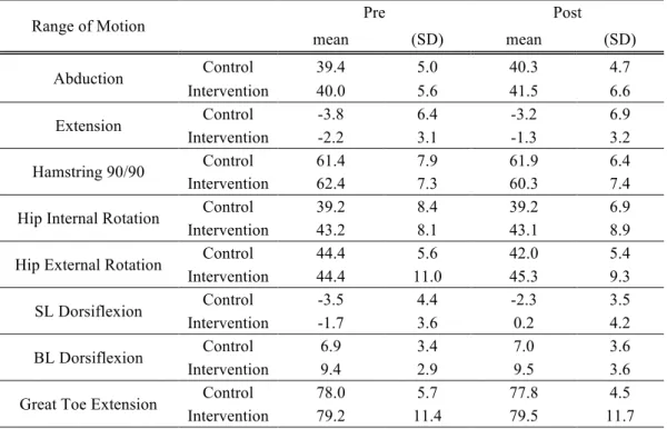
Table 2: Raw Mean Values of Range of Motion Pre and Post Intervention for Intervention and Control Groups
Range of Motion Pre Post
mean (SD) mean (SD)
Abduction Control 39.4 5.0 40.3 4.7
Intervention 40.0 5.6 41.5 6.6
Extension Control -3.8 6.4 -3.2 6.9
Intervention -2.2 3.1 -1.3 3.2
Hamstring 90/90 Control 61.4 7.9 61.9 6.4 Intervention 62.4 7.3 60.3 7.4
Hip Internal Rotation Control 39.2 8.4 39.2 6.9 Intervention 43.2 8.1 43.1 8.9
Hip External Rotation Control 44.4 5.6 42.0 5.4 Intervention 44.4 11.0 45.3 9.3
SL Dorsiflexion Control -3.5 4.4 -2.3 3.5 Intervention -1.7 3.6 0.2 4.2
BL Dorsiflexion Control 6.9 3.4 7.0 3.6 Intervention 9.4 2.9 9.5 3.6
REFERENCES
Atha, J., & Wheatley, D. W. (1976). Joint mobility changes due to low frequency vibration and stretching exercises. British Journal of Sports Medicine, 10(1), 26–34. Retrieved from http://articles.sirc.ca/search.cfm?id=18938%5Cnhttp://ezproxy.library.usyd.edu.au/login?url =http://search.ebscohost.com/login.aspx?direct=true&db=sph&AN=SPH18938&site=ehost-live%5Cnhttp://www.bmjpg.com
Chance-Larsen, K., Littlewood, C., & Garth, A. (2010). Prone hip extension with lower
abdominal hollowing improves the relative timing of gluteus maximus activation in relation to biceps femoris. Manual Therapy, 15(1), 61–65.
https://doi.org/10.1016/j.math.2009.07.001
Cheatham, S. W., Kolber, M. J., Cain, M., & Lee, M. (2015). The effects of self-myofascial release using a foam roll or roller massager on joint range of motion, muscle recovery, and performance: a systematic review. Int J Sport. Phys Ther, 10(6), 827–838.
Dupuy, O., Douzi, W., Theurot, D., BOSQUET, L., & DUGUE, B. (2018). An evidence-based approach for choosing post-exercise recovery techniques to reduce markers of muscle damage, soreness, fatigue and inflammation: a systematic review with meta-analysis. Frontiers in Physiology, 9(April), 403. https://doi.org/10.3389/FPHYS.2018.00403
Herbert, R. D., & Gabriel, M. (2002). Soreness and Risk of Injury : Systematic Review. British Medical Journal, 325(August), 1–5. https://doi.org/10.1136/bmj.325.7362.468
Mohr, A. R., Long, B. C., & Goad, C. L. (2014). Effect of Foam Rolling and Static Stretching on Passive Hip-Flexion Range of Motion. Journal of Sport Rehabilitation, 23(4), 296–299. https://doi.org/10.1123/JSR.2013-0025
Pearcey, G. E. P., Bradbury-Squires, D. J., Kawamoto, J.-E., Drinkwater, E. J., Behm, D. G., & Button, D. C. (2015). Foam Rolling for Delayed-Onset Muscle Soreness and Recovery of Dynamic Performance Measures. Journal of Athletic Training, 50(1), 5–13.
https://doi.org/10.4085/1062-6050-50.1.01
CHAPTER V Statistical Analysis
Each range of motion was taken five times at each time point. An average was taken of all five trials to determine an average for each time point. A three by two repeated measures ANOVA was used to determine the differences in range of motion between a control group and a corrective exercise intervention group over three time points: pre-fatigue, immediately post-fatigue, 24 hours post-fatigue. Statistical significance was set at α<0.05. A Tukey post hoc analysis was used for significant results. All data were analyzed using SPSS 24.0 statistical software (SPSS, Inc., Chicago, IL).
Results
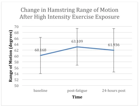
There was a significant difference within time points for hamstring range of motion (p = .02) (Table 3). Specifically there was a decrease in hamstring range of motion from baseline, post-0 fatigue and post-24 fatigue. Tukey Post Hoc analyses were performed and demonstrated that hamstring range of motion immediately post fatigue was significantly reduced compared to pre-fatigue (p = .002) (Graph 1). However, there were no differences between twenty four hours post fatigue and pre-fatigue measures (p =.137) or immediately post fatigue (p = .253) measures.
Discussion
Reliability was established between two investigators (Table 4, 5). Although two different researchers took range of motion measurements inter rater reliability was established (Table 6). Thus, there should be no difference in range of motion outcomes based on which investigator collected the data.
We observed a decrease in knee extensibility and hamstring range of motion immediately post-fatigue but not twenty-four hours post-fatigue. Overall, we did not observe a change across muscle groups. This could potentially be related to viscoelasticity increasing due to tissue warming (McHugh, Magnusson, Gleim, & Nicholas, 1992). This thixotropic increase may have offset any other changes from happening (Proske, Morgan, & Gregory, 1993). There were most likely minimal changes for group by time and time for range of motion based on the baseline values for subjects across the board. The mean averages of subjects at baseline were lower than what normative values are. Based on this, from pre-fatigue to post-fatigue and twenty-four hours post fatigue there is no room for range of motion to decrease more. Further research should establish normative range of motion values for athletes. A possible explanation for why we saw a change in only hamstring range of motion would be how many muscles were targeted in in the fatigue protocol that cross the knee joint. The amount of running and then jump landings and split squats performed would cause an increase in the stretch shortening cycle and both eccentric and concentric contractions of the hamstrings multiple times, which create a decrease in
motion and knee extensibility (Shamus & Email, 2003). Further research should focus on how this fatigue protocol could be modified to target other muscles of the body to include the gastrocsoleus complex, hip internal and external rotators, hip flexors, and hip adductors.
TABLES
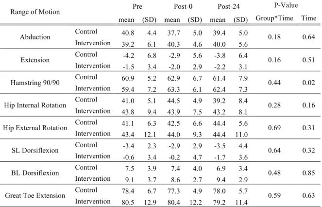
Table 3: Change in Range of Motion over Three Time Points (Baseline, Post-0 Fatigue, Post-24 Fatigue) Within Groups
Range of Motion Pre Post-0 Post-24 P-Value mean (SD) mean (SD) mean (SD) Group*Time Time
Abduction Control 40.8 4.4 37.7 5.0 39.4 5.0 0.18 0.64 Intervention 39.2 6.1 40.3 4.6 40.0 5.6
Extension Control -4.2 6.8 -2.9 5.6 -3.8 6.4 0.16 0.51 Intervention -1.5 3.4 -2.0 2.9 -2.2 3.1
Hamstring 90/90 Control 60.9 5.2 62.9 6.7 61.4 7.9 0.44 0.02 Intervention 59.4 7.2 63.3 6.1 62.4 7.3
Hip Internal Rotation Control 41.0 5.1 44.5 4.9 39.2 8.4 0.28 0.16 Intervention 43.8 9.4 43.9 7.5 43.2 8.1
Hip External Rotation Control 41.1 6.3 42.5 6.6 44.4 5.6 0.69 0.31 Intervention 43.4 12.1 44.0 9.3 44.4 11.0
SL Dorsiflexion Control -3.4 2.3 -2.9 2.9 -3.5 4.4 0.64 0.32 Intervention -0.6 3.4 -0.2 4.7 -1.7 3.6
BL Dorsiflexion Control 7.5 3.9 7.4 4.0 6.9 3.4 0.48 0.85 Intervention 9.1 3.7 8.6 2.7 9.4 2.9
Great Toe Extension Control 78.4 6.7 77.3 4.9 78.0 5.7 0.59 0.63 Intervention 80.5 12.9 80.4 12.2 79.2 11.4
Table 4: Intra Rater Reliability for Investigator #1
Assessment ICC value Sdpre Sdpost Pooled SD SEM
Abduction 0.984 7.6 7 7.31 0.92
Hip Extension 0.984 4.8 4 4.42 0.56
Hamstring 90/90 0.958 10.8 9.7 10.26 2.10
Hip Internal Rotation 0.982 10 11 10.51 1.41
Hip External Rotation 0.918 6.5 6.1 6.30 1.80
Straight Leg Dorsiflexion 0.988 3.6 4.1 3.86 0.42
Bent Leg Dorsiflexion 0.86 3.6 3.1 3.36 1.26
Table 5: Intra Rater Reliability for Investigator #2
Assessment ICC value Sdpre Sdpost Pooled SD SEM
Abduction 0.912 3.5 3 3.26 0.97
Hip Extension 0.877 2.5 3 2.76 0.97
Hamstring 90/90 0.887 5.7 6.2 5.96 2.00
Hip Internal Rotation 0.947 8.9 8.6 8.75 2.01
Hip External Rotation 0.898 6 4.4 5.26 1.68
Straight Leg Dorsiflexion 0.979 4 4.2 4.10 0.59
Bent Leg Dorsiflexion 0.992 4.1 4.6 4.36 0.39
Great Toe Extension 0.928 9.4 9.1 9.25 2.48
Table 6: Inter Rater Reliability Between Investigator #1 and #2
Assessment ICC value Sdpre Sdpost Pooled SD SEM
Abduction 0.843 5.7 4.9 5.32 2.11
Hip Extension 0.848 2.5 2.3 2.40 0.94
Hamstring 90/90 0.95 5.1 5.7 5.41 1.21
Hip Internal Rotation 0.93 7.7 8.9 8.32 2.20
Hip External Rotation 0.912 6.5 6.8 6.65 1.97
Straight Leg Dorsiflexion 0.879 3.4 4 3.71 1.29
Bent Leg Dorsiflexion 0.935 2.8 2.9 2.85 0.73
Great Toe Extension 0.83 6.9 11.8 9.67 3.99
GRAPHS
Graph 1: Change in Hamstring Range of Motion After High Intensity Exercise Exposure
60.168 63.109 61.936 50 52 54 56 58 60 62 64 66 68 70
baseline post-fatigue 24-hours post
R an ge o f M ot io n ( d egr ee s) Time
REFERENCES
Atha, J., & Wheatley, D. W. (1976). Joint mobility changes due to low frequency vibration and stretching exercises. British Journal of Sports Medicine, 10(1), 26–34. Retrieved from http://articles.sirc.ca/search.cfm?id=18938%5Cnhttp://ezproxy.library.usyd.edu.au/login?url =http://search.ebscohost.com/login.aspx?direct=true&db=sph&AN=SPH18938&site=ehost-live%5Cnhttp://www.bmjpg.com
Beynnon, B. D., Vacek, P. M., Newell, M. K., Tourville, T. W., Smith, H. C., Shultz, S. J., … Johnson, R. J. (2014). The Effects of Level of Competition, Sport, and Sex on the Incidence of First-Time Noncontact Anterior Cruciate Ligament Injury. The American Journal of Sports Medicine, 1–8. https://doi.org/10.1177/0363546514540862
Boden, B. P., Sheehan, F. T., Torg, J. S., & Hewett, T. E. (2010). Noncontact anterior cruciate ligament injuries: mechanisms and risk factors. The Journal of the American Academy of Orthopaedic Surgeons, 18(9), 520–7. https://doi.org/10.1016/j.drudis.2011.09.009
Borotikar, B. S., Newcomer, R., Koppes, R., & McLean, S. G. (2008). Combined effects of fatigue and decision making on female lower limb landing postures: Central and peripheral contributions to ACL injury risk. Clinical Biomechanics, 23(1), 81–92.
https://doi.org/10.1016/j.clinbiomech.2007.08.008
Caine, D., Maffulli, N., & Caine, C. (2008). Epidemiology of Injury in Child and Adolescent Sports: Injury Rates, Risk Factors, and Prevention. Clinics in Sports Medicine, 27(1), 19– 50. https://doi.org/10.1016/j.csm.2007.10.008
Chance-Larsen, K., Littlewood, C., & Garth, A. (2010). Prone hip extension with lower
abdominal hollowing improves the relative timing of gluteus maximus activation in relation to biceps femoris. Manual Therapy, 15(1), 61–65.
https://doi.org/10.1016/j.math.2009.07.001
Cheatham, S. W., Kolber, M. J., Cain, M., & Lee, M. (2015). The effects of self-myofascial release using a foam roll or roller massager on joint range of motion, muscle recovery, and performance: a systematic review. Int J Sport. Phys Ther, 10(6), 827–838.
Christina, K. A., White, S. C., & Gilchrist, L. A. (2001). Effect of localized muscle fatigue on vertical ground reaction forces and ankle joint motion during running. Human Movement Science, 20(3), 257–276. https://doi.org/10.1016/S0167-9457(01)00048-3
Conn, J. M., Annest, J. L., & Gilchrist, J. (2003). Sports and recreation related injury episodes in the US population, 1997-99. Injury Prevention, 9(2), 117–123.
https://doi.org/10.1136/ip.9.2.117
https://doi.org/10.1136/bjsm.2007.037937
Dill, K. E., Begalle, R. L., Frank, B. S., Zinder, S. M., & Padua, D. A. (2014). Altered knee and ankle kinematics during squatting in those with limited weight-bearing-lunge
ankle-dorsiflexion range of motion. Journal of Athletic Training, 49(6), 723–732. https://doi.org/10.4085/1062-6050-49.3.29
Dupuy, O., Douzi, W., Theurot, D., BOSQUET, L., & DUGUE, B. (2018). An evidence-based approach for choosing post-exercise recovery techniques to reduce markers of muscle damage, soreness, fatigue and inflammation: a systematic review with meta-analysis. Frontiers in Physiology, 9(April), 403. https://doi.org/10.3389/FPHYS.2018.00403
Favre, J., Clancy, C., Dowling, A. V., & Andriacchi, T. P. (2016). Modification of Knee Flexion Angle Has Patient-Specific Effects on Anterior Cruciate Ligament Injury Risk Factors During Jump Landing. The American Journal of Sports Medicine, 0363546516634000-. https://doi.org/10.1177/0363546516634000
Fernandez, W. G., Yard, E. E., & Comstock, R. D. (2007). Epidemiology of Lower Extremity Injuries among U.S. High School Athletes. Academic Emergency Medicine, 14(7), 641–645. https://doi.org/10.1111/j.1553-2712.2007.tb01851.x
Fong, C.-M., Blackburn, J. T., Atc, À., Norcross, M. F., Atc, À., Mcgrath, M., & Padua, D. A. (2011). Ankle-Dorsiflexion Range of Motion and Landing Biomechanics, 46(1), 5–10. https://doi.org/10.4085/1062-6050-46.1.5
Frank, B. S. (2016). THE INFLUENCE OF MOVEMENT PROFILE ON THE FEMALE ATHLETE’S BIOMECHANICAL RESILIENCE & TRAINING LOAD RESPONSE TO CONTROLLED EXERCISE EXPOSURE Barnett.
Gianotti, S. M., Marshall, S. W., Hume, P. A., & Bunt, L. (2009). Incidence of anterior cruciate ligament injury and other knee ligament injuries: A national population-based study. Journal of Science and Medicine in Sport, 12(6), 622–627.
https://doi.org/10.1016/j.jsams.2008.07.005
Gomes, J. L. E., de Castro, J. V., & Becker, R. (2008). Decreased Hip Range of Motion and Noncontact Injuries of the Anterior Cruciate Ligament. Arthroscopy - Journal of Arthroscopic and Related Surgery, 24(9), 1034–1037.
https://doi.org/10.1016/j.arthro.2008.05.012
Herbert, R. D., & Gabriel, M. (2002). Soreness and Risk of Injury : Systematic Review. British Medical Journal, 325(August), 1–5. https://doi.org/10.1136/bmj.325.7362.468
Hewett, T. E. (2005). Biomechanical Measures of Neuromuscular Control and Valgus Loading of the Knee Predict Anterior Cruciate Ligament Injury Risk in Female Athletes: A
Hewett, T. E., Ford, K. R., Hoogenboom, B. J., & Myer, G. D. (2010). Understanding and preventing acl injuries: current biomechanical and epidemiologic considerations - update 2010. North American Journal of Sports Physical Therapy : NAJSPT, 5(4), 234–251. Hootman, J. M., Dick, R., & Agel, J. (2007). Epidemiology of Collegiate Injuries for 15 Sports :
Summary and ... Journal of Athletic Training, 42(2), 311–319.
Jonhagen, S., Nemeth, G., & Eriksson, E. (1994). Hamstring injuries in sprinters. The role of concentric and eccentric hamstring muscle strength and flexibility. American Journal of Sports Medicine, 22(2), 262–266. Retrieved from
http://www.ncbi.nlm.nih.gov/pubmed/8198197
Leppänen, M., Pasanen, K., Kujala, U. M., Vasankari, T., Kannus, P., Äyrämö, S., … Parkkari, J. (2016). Stiff Landings Are Associated With Increased ACL Injury Risk in Young Female Basketball and Floorball Players. The American Journal of Sports Medicine,
0363546516665810-. https://doi.org/10.1177/0363546516665810
Lohmander, L. S., Englund, P. M., Dahl, L. L., & Roos, E. M. (2007). The Long-term
Consequence of Anterior Cruciate Ligament and Meniscus Injuries. The American Journal of Sports Medicine, 35(10), 1756–1769. https://doi.org/10.1177/0363546507307396
MacDonald, G. Z., Penney, M. D. H., Mullaley, M. E., Cuconato, A. L., Drake, C. D. J., Behm, D. G., & Button, D. C. (2013). An Acute Bout of Self-Myofascial Release Increases Range of Motion Without a Subsequent Decrease in Muscle Activation or Force. Journal of Strength and Conditioning Research, 27(3), 812–821.
https://doi.org/10.1519/JSC.0b013e31825c2bc1
Mather, R. C., Koenig, L., Kocher, M. S., Dall, T. M., Gallo, P., Scott, D. J., … Spindler, K. P. (2013). Societal and economic impact of anterior cruciate ligament tears. The Journal of Bone and Joint Surgery. American Volume, 95(19), 1751–9.
https://doi.org/10.2106/JBJS.L.01705
McHugh, M. P., Magnusson, S. P., Gleim, G. W., & Nicholas, J. A. (1992). Viscoelastic stress relaxation in human skeletal muscle. Medicine and Science in Sports and Exercise, 24(12), 1375—1382. Retrieved from http://europepmc.org/abstract/MED/1470021
McLean, S. G., Huang, X., & Van Den Bogert, A. J. (2005). Association between lower extremity posture at contact and peak knee valgus moment during sidestepping: Implications for ACL injury. Clinical Biomechanics, 20(8), 863–870.
https://doi.org/10.1016/j.clinbiomech.2005.05.007