Original Research Article
Effect of vitamin E on biochemical and ultrastructural changes in
acrylamide-induced renal toxicity in rats
Hoda Atef
1, Ghalia M. Attia
1,2,
Hassan M. Rezk
3,4*, Mohamed El-Shafey
3INTRODUCTION
Acrylamide (ACR) is an odorless, white crystalline, reactive and highly water-soluble monomer. These properties facilitate its rapid absorption and distribution through the body.1 It is solid at room temperature with a molecular formula of C3 H5 NO.2 It is present in plants
like potatoes, carrots, radish, lettuce, Chinese cabbage, parsley, onions, spinach and rice also in sugar and olives.3,4 ACR represents a chemical widely used in industry and manufacturing of polyacrylamides which are common in personal care products as lotions, deodrants and cosmetics, also in paper industry.5 It was also highly detected in tobacco and cigarette smoke.6
ABSTRACT
Background: Acrylamide (ACR) is a widely used chemical in industry and it accounts for major health problems as
it has been detected in highly consumed food items, carbohydrate-rich food items cooked at high temperature. Accordingly the population is highly exposed to ACR. The aim of the study was to assess the effect of vitamin E on biochemical and ultrastructure changes in acrylamide-induced renal toxicity in wistar albino rats.
Methods: Three groups of adult albino wistar rats weighing about (200-250 gm) were used in this study to investigate the effect of vitamin E on acrylamide induced renal toxicity; 10 rats in each group; Group I: Control group, Group II: Acrylamide treated and Group III: Acrylamide-Vit E treated group. Blood samples were collected for estimation of serum creatinine, blood urea nitrogen (BUN), lactic dehydrogenase (LDH) and albumin. Kidneys specimens were processed for light and electron microscopic studies. Kidney sections were stained with H&E, MT, PAS and immuno-histochemical stains for detection of NF-kβp65 and Bcl-2. Morphometric study was done followed by biochemical and morphometric statistical analysiss.
Results: Acrylamide treated rats showed degeneration of cells lining PCT and DCT, atrophy of glomeruli and fibrosis. Ultrastructurally; tubules lining cells showed loss of microvilli, basal membrane in foldings and mitochondrial changes. Podocytic changes include: Vacuolation, irregularity and disorientation of their processes and thinning of glomerular basement membrane. Significant increase in the mean number of NF-kβp65 positive cells and decrease in the mean area% of Bcl-2 immuno-expression, increase in the mean area % of collagen fibers and urinary space diameter and decrease in thickness of epithelial cells of PCT and DCT were also observed. Serum creatinine, BUN anLDH were increased whereas serum albumin was decreased. Vitamin E co-administration with ACR improve all these histological, ultrastructural and biochemical changes.
Conclusions: Acrylamide induced renal toxicity could be ameliorated by vitamin E co-administration.
Keywords: Acrylamide, Kidney, Vitamin E, NF-kβp65, Bcl-2, Ultarstructure
1Department of Histology, Faculty of Medicine, Mansoura University, Egypt; 2 Department of Anatomy, Faculty of
Medicine, Taibah University, AlMadinah Al Monawarrah, KSA; 3Department of Anatomy and Embryology, College of Medicine, Mansoura University, Egypt; 4Department of Anatomy and Embryology, Batterjee Medical College, Jeddah, KSA
Received: 07 April 2017
Revised: 20 April 2017
Accepted: 26 April 2017
*Correspondence:
Dr. Hassan M. Rezk
E-mail: [email protected]
Copyright: © the author(s), publisher and licensee Medip Academy. This is an open-access article distributed under the terms of the Creative Commons Attribution Non-Commercial License, which permits unrestricted non-commercial use, distribution, and reproduction in any medium, provided the original work is properly cited.
ACR monomer may form in foods cooked at very high temperatures.7 It is present at high concentrations in carbohydrate rich food as potato and grain-base foods prepared at high temperature such as French fries and potato chips. Since ACR is formed from natural chemicals in food while cooking, its levels in organic foods are similar to inorganic ones.8 It has become a major public health concern as it is detected in highly consumed food.9
ACR toxicity has been reported to various body organs; neurotoxicity is the main effect, reproductive system toxicity.10,11 It has a significant binding capacity to liver, kidney, erythrocyte and brain.12 The US EPA and IARC have classified acrylamide as B2, a carcinogen and as 2B a human carcinogen, respectively.13 The toxicity of ACR is due to its biotransformation to a more potent molecule that initiates cellular toxicity. Therefore, the significant pathogenic pathway is the oxidative biotransformation of ACR by cytochrome P450 2E1 (CYP2E1).14 The resulting metabolite is an epoxide glycidamide, which is more reactive towards DNA and proteins than ACR.15,16 Once absorbed, ACR may be conjugated by glutathione-S-transferase (GST) to N-acetyl- S-(3-amino-3-oxopropyl) cysteine or it reacts with cytochrome P450 (CYP450) to produce glycidamide. Several metabolic studies have been conducted that focused on the interaction of ACR with CYP450 and GST in rats and mice. The results of these studies indicated that liver, brain, kidney and erythrocyte GST have high binding capacity with ACR.17,18
ACR inducedoxidative stress, resulting in generation of reactive oxygen species ROS which plays a critical role in the initiation and progression of fibrotic diseases. Two important markers for ROS Nicotinamide adenine dinucleotide phosphate (NADPH) oxidase and advanced oxidation protein products (AOPPs). NADPH oxidase is recognized as a key mediator of cell proliferation and matrix accumulation in renal disease. AOPPs are not only markers of oxidative stress but also cause renal injury. Inhibition of NADPH oxidase and/or reducing AOPPs production might be a corner stone for the therapeutic intervention of variety of fibrotic kidney disorders.19 AOPPs induce vascular endothelial and smooth muscle cell dysfunction, promote podocyte depletion and apoptosis, upregulate the expression of fibronectin and collagen IV in mesangial cells.20-22
Vitamin E, as an important antioxidant, plays a role in inhibition of mutagen formation and repair of membranes and DNA.23 So vitamin E is a non-enzymatic antioxidant which can act to reduce oxidative stress. Studies have reported that vitamin E can reduce lipid peroxidation caused by toxic substances.24,25 Vitamin E supplementation in cancer patients showed that it has an important cytoprotective effect.26,27
Since ACR is present in many commonly consumed foods and industries, the general population is highly
exposed to it. The aim of this study is to investigate renal toxicity of acrylamide ACR and the ameliorative effect of vitamin E on the toxicity.
METHODS
Animals and housing
The experiment was conducted on thirty adult albino wistar rats, weighing about 200-250 gm. The animals were acclimated for about one week and housed in plastic cages at room temperature prior to experiment in Mansoura Medical Research Center (MRC). Throughout the experiment, the animals were housed under standard laboratory conditions. Care and treatment of animals were approved and practices were performed according to approval of ethics regulation at Mansoura University.
Chemicals
Acrylamide (99.50%) and vitamin E pure were purchased from Hi Media Laboratories Pvt. Limited, Mumbai. All other required chemicals were used of highest quality available.
Experimental design
Experiment was designed to study toxicity of acrylamide on kidney for the duration of 4 weeks. The rats were assigned to one of the three groups, with ten rats each as follows:
Group I (Control group): given distilled water for 4 weeks by oral gavage tube.
Group II (Acrylamide treated group): treated orally with acrylamide dissolved in distilled water at a dose of 20 mg/kg body weight once daily for 4 weeks.28
Group III (Acrylamide –Vitamin E treated group): Was treated orally with acrylamide dissolved in distilled water at a dose of 20 mg/kg body weight daily with vitamin E at a dose of 50 IU/kg body weight once daily for 4 weeks.28
Blood samples and kidney tissue-specimens collection
At the end of the experiment after 4 weeks, rats of all groups were anesthetized by intraperitoneal injection of sodium thiopental (40 mg/kg of body weight) (Sigma Chemical Co., ST Louis, MO, USA). Blood samples and renal specimens were collected.
Biochemical study
Light microscopic (LM) study
kidneys were excised and fixed in 10% neutral buffered formalin; then routinely processed to obtain paraffin blocks which were cut into sections of 5µm thickness, mounted on glass slides, deparaffinized in xylene and stained by: hematoxylin and eosin stain (H&E), Masson trichrome )MT), periodic acid schiff(PAS) reagent stains, immuno-histochemical stains for detection of NF-kβ and Bcl2 using avidin–biotin–peroxidase complex (ABC) techniques (The primary anti- Bcl-2 and anti- NF-kBp65 antibodies were rat anti-mouse Bcl-2 monoclonal antibody (Genemed Biotechnologics Inc., South San Francisco, CA 94080, USA) and rabbit polyclonal NF-kβp65 antibody (dilution 1:100; Santa Cruz Bio-technology, Santa Cruz, CA, USA) respectively.29-31 Sections were then examined and observed under light microscope.
Electron microscopic (EM) study
Small kidney specimens (1 mm³) of kidneys were immediately fixed in glutaraldehyde (2.5%), and then post fixed in osmium tetroxide (1.0%), dehydrated, and embedded in epoxy resin. Semithin sections (1 um thickness) were prepared and stained with Toluidine blue. Ultrathin sections were cut at thickness of 80nm mounted on copper grids and stained with Uranyl acetate 5% for 15 min followed by lead citrate for 8 min.32 Sections were examined by transmission electron microscope (JEOL2100 electron microscope, Tokyo, Japan) at Electron Microscopy Unit, Faculty of Agriculture, Mansoura -University, Egypt.
Morphometric study
From H&E stained sections (x400); the epithelial height of PCT and DCT and the width of urinary space (the distances between the parietal and visceral layers) of renal corpuscles were measured). From MT stained sections (x400); area% of collagen fibers deposition in the kidney tissues were measured. From immune-stained sections (x400); number of NF-kBp65 positive cells (nuclear reaction) and the area % of Bcl-2 immuno-expression were measured. For each mentioned stain measured parameter, five non-overlapping fields/rat sections were examined (The total was 50 measurements /group) and photographed using color video camera (digital camera CH- 9435 DFC 290). The photographs were analyzed using Leica Qwin 500 (Imaging System, Cambridge, UK) within a frame area of 293.4288 μm2
. Morphometry was carried out at the Image Analysis Unit, Anatomy Department, Faculty of Medicine, Taibah University, Al Madinah Al Monawarrah, KSA.
Statistical analysis
All data were expressed as mean ± SEM. Statistical analysis was performed using IBM SPSS software version 21.00 (Chicago, Illinois, USA). One-way analysis
of variance (ANOVA) (data were normally distributed and variances of populations were equal), post-hoc and least significant difference (LSD) were performed for inter-group comparison. P >0.05, P ≤0.05 and P ≤0.001 were considered non-significant, significant and highly significant, respectively.33
RESULTS
Light microscopic results
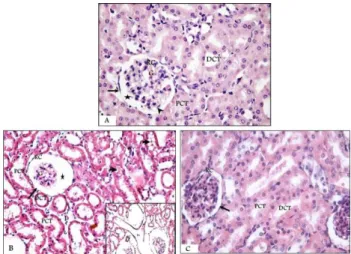
Acryl group showed disturbed architecture of renal cortex with corpuscle shrinkage up to partial or complete loss with congestion of the glomerulus and an apparent widening of the urinary space. Some PCT and DCT showed disturbance of their shape with degeneration, and vacuolation of their lining cells. Other tubules show marked dilatation and their cells appear small with pyknotic nuclei and an apparent increase in the interstitial tissue in between is observed (Figure 1B).
Figure 1: Photomicrographs of H&E stained sections in the renal cortex of A): Control group, showing
normal renal cortex with glomeruli, parietal and visceral (arrow head) layers of Bowman’s capsule,
urinary space, PCT and DCT. B): Acryl group showing RC shrinkage, congestion of the glomerulus up to its complete loss (Inset), partial loss of parietal layer and widening of the urinary space. PCT and DCT show degeneration and vacuolation (arrow head)
of lining cells with pyknotic nuclei. C): Acryl-Vit E group, with preserved normal renal architecture.
(H&E stain; A and CX400; B and Inset BX200).
RC: renal corpuscle, G: glomeruli, Arrow: parietal layer of Bowman’s capsule, Star: urinary space, PCT: proximal convoluted tubules, DCT: distal convoluted tubules, I: interstitial tissue, Red arrows: pyknotic nuclei.
Masson trichrome stained sections of Acryl group, increasing amount of collagen fibers deposition among glomerular capillaries, however the renal tubules (RT) were still surrounded with minimal amount of collagen.
Moreover, areas of loss of RC or RT and their replacement with fibrous tissue were also detected (Figure 2B and C). Acryl-Vitamin E group showed an apparent decrease in the deposition of collagen fibers around the wall of RC and among glomerular capillaries. RT and blood vessels were also surrounded with minimal amount of collagen fibers when compared with Acryl group (Figure 2D).
Figure 2: Photomicrographs of Masson trichrome stained sections in the renal cortex of A): Control group: minimal collagen fibers (arrow heads). B): Acryl group: increasing amount of collagen fibers deposition among glomeruli with renal tubules (RT)
surrounded with minimal collagen around renal tubules. C): Acryl group, show area of loss of RC or
RT and their replacement by fibrous tissue (F). D): Acryl-Vit E group shows decreased fibrous tissue (arrow). (Masson trichrome stain; AX200; B, C and
DX400).
G: glomeruli, RT: renal tubules, RC: renal corpuscle, F: fibrous tissue, Arrow heads: collagen fibres, Bv: blood vessels.
PAS stained sections of Acryl group, the RC, PCT and DCT showed partially lost and ill-defined weak PAS positive basement membranes and PCT showed partial to complete loss of their apical brush borders (Figure 3B). Acryl-Vitamin E group appeared with PAS positive and intact basement membranes of RC, PCT and DCT and the
brush borders of cells of PCT (Figure 3C). Anti-NF-kβp65 immunostained sections of control
kidney showed NF-kβ negative immunoreaction in RC components and renal tubules epithelium (Figure 4A). Acryl group exhibited strong positive NF-kβ immunoreaction in the epithelial cells of RT and cells of glomerulus (Figure 4B), whereas in Acryl- Vitamin E group, weak positive NF-kβ immunoreaction was detected in few numbers of cells of glomerulus and renal tubules (Figure 4C).
Figure 3: A photomicrograph of PAS stained sections in the renal cortex of A): Control group shows renal corpuscle (RC), proximal (PCT) and distal convoluted
tubules (DCT) with PAS positive well defined basement membranes (thick arrows). PCT epithelial cells have prominent PAS positive apical thick brush borders (thin arrows). B): Acryl group shows weak
PAS positive basement membranes (arrow heads) with partial to complete loss of apical brush borders of PCT (arrows). C): Acryl-Vit E group appears with
PAS positive (arrows) areas. (PAS stain; A, B and C 400 X).
Figure 4: A photomicrograph of Anti-NF-kβp65 immune-stained sections of renal cortex (A, B and C)
and Anti-BCL2 immuno-stained sections of renal cortex (D, E and F).
Anti-BCL2 immuno-stained sections of Control renal cortex exhibited strong Bcl2 positive cytoplasmic immune reaction which was detected in cells of nearly all renal tubules (Figure 4D). Acryl group showed very weak Bcl2 immunoreaction in few numbers of cells of renal tubules and glomerulus (Figure 4E). On the other hand, Acryl- Vitamin E group showed strong Bcl2 positive reaction in the epithelial cells of the large number of renal tubules (Figure 4F).
Electron microscopic results
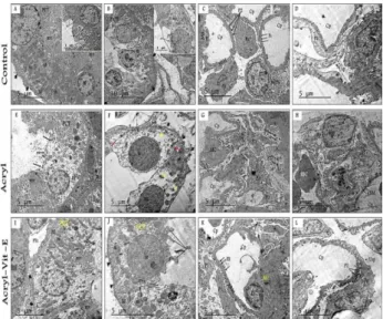
In Acryl group, both DCT and PCT showed signs of degeneration of its cells in the form of PCT cells loss of brush borders, degeneration and loss of normal orientation of mitochondria and separation of cells with loss of lateral border interdigitaions in cells of DCT. Glomerulus showed abnormal shape and orientation and marked irregularity of secondary feet processes of podocytes with extensive vacuolations of their cytoplasm. Lumen of glomerulus capillary contained eosinophil cell with its characteristic cytoplasmic ellipsoid granules. Apparent thinning of glomerular basement membrane was evident (Figure 5E-H).
Figure 5 (A-D): Electron micrographs of control kidney.
A): PCT lining cell surrounded with intact basement membrane (arrow head) with show basal infolding (BF) in between them longitudinally arranged mitochondria (m). The cells contain basal rounded nuclei (N), apical endocytic vacuoles (Ev) with microvilli (mv) forming the characteristic brush border of PCT (Inset). B): DCT lining cell surrounded with intact basement membrane (arrow head) which show basal infolding (BF) with longitudinally arranged mitochondria (m) in between. The lining cells contain rounded nuclei (N) with apical microvilli (mv) (Inset). C): glomerular capillary loops (Cp) invested with 2ry feet (P2) which extend from 2ry processes (P1) of podocytes (Po).Capillaries lined by endothelial cells (E) which show fenestrae (Fs) and rest on glomerular basement membranes (gBM). D): Capillary loop (Cp), 2ry processes (P1) of podocytes, mesangial cell (Mc) and urinary space (Usp). Acryl group (E,F,G and H); E): PCT degenerated of cells with loss of
brush borders (arrows), focal loss of cytoplasm (asterisks) and degeneration of mitochondria(m). F): Degenerated DCT with thin and irregular basement membrane (BM), nucleur lysis (N) and loss of lateral interdigitaions (red arrows). G): Irregular of capillary loops (Cp), irregular 2ry feet (P2) of vaculated (V) podocytes. H): Glomerular capillary (Cp) with an eosinophil (Eo) with ellipsoid granules. Thin glomerular basement membrane (gBM). Acryl –Vit E group (I, J, K and L); I) preserved PCT structure. J): preserved DCT cells with basement membrane (arrow heads), lateral cell borders interdigitations (arrows). K and L): Glomerular capillaries with normal 1ry (P2) and 2ry feet (P1) processes of podocytes. (Uranyl acetate and Lead citrate X 2000, Print Mag. X 11700).
In Acryl –Vitamin E group, the PCT and DCT appeared of nearly normal shape with their lining cells showed preservation of apical microvilli of PCT, basement membrane with basal infoldings and longitudinally oriented mitochondria, lateral interdigitations and apical border stubby microvilli of DCT. Glomerular capillaries appeared of normal shape and showed normal shape and orientation of primary and secondary feet processes of podocytes which invested the glomerular basement membranes (Figure 5I-L).
Biochemical and morphometric statistical results
A highly significant increase in the mean concentration of serum Creatinine, BUN and LDH, and a highly significant decrease in the mean concentration of serum albumin was detected in group II when it was compared to Control group. On the other hand, A highly significant decrease in the mean concentration of serum Creatinine, BUN and LDH and a highly significant increase in the mean concentration of serum albumin was observed in group III when it was compared to group II (Table 1). A highly significant decrease in the mean epithelial thickness of PCT and DCT and a highly significant increase in the mean diameter of urinary space in Group II when it was compared to control group. Group III showed a highly significant increase in the mean epithelial thickness of PCT and DCT and a highly significant decrease in the mean diameter of urinary space when it was compared to group II (Figure 6). Regarding the main area percentage of collagen fibers deposition in the renal tissue, it showed a highly significant increase in group II when it was compared to control and a highly significant decrease in group III when it was compared to group II (Figure 7A).
Figure 6: Clustered bars show a highly significant decrease in the mean epithelial thickness of PCT&DCT and a highly significant increase in the diameter of urinary space in Acryl groups when it is compared to control group. Acryl-Vit E group show a
highly significant increase in the mean epithelial thickness of PCT and DCT and a highly significant
decrease in the diameter of urinary space in Acryl groups when compared to Acryl group. Values are expressed as mean ± standard error of mean (SE).P1<
0.001, P3< 0.001. (P1; Acryl vs Control. P3; Acryl vs
Acryl-VitE).
Figure 7: Bars morphometry of A) Mean area % of collagen fibers deposition, B) Mean number of NF-kβ
immune-positive cells C): Mean area% of Bcl-2 immuno-expression in the renal tissue of different
groups. Values are expressed as mean ± standard error of mean (SE). P1; Acryl vs Control, P3; Acryl vs
Acryl-VitE.
Table 1: Mean concentration of creatinine, BUN, LDH and Albumin in the serum (mg/dL).
Creatinine (mg/dL) BUN
(mmol/L)
LDH (U/L)
Albumin (g/dL) Group I (Control)
Mean± SE 0.66± 0.32 37.28 ± 1.24 462.48± 9.76 4.25 ± 0.056
Group II (Acryl group)
Mean ± SE 1.48± 0.11 112.47 ± 3.96 1397.25 ± 30.50 3.15 ± 0.061
P value 1 < 0.001*** < 0.001*** < 0.001*** < 0.001***
Group III (Acryl-Vit E Group)
Mean± SE 0.86 ± 0.04 48.54 ± 1.80 570.87 ±15.63 3.85 ± 0.109
P value 2 0.063 (non-significant) 0.005** 0.001*** 0.001***
P value 3 < 0.001*** < 0.001*** < 0.001*** < 0.001***
Data are expressed as Mean ± SE: (SE :Standard error of mean), P value 1; Acryl group compared to control group, P value 2; Acryl-Vit E , compared to control group, P value 3; Acryl-Acryl-Vit E group compared to Acryl group, Non-significant, P value >0.05, ** Significant P value ≤0.01,***highly significant, P value ≤ 0.001.
DISCUSSION
In this research, our aim was to study the effects of ACR and vitamin E on the kidney. In group II (ACR-treated group) showed shrinked renal corpuscle with loss of parietal layer of Bowman’s capsules and wide urinary space. The cells of renal tubules showed vacuolations and pyknotic nuclei. Other research used several doses of ACR (0.5, 5, 50, 100 mg/kg ACR per day for 11 days intraperitoneally) to investigate the nephrotoxic effect of ACR. The H&E technique of the low doses (0.5 and 5 mg/kg) showed normal renal cortex while, the high doses of (50 and 100 mg/kg) showed proliferative glomerulonephritis. It had been concluded that, the ACR induced acute nephrotoxicity.34 These findings were in harmony with another research that was studied the
effects of several doses of ACR on the kidney which showed degeneration of the glomerular tuft with lymphocytic infiltration. The cells lining renal tubules showed cytoplasmic vacuolations and loss of brush borders.35,36
interstitial hypercellularity, matrix accumulation, and atrophy of epithelial structures, resulting in loss of normal function and ultimately organ failure.37
The PAS results of this research were similar to another research that was done to study the effect of cisplatin (CP) on the kidney, revealed, weak positive PAS reaction in the brush border and basement membranes of the healthy PCT and DCT in CP-treated rats.38
All the above mentioned apoptotic figures were confirmed by strong positive cytoplasmic and nuclear NF-kβ immunoreaction in the epithelial cells of the renal cortex compared with very weak Bcl2 immunoreaction. These results were approved statistically and by another study which concluded that the ROS activates NF-κB, which then ultimately leads to activation of ERK1/ERK2 pathway that lead to fibronectin expression.37,39 In a previous study, there was downregulation of Bcl2 in gentamicin induced nephrotoxicity. The NF-kB controlled through interaction with an inhibitor protein called IκB. In normal cells, NF-kB is inactive in cytoplasm through its binding with its inhibitors, p105 and IkBα-like proteins. The increased ROS in rat kidney causes the degradation of its inhibitor IkB-alpha or Proteolytic cleavage of p105, and free NF-kB dimers translocate to nucleus and activate the target antinflamatory genes.40
The histopathological results of the effect of ACR on the kidney (Group II) were approved by electron microscopic study of the effect of ACR on the RC, PCT and DCT. These findings agreed with ultrastructure findings of the effect of cisplatin (CP) on the kidney revealed that the renal corpuscles showed wide capsular space, fused foot processes of podocytes, dilated congested capillary loops and irregular capillary basement membrane. The PCT of the same study revealed irregular thick basement membrane with loss of its basal infoldings. However, the DCT of CP-treated rats showed apoptotic nuclear changes with fragmented nuclear envelop, thick basement membrane with few basal infoldings, small sized degenerated mitochondria, many lysosomes and cytoplasmic vacuoles.38
The harmful effects of ACR were also approved by the biochemical results which showed high significant increase in the mean concentration of serum creatinine, BUN and LDH, and a high significant decrease in the mean concentration of serum albumin.
In this study ACR was used in a dose of 20 mg/kg body weight once daily for 28 days.28Other researchers used ACR only in three doses (2, 10 and 30 mg/Kg/day for eight weeks). They found no significant differences in s. creatinine level between the low dose ACR, but it showed a significant decrease in both mild and high dose groups associated with significant decrease in albumin showed in alltreated groups.35
On the other hand, some authors reported that the BUN and creatinine levels significantly increased in the ACR treated group (40 mg/kg/day intraperitoneally). They explained these results as the higher dosage of ACR with a different route of administration could have increased the absorption of the toxin and in turn increased the damage.40
Other researchers used different doses of ACR (10, 30, 60 and 90 mg/kg/day for 6 weeks) followed by Recovery period was adopted thereafter for 4 weeks. They found that the albumin in rats fed on 10 mg/kg dietary ACR was significantly decreased, but there was non-significant change in albumin level in rats rats fed on 90 mg/kg dietary ACR. While in the recovery period, there were no significant differences in the concentrations of the albumin between the groups of their experiment. The hypoproteinaemia in rats fed on different concentrations of ACR might have resulted from hepatocellular dysfunction.14
Glycidamide has a close relationship to the toxic mechanism of ACR. In order to explore the toxic mechanism of ACR, some authors discussed the effects of intragastric and intraperitoneal administration of glycidamide-induced toxicity by determining the LDH, BUN and creatinine. They found that the same dose of glycidamide had more toxic effects and damage effects to the mice compared to the ACR. It could markedly increase the level of LDH and BUN.41
On the other hand, vitamin E is the main endogenous antioxidant which reacting with oxygen radical prevents the chain reaction of free radicals, protecting thus the membranes. However, the endogenous antioxidants reserves, such as vitamin E, gradually decrease in reactions with free radicals.42
In group III (ACR-Vitamin E treated group), showed nearly preserved renal cortex with significant decrease in both the width of urinary space. In two different studies used Vitamin E. to minimize the Nickle and Colistin Methane Sulfonate induced nephrotoxicity, the histopathological studies showed remarkable reduction in the nickel-induced PCT degeneration and tubular necrosis, multiple foci of hemorrhage and inflammation with the presence of lymphocytes in interstitial tissue by the administration of vitamin E. There was a relatively normal appearing urinary space area of the RC.43-45 The results from the same (group III) showed also collagen fibers deposition in the renal tissue. These tubules were also surrounded with minimal amount of collagen fibers with absence of fibrosis. In another research used vitamin E against aflatoxin induced nephrotoxicity, the Masson’s trichrome stain sectioned revealed few collagen fibers were be detected nearly similar to the control ones.45
basement membranes of renal corpuscle RC, PCT and DCT and the brush borders of cells of PCT. In PAS-stained sections to investigate the antioxidant effect of aged garlic extract against cisplatin induced nephrotoxicity, strong positive PAS reaction was observed in the brush border of proximal convoluted tubules (PCT), basement membrane of both PCT and distal convoluted tubules (DCT), and parietal layer of Bowman’s capsule.38
There was also weak positive cytoplasmic and nuclear NF-kβ immunoreaction with strong Bcl2 positive reaction. Several agents antagonize NF-kB, such as drugs in clinical use for treatment of renal disease, e.g. steroids, statins, and vitamin D receptor activators. During recovery from renal injury, there was downregulation of NF-kB and upregulation of anti-inflammatory genes. There is experimental evidence supporting the critical role of NF-kB activation in the pathogenesis of renal inflammation.46
The ultrastructure results from group III (ACR-Vitamin E treated group) revealed glomerular capillaries normal shape and showed normal shape and orientation of primary and secondary feet processes of podocytes. The ultrastructure study of PCT and DCT appeared of nearly normal shape with their lining cells showed preservation of apical microvilli of PCT, nuclei with prominent nucleolus. The basement membrane showed normal basal infolding and longitudinally oriented mitochondria, lateral cell borders interdigitations and apical border stubby microvilli of DCT. In another research to study the ultrastructure antioxidant effect of vitamin -E against Aflatoxin induced nephrotoxic effect, it showed nuclei of the podocytes and mesangial cells appeared vesicular nearly similar to those of control. It also showed, PCT were lined by cells having relatively normal appearing mitochondria, extensive microvilli at their apical border and euchromatin inside nucleus. The DCT were also lined by mildly affected cells containing many normal shaped mitochondria mostly infranuclear similar to those in the control and mildly affected basal in foldings.45 The protective effects of Vitamin E on the ACR-treated rats were also confirmed in our study by the significant decrease in the mean concentration of serum creatinine, BUN and LDH and a highly significant increase in the mean concentration of serum albumin.
CONCLUSION
Apoptosis plays a central role in inflammation as well as in many renal diseases and drug induced nephrotoxicity.46 Bcl-2 acts as an anti-apoptotic protein. Bcl-2 binding to the mitochondrial outer membrane inhibits cytochrome c activation.47 The ACR-treated rats displayed the upregulation of NF-kB and down regulated Bcl-2
expression. These results further suggested that ACR administration induces apoptosis as well as inflammation, resulting in tubular necrosis in kidney tissues. Vitamin E
supplementation with ACR significantly protected and prevented injuries associated with renal tubular apoptosis and minimized the renal protein expression of NF-κB, thereby inhibiting renal tubular apoptosis associated with ACR-induced nephrotoxicity alone. The results from this study corroborate previous findings [20, 29, 30 &31]. Our results indicate that Vitamin E may be an appropriate target molecule for renal protection from ACR-induced nephrotoxicity.
Funding: No funding sources Conflict of interest: None declared
Ethical approval: The study was approved by the institutional ethics committee
REFERENCES
1. Mannaa, F, Abdel-Wahhab MA, Ahmed HH, Park MH. Protective role of Panax ginseng extract standardized with ginsenoside Rg3 against acrylamide-induced neurotoxicity in rats. J Appl Toxicol. 2006;26:198-206.
2. Grivas S, Jagerstad M, Lingnert H, Skog K, Törnqvist M, Åman P. Acrylamide in food, mechanisms of formation and influencing factors during heating of foods. Sweden: Swedish National Food Administration. 2002;46(4):159-72.
3. Schultzova K, Tekel J. Acrylamide monomer occurrence in sugar. Deutsche Lebensmittel Rundschau. 1996;92(9):281-2.
4. Friedman M. Chemistry, biochemistry, and safety of acrylamide- A review. J Agricult Food Chem. 2003;51:4504-26.
5. Exon JH. A review of the toxicology of acrylamide. J Toxicol Environ Health B Crit Rev. 2006;9:397-412.
6. Tareke E, Rydberg P. Analysis of Grape seeds. 2005;(17):4998-5006.
7. Surdyk, N, Rosen J, Andersson R, Aman P. Effects of asparagine, fructose and baking conditions on acrylamide content in yeast-leavened wheat bread. J Agric Food Chem. 2004;52:2047-51.
8. Pruser KN, Flynn NE. Acrylamide in health and disease. Front Biosci. (Scholar edition). 2011;3:41-51
9. Guyton AC, John E. Medical Physiology. 11th Edition. Elsevier Inc; 2006: 1-1115.
10. Seale SM, Feng Q, Agarwal AK, El-Alfy AT. Neurobehavioral and transcriptional effects of acrylamide in juvenile rats. Pharmacol Biochem Behav. 2012;101:77-84.
11. Ma Y, Shi J, Zheng M, Liu J, Tian S, He X, et al. Toxicological effects of acrylamide on the reproductive system of weaning male rats. Toxicol Ind Health. 2011;27:617-27.
13. Friedman MA, Dulak LH, Stedham MA. A lifetime oncogenicity study in rats with acrylamide. Fundam Applied Toxicol. 1995;27:95-105.
14. Hammad AY, Osman ME, Abdelgadir WS. Effects of acrylamide toxicity on growth performance and serobiochemisty of Wistar rats. Br J Pharmacol Toxicol. 2013;4:163-8.
15. El-Mottaleb EMA, Rashed AYM. Some studies on acrylamide intoxication in male Albino rats. Egypt J Comp Path Clin Path. 2008;21:222-45.
16. El-Bohi KM, Moustafa GG, El sharkawi NI, Sabik LME. Genotoxic effects of acrylamide in adult male Albino rats liver. J Am Sci. 2011;7:1097-108. 17. Alturfan EI, Beceren A, Sehirli AO. Demiralp ZE,
Sener G, Omurtag GX. Protective effect of Nacetyl-L-cysteine against acrylamide induced oxidative stress in rats Turk. J Vet Anim Sci. 2011;36:438-45. 18. Rawi SM, Marie MA, Fahmy SR, El- Abied SR. Hazardous effects of acrylamide on immature male and female rats. Afr J Pharm Pharmacol. 2012;6:1367-86.
19. Nie J, Hou FF. Role of reactive oxygen species in the renal fibrosis. Chin Med J (Engl). 2012;125(14):2598-602.
20. Guo ZJ, Niu HX, Hou FF, Zhang L, Fu N, Nagai R, et al. Advanced oxidation protein products activate vascular endothelial cells via a RAGE-mediated signaling pathway. Antioxid Redox Signal. 2008;10:1699-1712.
21. Zhou LL, Hou FF, Wang GB, Yang F, Xie D, Wang YP, et al. Accumulation of advanced oxidation protein products induces podocyte apoptosis and deletion through NADPHdependent mechanisms. Kidney Int. 2009;76:1148-60.
22. Wei XF, Zhou QG, Hou FF, Liu BY, Liang M. Advanced oxidation protein products induce mesangial cell perturbation through PKC-dependent activation of NADPH oxidase. Am J Physiol Renal Physiol. 2009;296:427-37.
23. Aluntas I, Delibas N, Sutcu R. The effects of organophosphate insecticide methidathion on lipid peroxidation and anti-oxidant enzymes in rat erythrocytes: Role of vitamins E and C. Hum Exp Toxicol. 2002;21(12):681-5.
24. Ayala A, Mario FM, Arguelles S. Lipid peroxidation: production, metabolism, and signaling mechanisms of malondialdehyde and 4-hydroxy-2-nonenal. Oxidative Med Cell Longevity. 2014:1-31. 25. Serbecic N, Beutelspacher SC. Anti-oxidative
vitamins prevent lipid-peroxidation and apoptosis in corneal endothelial cells. Cell Tissue Res. 2005;320(3):465-75.
26. Al-Serwia RH, Ghoneim FM. The impact of vitamin E against acrylamide induced toxicity on skeletal muscles of adult male albino rat tongue: Light and electron microscopic study. J Microsc Ultrastruct. 2015;3(3):137–47.
27. Pace A, Savarese A, Picardo M, Maresca V, Pacetti U, Del Monte G, et al. Neuroprotecteve effect of Vitamin E supplementation in patiens treated with
cisplantin chemotherapy. J Clin Oncol. 2003;21:927-31.
28. Rahangadale S, Kurkure N, Prajapati B, Hedaoo V, Bhandarkar AG. Neuroprotective effect of vitamin e supplementation in wistar rat treated with acrylamide. Toxicol Int. 2012;19(1):1-8.
29. Bancroft JD, Kim S, Christopher L. Theory and Practice of Histopathological Technique, 7th edition. Churchill, Livingstone, New York; 2012. 30. Kiernan J. Histological and histochemical methods,
theory and practice. 3rd edition. Oxford: Butterworth-Heinemann; 2000: 320-390.
31. Elias JM, Newgard OF, Schorck TL. Sensitivity and efficiency of the peroxidase anti peroxidase (PAP), avidin-biotin peroxidase complex (ABC) and peroxidase-labeled avidin-biotin (LAB) method. Am J Pathol. 1989:92:62-9.
32. Glauert AM, Ewis PR. Biological specimen preparation for transmission electron microscopy. London: Portland Press; 1998: 17.
33. Dean J, Dean AJ, Coloumbier D. Epi- Info. Software computer package was on microcomputer for epidemiology, statistics and data processing. 2004, CDC, USA.
34. Jamshidi K. Acrylamide - induced acute nephrotoxicity in Rats. Int J Sci Res Sci Technol. 2015;1(5):286-93.
35. Mahmood SAF, Amin KAM, Salih SFM. Effect of Acrylamide on Liver and Kidneys in Albino Wistar Rats. Int J Curr Microbiol App Sci. 2015;4(5):434-44.
36. Ghorbel I, Maktouf S, Fendri N, Jamoussi K, Chaabouni ES, Boudawara T, et al. Co-Exposure to Aluminum and Acrylamide Disturbs Expression of Metallothionein, Proinflammatory Cytokines and Induces Genotoxicity: Biochemical and Histopathological Changes in the Kidney of Adult Rats. Environ Toxicol. 2016;31(9):1044-58.
37. Barnes JL, Gorin Y. Myofibroblast differentiation during fibrosis: role of NAD(P)H oxidases. Kidney Int. 2011;79:944-56.
38. Nasr AY, Saleh HA. Aged garlic extract protects against oxidative stress and renal changes in cisplatin-treated adult male rats. Cancer Cell Int. 2014;14(1):92.
39. Reich H, Tritchler D, Herzenberg AM, Kassiri Z, Zhou X, Gao W, et al. Albumin activates ERK via EGFR in human renal epithelial cells. J Am Soc Nephrol. 2005;16:1266-78.
40. Ansari MA, Raish M, Ahmad A, Ahmad SF, Mudassar S, Mohsin K. Sinapic acid mitigates gentamicin-induced nephrotoxicity and associated oxidative/nitrosative stress, apoptosis, and inflammation in rats. Life Sci. 2016;165:1-8. 41. Wang ET, Chen DY, Liu HY, Yan HY, Yuan Y.
Protective effect of allicin against glycidamide-induced toxicity in male and female mice. Gen Physiol Biophys. 2015;34,177–87.
gentamycin-induced acute renal failure in rats. Med Biol. 2014;16(2):61-6.
43. Ghlissi Z, Hakim A, Mnif H, Zeghal K, Rebai T, Sahnoun Z. Evaluation of the Protective Effect of Vitamins E and C on Acute Tubular Damage Induced by Colistin in Rat Model. Am J Phytomedici Clini Therap. 2015:3(1):43-53.
44. Dahdouh, F, Attalah S, Djabar MR, Kechrid Z. Effect of the joint supplementation of vitamin C and vitamin E on nickel heamatotoxicity and nephrotoxicity in male swiss albino mice. Int J Pharm Sci. 2016;8(6):234-9.
45. Abdel-Hamid AA, Firgany Ael-D. Vitamin E supplementation ameliorates aflatoxin B1-induced
nephrotoxicity in rats. Acta Histochem. 2015;117(8):767-79.
46. Sanz AB, Sanchez-Nino MD, Izquierdo MC, Jakubowski A, Justo P, Blanco-Colio LM, et al. TWEAK activates the non-canonical NFkappaB pathway in murine renal tubular cells: modulation of CCL21. PLoS One. 2010;5(1):e8955.