EFFECT OF FATIGUING AND PREHEATING ON THE MECHANICAL PROPERTIES OF BULK-FILL VERSUS CONVENTIONAL COMPOSITE RESIN
Awab Abdulmajeed
A thesis submitted to the Faculty of The University of North Carolina at Chapel Hill in partial fulfillment of the requirements for the degree of Master of Science in the Department of Restorative Sciences of the School of Dentistry at the Division of Operative Dentistry and
Biomaterials
Chapel Hill 2018
Approved by:
Ryan Cook
Terence E. Donovan
iii ABSTRACT
Awab Abdulmajeed: Effect of fatiguing and preheating on the mechanical properties of bulk-fill versus conventional composite resin
(Under the direction of Terence E. Donovan)
Objective: To evaluate the effect of fatiguing and preheating on mechanical properties of bulk-fill composite resin and compare it to a conventional composite resin counterpart. Materials and Methods: Hundred and eighty specimen of Filtek One Bulk Fill Restorative (Bulk-Fill) and Filtek Supreme Ultra (Conventional) were prepared for each of the following tests: Fracture Toughness (ISO 6872), Diametral Tensile Strength (No. 27 of ANSI/ADA), Flexural Strength and Elastic Modulus (ISO standard 4049). Specimens in the preheated group were heated
to 68º for 10 minutes and in the fatiguing group were cyclically loaded and thermocycled for 600000 cycles and then tested. Statistical analysis was performed using analysis of variance (ANOVA) and Tukey’s multiple comparison tests for pairwise comparisons.
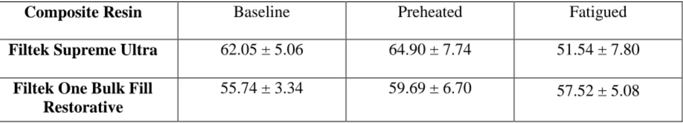
Results: Preheating and fatiguing had a significant effect on the properties of both FSU and FOBR. Fracture toughness was increased for FOBR when specimens preheated and decreased
when fatigued, FSU was not affected. Diametral tensile strength was decreased significantly after fatiguing for the FSU. FOBR had lower tensile strength for all groups when compared to FSU. Fatiguing significantly reduced flexural strength of both FSU and FOBR with latter significantly
different than FSU. Preheating had no effect on the flexural strength of both. Preheating and fatiguing significantly decreased the elastic modulus of both composite resins equally.
iv
“In the name of God, the Most Gracious, the Most Merciful”
v
ACKNOWLEDGEMENTS
I thank GOD for blessing me with health, knowledge, determination and support of the people around me to accomplish my goals.
I would like to express my profound gratitude to my mentor and supervisor Dr. Terence E. Donovan, and my thesis committee members, Drs. Ryan Cook and Taiseer Sulaiman. Your
guidance and help throughout this project has been invaluable.
I would also like to thank Mr. Brandon Rogers and Dr. Tarek Alsahafi for all of the help and support they genuinely offered in the laboratory. Without their help this project would have
been impossible. Dr. Ceib Phillips and Dr. Ali Altitinchi deserve exceptional gratitude for their extensive help with the statistical analysis of my data.
Special thanks to Drs. Lee Boushell, Harald H. Heymann, Gustavo Oliveira, Al Wilder, Patricia Miguez, Mauro Nunes, John Sturdevant, Ricardo Walter and Sumitha Ahmed. It has been an honor and a privilege to be part of the UNC Operative Dentistry Family.
I thank the staff of the Division of Operative Dentistry, Mrs. Shannon Tate, Mrs. Melissa Bennett, Mrs. Barbara Walton, Mrs. Kimberly Stoen and Mr. Jose Negron for all your devotion and commitment.
vi
I would like to thank all of my co-residents in the Graduate Operative Program for making
this an awesome journey in bitter and sweet. Drs. Eduard Epure, Mohammad Atieh, Carolina Nguyen Ngoc, Leslie Trippe, Elizabeth Griffis, Savita Gupta, Bassam Alrawi, Gustavo Mahn, Art Valeri, Zaid Badr, Karina Irusa, Islam Abd Raheem and Basheer Alsayed. You have given me
wonderful memories, thank you all!
Special thanks to my parents, Mrs. Wafaa Hameed and Dr. Abdulhaq Suliman for their
endless support, love, care, prayers and sacrifice. I would not be who I am without you two, the greatest parents of all time. Special thanks to my sister, co-resident, and best friend Dr. Sama. Sama, I love you more than anything. I thank my brothers Drs. Taiseer and Aous for being always
there and setting such a wonderful example of how successful people are. I am blessed and thankful to have such wonderful two brothers, you guys are my wings. I would also like to thank my beloved fiancée Dr. Amna Altak for the support, prayers, love and faith she has in me. Thank you all, the
vii
TABLE OF CONTENTS
LIST OF TABLES ... x
LIST OF FIGURES ... xi
LIST OF ABREVIATIONS AND SYMBOLS ... xii
CHATER 1: REVIEW OF THE LITERATURE ... 1
1. Introduction ... 1
2. Literature review ... 2
2.1. Evolution of Dental Composite Resins ... 2
2.1.1. Resin Matrix... 4
2.1.2. Filler Particles ... 4
2.1.3. Coupling Agents ... 6
2.1.4. Initiator-accelerator System ... 6
2.2. Polymerization Shrinkage Stress ... 7
2.3. Incremental placement ... 9
2.4. Bulk-Fill Composite Resins ... 9
2.4.1. Depth of Cure of Bulk-Fill Composite Resins ... 10
viii
2.4.3. High Viscosity Bulk-Fill Composite Resins ... 12
2.5. Mechanical Properties of Composite Resins ... 14
2.5.1. Flexural Strength and Elastic Modulus ... 15
2.5.2. Fracture Toughness ... 16
2.5.3. Diametral Tensile Strength Testing ... 17
2.6. Preheating of Composite Resins ... 18
2.7. Laboratory Fatigue Testing ... 20
REFERENCES ... 23
CHAPTER 2: MANUSCRIPT ... 31
1. Introduction ... 31
2. Materials And Methods... 33
2.1. Materials: ... 33
2.2. Specimens Distribution and Groups Description:... 33
2.3. Fracture toughness ... 34
2.4. Diametral Tensile Strength ... 37
2.5. Flexural Strength and Young’s Elastic Modulus: ... 39
3. Results ... 41
3.1. Fracture Toughness ... 41
3.2. Diametral Tensile Strength ... 42
ix
3.4. Young’s Elastic Modulus ... 43
4. Discussion ... 46
x
LIST OF TABLES
Table 2.1: Composite Resin Materials Used in the Study... 41
Table 2.2: Mean (MPa.m1/2) and Standard Deviation of Fracture Toughness per Group... 42
Table 2.3: Mean (MPa) and Standard Deviation of Diametral Tensile Strength per Group... 42
Table 2.4: Mean (MPa) and Standard Deviation of Flexural Strength per Group... 43
xi
LIST OF FIGURES
Figure 1.1: Classification of Composite Resins Based on Filler Particles Size………….……… 5
Figure 1.2: Potential Clinical Complication Due to Polymerization Shrinkage….……….…….. 8
Figure 1.3: Placement of Sample for Dimetral Tensile Testing……….…….. 18
Figure 1.4: Calset Composite Warmer………..……….…….. 19
Figure 1.5: Heatsync Composite Warmer……….………. 19
Figure 1.6: SD Mechatronik Chewing Simulator………..……….……….. 21
Figure 2.1: Schematic Illustration of Fracture Toughness Specimen………….…….….……… 36
Figure 2.2: Schematic Illustration of Specimen Placement for Dimetral Tensile Testing... 38
Figure 2.3: Schematic Illustration of Specimen Placement Flexural Strength Testing... 40
Figure 2.4: Mean of Fracture Toughness per Group………... 44
Figure 2.5:Mean of Diametral Tensile Strength per Group………... 44
Figure 2.6: Mean of Flexural Strength per Group………...………… 45
xii
LIST OF ABREVIATIONS AND SYMBOLS
°C Degree of celsius
10-MDP 10-methacryloyloxydecyl dihydrogen phosphate
4-META 4-methacryloxyethyl trimellitate anhydrate
ADA American Dental Association
AFM Addition fragmentation monomer
APS Average particle size
AUDMA Aromatic urethane dimethacrylate
BHT Butylated hydroxytoluene
Bis-EMA Ethoxylated bisphenol A glycol dimethacrylate
Bis-GMA Bisphenol A glycidyl methacrylate
DDDMA 1, 12-Dodecanediol dimethacrylate
DEAEMA diethyl-amino-ethyl-methacrylate
DTS Diametral tensile strength
FOBFR Filtek one bulk fill restorative
FSU Filtek supreme ultra
HEMA 2-hydroxyethyl methacrylate
KIC Fracture toughness
xiii
MPa MegaPascal
N Newton
nm Nanometer
PEGDMA Polyethylene glycol dimethacrylate
PVS Polyvinylsiloxane impression material
S.D. Standard deviation
SDR Stress decreasing resin, smart dentin replacement
SENB Single edge notch-3-point bending
SEVNB Single edge “v” notch beam
TEGDMA Triethylene glycol dimethacrylate
UDMA Urethane dimethacrylate
vol. Volume
wt. Weight
1
CHAPTER 1: REVIEW OF THE LITERATURE 1. Introduction
Composite resins have become one of the most important materials in dentistry since they were first introduced in 1957. They have a wide range of applications and are considered a
mainstream for contemporary dentistry. Composite resin restorations are technique sensitive and require very meticulous attention to detail in-order to achieve optimal results.
One of the major problems associated with composite resin restoration is polymerization shrinkage and its resultant stress on the tooth structure. To minimize the effect of polymerization these materials needs to be placed in small increments of 2 mm or less. This will ensure proper
polymerization with reduced stress. This incremental placement technique is time consuming and technique sensitive. Therefore, bulk-fill composite resin materials were introduced to address these
problems. Bulk-fill composite resins are meant to minimize polymerization shrinkage and reduce the application time of the material by eliminating the incremental placement technique. These materials can be theoretically applied in 4-6 mm thicknesses and still achieve an adequate
polymerization.
Preheating composite resin has beneficial effects on their physical properties. It enhances the flowability of the composite resin which results in better wall adaptation and reduced void
formation.
Laboratory testing of mechanical properties is a valid approach to collect some information
2
mechanical properties do not necessarily represent what happens in clinical conditions. However,
to maximize the benefit of those tests, composite resins can be fatigued by dynamic mechanical loading accompanied with thermocycling to simulate the oral environmental challenge.
Little is known about the effect of preheating and fatiguing on the mechanical properties
of composite resins, both conventional and bulk-fill. Therefore, the purpose of this study is to assess and compare the mechanical properties of conventional composite resin versus bulk-fill
composite resin taking into an account the effect of fatiguing and preheating.
2. Literature review
2.1. Evolution of Dental Composite Resins
Composite resin is defined by the Glossary of Prosthodontic Terms as a highly cross-linked polymeric material reinforced by a dispersion of amorphous silica, glass, crystalline, or organic resin filler particles and/or short fibers bonded to the matrix by a coupling agent.1 Composite resin
was invented and introduced to dentistry by Rafael Bowen in 1957, a year after Michael Buonocore introduced the concept of acid-etching enamel.2 Later Bowen created a formula of composite resin
which contained Bisphenol A glycidyl methacrylate (Bis-GMA) also known as Bowen’s resin. Bowen patented his formula which later became the major monomer used in most of the modern resin matrices.3 Bowen’s contribution did not end there. It extended to developing organic coupling
agents (Silanes) which are chemical agents that bond filler particles to the highly polymerized resin matrix, which greatly improved the physical properties of the material.4
The introduction of composite resin to dentistry was highly appreciated and welcomed by the dental community. It is considered the first durable direct esthetic restorative material. Silicate
3
esthetics. Therefore, their clinical service time was limited to 3-5 years. Poly methyl methacrylate
(PMMA) was also used as a direct restorative material. They were esthetic and highly polishable. Their use was discontinued due to their poor color stability, increased thermal expansion/contraction and polymerization shrinkage. Their polymerization shrinkage behavior led
to open margins, microleakge, post-operative sensitivity and secondary caries.5 Composite resin soon became a successful substitute of silicate cements and PMMA since they overcame the
majority of problems associated with these two restorative materials. However, composites resin still have some deficiencies that have not been resolved.
Composite resin materials have been the subject of extensive research. A simple search of “dental composite resins” electronically on the MEDLINE database search engine “www.ncbi.nlm.nih.gov/pubmed/” reveals more than twenty thousand published articles. This
extensive research led to their improvement throughout the years, and allowed them to be one of
the most popular current restorative materials.
All composite resins have the same basic composition, yet use chemically different
constituents. The three main constituents of composite resins are: 1) a highly cross-linked polymeric resin matrix which represents the continuous phase, 2) glass, mineral or resin filler
particles which are considered the dispersed phase and 3) coupling agents, bonding the filler to the resin matrix. Current composite resin materials contain an activator-initiator system. This system is required to initiate and complete polymerization of the matrix. Composite resins have pigments
to alter color and opacity, ultraviolet absorbers and other additives to improve color stability. They also contain polymerization inhibitors which are needed to prevent spontaneous polymerization,
4 2.1.1. Resin Matrix
The resin matrix is the primary component of composite resin. It acts as scaffold in which all of the other constituents exist and interact together. It is chemically active and presents initially as a fluid monomer that later converts into a rigid highly cross-linked polymer through radical
addition reaction. The resin matrix can be polymerized through chemical activation (polymerization reaction begins when two pastes are mixed together) or light activation
(polymerization reaction begins when exposed to curing light at a certain wavelength).
The main monomer used in commercial dental composites is Bis-GMA. It is derived from the reaction of bisphenol-A and glycidyl-methacrylate. It has a high molecular weight (512g/mol)
and high viscosity which necessitate dilution with other monomers of lower molecular weight to obtain a workable consistency. Triethylene glycol dimethacrylate (TEGDMA) is the most common monomer diluent used in composite resin. It has molecular weight of (286g/mol).7 The addition of
TEGDMA addition has the disadvantage of considerably increasing polymerization shrinkage.
Urethane dimethacrylate (UDMA) is another common monomer used in composite resins.
It behaves like Bis-GMA and has a high molecular weight (470g/mol) and high viscosity which also requires dilution with a low molecular weight monomer as well. Recently, the silicone-epoxy
monomer (Silorane) restorative system represents a relatively new category of composite resin. These materials are marketed commercially as low shrinkage composite resin with and increased hydrophobicity and stability.8
2.1.2. Filler Particles
Filler particles are added to the organic resin to increase its strength, improve its wear
5
different materials such as quartz, silicates, ytterbium fluoride and glasses of lithium, barium,
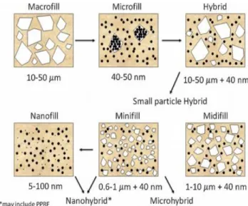
zirconium, strontium and zinc. Filler particles can be spherical or irregular in shape. They range in sizes from 0.005 to 100μm. Classification of contemporary composite resins is based on filler
particles size, or specifically, average particle size (APS) and size distribution. They can be
classified as macrofill, microfill, hybrid, nanofill, minifill and midfill (Figure 1.1). 83
Figure 1.1: Classification of Composite Resins Based on Filler Particles Size
Adding filler particles to composite resin results in significant benefits and enhanced physical properties of the material. However, there is an upper volumetric limit to how much filler
can be added. Filler load above 80% results in composite pastes that are unworkable.7 The size and percentage of filler particles control the characteristics of composite resins. Increasing filler
6 2.1.3. Coupling Agents
Regardless of the filler particle volume, they cannot contribute to improved clinical performance of composite resins unless they are properly bonded to the resin matrix. Coupling agents are chemicals responsible for bonding filler particles to the resin matrix. They contain two
functional groups at the ends of their molecular back bone, which connect the unpolymerized resin matrix and inorganic filler particles. Organosilanes, such as γ-methacryloxypropyl
trimethoxysilane, are the most commonly used coupling agents in composite resins.
2.1.4. Initiator-accelerator System
Composite resins undergo an addition reaction polymerization reaction. This reaction is
induced by free radicals which in turn can be generated either through chemical activation or activation from a light source of a specific wavelength. The majority of current composite resins are light polymerized with visible blue light within the wavelength of 420-560 nm. Accelerators
are used to control the rate at which the polymerization reaction occurs.
Chemically polymerized (Auto-cured) composite resins are dispensed in two paste
systems. One paste contains a tertiary amine activator. The other paste has a benzoyl peroxide initiator. Mixing the two pastes results in tertiary amine activator react with the benzoyl peroxide
to form free radicals that initiate an addition polymerization reaction. Mixing chemical cured composite resins is technique sensitive. Accurate amounts have to be mixed correctly without entrapping air into the mix. Air voids may compromise the integrity and strength of the
polymerized resin. Lacking control over the working time is another disadvantage of chemically cured composite resins.
7
mixing step is eliminated which prevents the problem of air incorporation. Working time can be
controlled and is basically on demand. The polymerization reaction occurs more rapidly as the paste solidifies in a matter of seconds. Current light cured composites are sensitive to visible blue light. They have a diketone photoinitiator, such as camphorquinone (CQ), and an amine accelerator
such as diethyl-amino-ethyl-methacrylate (DEAEMA) that interact together to form free radicals which facilitate the polymerization reaction.10 Only a very small amount of a photoinitiator (about
0.2% by weight or less) is required to control the polymerization reaction.11
One of the main disadvantages of conventional light polymerized composite resin is that light cannot penetrate through composite resin increments of more than 2-mm thickness.
Therefore, the material has to be placed in multiple increment to insure optimum photo-polymerization. This makes the placement procedure time consuming, technique sensitive and imposes a chance for inherent problems. Light cured composite resins may polymerize when
exposed to ambient light or during storage. Hence, inhibitors are added in a very minimal amount to prevent this undesired polymerization. Butylated hydroxytoluene (BHT) is a typical commonly
added inhibitor in current composite resins. It has strong reactivity potential with free radicals and reacts with them faster than the radicals can react with monomers. It is quite useful in controlling the rate of the polymerization reaction of light-cured composite resins.12 Thus directly affecting
polymerization shrinkage stress which is considered as one of the most complicated and significant problems with composite resins.
2.2. Polymerization Shrinkage Stress
Despite the fact that conventional composite resins have many desirable properties, they
8
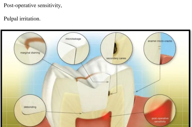
polymerization of the resin component is characterized by transforming monomers into
polymers, which is accompanied by reduction in the volume of the material and the ability of the material to flow.13 When this happens, while the material is restrained by cavity walls, the flow will be restricted and polymerization shrinkage stress will be the result. This type of stress was
first described by Bowen in 1967. 14 Many potential clinical complications (Figure 1.2) can occur as a result of polymerization shrinkage stress. This may include15-31:
Cusp deflection,
Fracture of enamel margins,
Debonding,
Micro-cracking of the shrinking composite,
Microleakage,
Post-operative sensitivity,
Pulpal irritation.
9 2.3. Incremental placement
Conventional composite resins should be placed in increments of 2 mm. This is indicated to insure adequate photo-polymerization, 35 optimal adaptation to cavity walls, 33 and to minimize polymerization shrinkage stress.34 Incremental placement maximizes the free surface in each
increment which allows for more stress relief by external flow.32 This placement technique produces predictable results but is technique sensitive and time consuming.
Placing composite resin incrementally can produce predictable results but, the main disadvantage is that this procedure is technique sensitive that requires a certain set of skills and instruments.
Bulk fill composite resins were introduced to the market, an effort to provide a material that is both efficient and offers similar qualities of conventional composite resins.
2.4. Bulk-Fill Composite Resins
To simplify and speed-up the placement of large posterior composite resin restorations, manufacturers have produced a range of materials which can be placed in single or deeper increments, known as “bulk-fill” composite resins. They have been marketed as materials that can
be placed and cured in increment depths between 4-8 mm. The placement of these larger
increments may reduce the time needed when placing posterior restorations and thereby reduce technique sensitivity. Bulk fill composite resins can reduce the working time of the restorations to approximately half of that with conventional composite.38 This reduction in working time and
technique sensitivity is a critical factor in the marketing of these materials. In order to allow bulk placement without negatively affecting marginal integrity and physical properties, these materials
10 2.4.1. Depth of Cure of Bulk-Fill Composite Resins
Bulk-fill composite resins must demonstrate adequate depth of cure to the proposed depth by the manufacturer to insure physical and mechanical properties consistent with the demands of the oral environment. Manufacturers have undertaken different modifications to increase the depth
of cure of bulk-fill materials. This may include adding photoactive monomers to the resin matrix,
36 new and more potent photoinitiator systems such as Ivocerin and increased translucency of the
materials. 37 In order to increase translucency and light scattering for deeper photopolymerization, filler particle size and shape had to be modified to have a refractive index similar to that of the resin matrix. 39 The majority of bulk-fill composite resins are more translucent for blue light than
conventional composite resins. 37 However, their increased translucency makes them less suitable for anterior restorations.
Many studies have been conducted to test the claimed depth of cure of bulk fill materials
but the results have been conflicting. Factors that might affect the results include; composite resin types, testing method and type of light curing units used. The standard method of testing depth of
cure of composite resins is according to the ISO 4049 method. A tube-shaped composite resin specimen is cured from one side; the uncured resin on the opposite side is then removed with a spatula, the depth of cure is half the length of the remaining hard specimen. 37 This technique was
found to be inappropriate for bulk fill composite resins because it overestimated curing depths. 40 Other methods have been used to assess depth of cure, such as the acetone-shake method 41
(unpolymerized resin is removed with acetone and hardness is tested on the opposite side of a composite resin cylinders). Results are varied and highly dependent on the method used. Generally
11
Bulk-fill composite resins can be categorized into two groups: low-viscosity bulk-fill
materials and high-viscosity bulk-fill materials. These materials differ greatly in both composition and use.
2.4.2. Low Viscosity Bulk-Fill Composite Resins
Low viscosity bulk-fill composite resins or “Flowable Bulk-Fill Composites” are low-viscosity materials with a reduced percentage of inorganic filler particles. They have higher
amounts of resinous components that results in a material consistency similar to flowable composite resins. They are theoretically preferred to be used in cavities in which wall adaptation is critical. 55
Due to lower filler load, these materials have high translucency, poor esthetics and low wear resistance. Therefore, their use is limited to that of a base, a concept known as “dentin replacement materials”. The majority of a large cavity can be filled with these materials, which
offer excellent wall adaptation due to ability to flow, and the top 2 mm of the cavity is capped with a conventional composite resin. This occlusal coverage by a conventional composite resin is
necessary for improved esthetics and wear resistance.
The physical and mechanical properties of low viscosity bulk-fill composite resins have
been moderately researched and compared to other types of composite resins. Despite the fact that they can be easily cured in thicker increments, their mechanical properties are significantly lower compared to other types of composite resins. 37 The elastic modulus of low viscosity bulk-fill
composite resins are either similar or lower than that of conventional composite resins. 46 Low viscosity materials are very susceptible to softening when stored in ethanol for 24 hours. This
12
those materials with a layer of conventional composite resins. Low viscosity bulk-fill composite
resins have flexural strength values that are less than other composite resins, but they fulfill the criteria of ISO 4049 that requires a flexural strength value minimum of 80 MPa. 46, 48, 49
As in any composite resin material, low viscosity bulk-fill composite resins undergo
polymerization volumetric shrinkage. The amount of this shrinkage has been studied in multiple studies and there was no large variation in their shrinkage.The mean percentage polymerization
shrinkage value ranges from 2.76 to 4.4. 45,50-52 This polymerization shrinkage is higher than with conventional composite resins which is around 2.4 percent. 45,52
Despite the fact that low viscosity bulk-fill composite resins have higher polymerization
shrinkage, the resultant stress they exhibit on the tooth is lower than that of conventional composite resins. 51-54 The mean polymerization stress values ranged from 1.07 MPa to 1.65 MPa. 51,53 Low
viscosity bulk-fill composite resins have a specific formulation to reduce the polymerization shrinkage stress, but little is known about them. SureFil SDR (Stress Decreasing Resin, Smart Dentin Replacement) flow, which was the first bulk-fill material to be introduced, contains
modified UDMA. It has a polymerization modulator that is chemically incorporated into the resin matrix.
2.4.3. High Viscosity Bulk-Fill Composite Resins
High viscosity bulk-fill composite resins have a paste like consistency and can be applied and sculpted with instruments similar to conventional composite resins. They have a high amount
of inorganic filler particles which results in physical properties, mechanical properties and wear resistance comparable to that of conventional hybrid composite resins. They are generally used in
13
composite resins. Filtek One Bulk Fill Restorative (3M ESPE; USA), for example, was introduced
recently clamming there is no need to use a capping layer as required for their flowable counterparts.
Because high viscosity bulk-fill composite resins have only been recently introduced, there
is a lack of literature that discusses their properties and clinical outcomes. However, the majority of laboratory studies agree that properties of this category of bulk-fill composite resins are similar
to those of conventional composite resins, which makes them suitable for posterior restorations.
46,50 A study found that the flexural strength of high viscosity bulk-fill composite resin, X-tra fil
(X-traF Voco Cuxhaven, Germany) was comparable with the flexural strength of conventional
composite resin. 47 Another two studies have found that high-viscosity materials also fulfil the ISO 4049 requirement for flexural strength. 41,46 Compressive strength and tensile strength values of high viscosity materials were found to be lower than those of conventional composite resins. 56
The clinical consequence of these lower strengths is not known.
Wear of high viscosity bulk-fill composite resins has not yet been evaluated. Theoretically
it should wear significantly less than low viscosity bulk-fill composite resins because of their higher filler load. However, some of these materials use larger filler particles than hybrid
composite resins materials. 37 This might result in unacceptable wear, a clinical problem that should be addressed and investigated.
Polymerization shrinkage of high viscosity bulk-fill composite resins has been assessed in
multiple studies and there was no wide variation in their findings. The mean percentage values were higher than that of conventional composite resins. 45,50-52 Polymerization shrinkage stress
14
There is a lack of clinical trials that test the clinical outcome of bulk-fill composite resins.
However, there is an abundance of in-vitro laboratory studies, the limitations of which are well known. There is an urgent need for well conducted randomized controlled clinical trials to validate the use of bulk-fill composite resins, especially since they are being used extremely in dental
practice.
2.5. Mechanical Properties of Composite Resins
Mechanical properties are the properties of the material which describe its behavior under loads. Any restorative material should have sufficient mechanical properties to function and service for a reasonable time in the oral cavity challenge.
There are key mechanical properties that are desired in composite resins. These proprties are directly related to their fracture behavior, deformation and wear resistance. These mechanical
properties are ranked according to priority 57:
1. Strength, Elastic Modulus, Fracture toughness, Fatigue, Indentation Hardness, Wear abrasion (third body) and Wear attrition (two body),
2. Toughness, Edge strength (chipping) and 3. Wear determined by toothbrush abrasion.
Studies of mechanical properties are considered to be highly relevant clinically. Premature failure of dental composite due to fracture is one of the main causes of failure. This fact supports the idea of clinical relevancy of the mechanical properties studies. 58 The mechanical properties of
15
For the purpose of this project the focus will be on Flexural Strength, Elastic Modulus,
Diamteral Tensile Strength and Fracture Toughness.
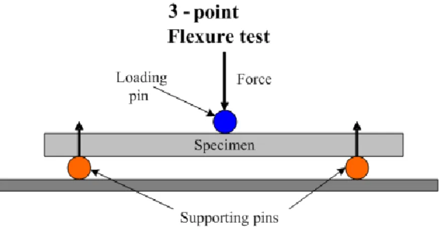
2.5.1. Flexural Strength and Elastic Modulus
Flexural strength is the mechanical property that determines the capacity of a material to
resist deformation/fracture under bending load. It is not an inherent property of the material. Elastic modulus (flexural modulus) is the mechanical property that measures the stiffness of a solid
material, high modulus indicates a rigid material.
Flexural strength and elastic modules are usually obtained from the same test. The elastic modulus is determined by applying a load to a composite resin specimen that is supported at each
end, measuring how much the specimen deflects. The value at which the specimen breaks is the flexural strength. There are multiple test methods that measures these properties 59:
1. Three-point bending test, 2. Four-point bending test and 3. Biaxial flexural strength
The most common used test is the three-point bending test. 60 The ISO standard 4049 describes sample preparation, sample dimension and the settings for the three-point bending flexural strength
test after 24 h storage in water. 61 The four-point bending test has a small difference in which the load of two points is separated by a specific distance. The benefit of this test is that stress is concentrated over a broader area of the beam, and this ensures that failure will occur within a
determined region of the beam. 57 Flexural strength values measured with three-point testing procedures are higher compared to a four-point testing procedure, but no difference in the elastic
16
Flexural strength is an important mechanical property of composite resins that may be
clinically relevant. A systematic review found direct correlation between flexural strength and clinical fracture of posterior composite resin restorations. 59 Another study found that flexural strength correlates moderately with clinical wear of composite resins. 63
2.5.2. Fracture Toughness
Fracture toughness is the mechanical property that measures the material’s ability to resist
crack propagation from a preexisting flaw. Due to the fact that almost all restorative materials contain flaws, fracture toughness is considered the most important mechanical proprties in determining resistance to clinical fracture. 67
There multiple laboratory tests that measure the fracture toughness property of composite resins 57:
1. Single Edge “V” notch beam (SEVNB), 2. Single edge notch-3-point bending (SENB), 3. Compact tension,
4. Double torsion and 5. Chevron notch
A very common method that is widely used to determine fracture toughness is the SEVNB. It is relatively easy test to perform compared to other test modalities, and can be used for ceramics and composites. The ISO standard 6872 describes sample preparation, sample dimension and
correct placement of the notch.68 The notch can be sharpened to produce tip radii ranging from 10-20 microns using razor blade.69 The most difficult step in fracture toughness testing is the creation
17
The clinical relevancy of fracture toughness has been established to some extent. One study
found that there is a good correlation between fracture toughness fracture failures with class IV composite resin restorations. 64 A clinical trial of composite resins in denture teeth, found that there was a correlation between marginal breakdown and fracture toughness. 65 Fracture toughness has
also been suggested an indicator of clinical wear. 66
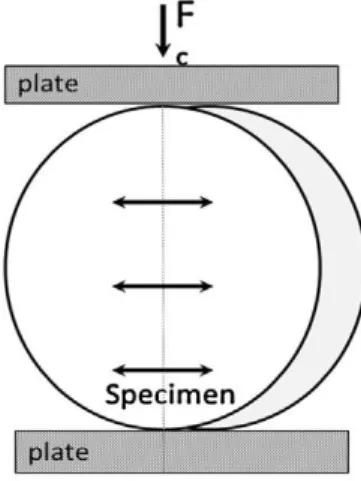
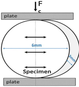
2.5.3. Diametral Tensile Strength Testing
Diametral tensile strength testing is a method of indirectly measuring the tensile strength of a brittle material with little or no plastic deformation.72 It was introduced to overcome the difficulties in performing a good uniaxial tensile strength test when the tested material is brittle,
which is applicable to composite resins. 70
This test involves the preparation of cylindrical specimens, ADA specification #27
describes how the specimens are prepared, stored and loaded. 71 The specimen is placed between two plates of a testing machine (Figure 1.3) and loaded with a compressive force in the diamteral plane.72 This technique insures that failure happens more due to tensile forces compared to not
compressive forces. This occurs only when the specimen is broken uniformly in the middle.47
The importance of this test lies in it is potential clinical relevance. The majority of clinical
18
Figure 1.3: Placement of Sample For Dimetral Tensile Testing
2.6. Preheating of Composite Resins
Preheating composite resin (warming) is a chairside method to increase flowability and reduce viscosity by changing the degree of conversion. 78 Warming composite resins to 37ºC or
58ºC may facilitate composite extrusion, improve adaptation to preparation walls and enhance contouring. 80
Preheating composite resins enhances maximal polymerization by insuring a highly cross-linked polymer network. This results from increased radical and monomer mobility. 79 This increase in polymerization may enhance mechanical and physical priorities of the material. One
study showed that preheating composite resins up to 60°C significantly increased surface hardness.
81 Another study found that preheating composite resins prior to polymerization reduced shrinkage
forces and improved the degree of conversion. 82
Preheating composite resins can be done by using composite warming devices. There are different designs and brands available. One of the oldest composite warmers available is Calset
19
literature.84 After reaching this temperature, it takes three minutes to warm the composite resin
material to working consistency. The device has interchangeable trays to meet the clinician's preference of composite dispensing method (Figure 1.4).
Figure 1.4: Calset Composite Warmer
The HeatSync composite warmer (Bioclear, Seattle, USA) is another example of a composite
warming device. Its design facilitates heating of two composite guns, two syringes and up to 6 extra
composite compules (Figure 1.5). It requires 15 minutes to warm up and reach 68°C. Other methods have been described to preheat composite resins up. Hand-holding the composite material for a few
minutes or using the light of dental unit chair may increase their temperature but not to a point required
to achieve the desired results.85
20 2.7. Laboratory Fatigue Testing
Fatigue can be defined as the weakening of a material caused by repeatedly applied cyclic loads. It is usually progressive with localized structural damage. Composite resins, as all other dental material, are challenged intraorally with thermal changes and cyclic loads not exceeding
fracture strength while functioning. Therefore, fatigue is one of the most important properties for composite resins. 73 Composite resin failure occurs mainly due to the propagation of pre-existing
cracks and flaws rather than catastrophic fracture, hence the importance of fatiguing.
Laboratory studies should assess the degradation of composite resins resulting from fatiguing in order to achieve clinical significance, thus acting as a predictor of clinical
performance. 74 Fatigue testing is very difficult to design and execute. It is expensive and requires a considerable resources. Therefore, it is not commonly used to assess strength and fracture resistance of composite resins.
There are two main methods that are used for composite fatigue testing. The first is called fatigue resistance in which a dumbbell shaped sample is cyclically loaded in tension at a certain
frequency until failure occurs. The second is called the staircase fatigue method in which a sample that survives a certain cyclic loading is replaced by another sample that is subjected to different
loading conditions.57
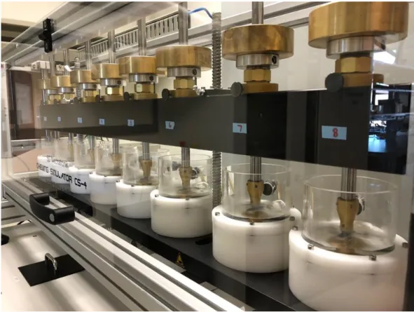
Chewing simulation is a relatively new laboratory testing technique in which restorative dental materials can be fatigued by emulating the intra oral environment. This works well as an
in-vitro predicator test prior to in-vivo studies. Chewing simulators (or emulators) are complex mechanical machines that perform a wide range of movements according to preset criteria,
21
Three of them have the capacity to move in both horizontal and vertical axes:
1. Willytec chewing simulator 75
2. SD Mechatronik chewing simulator (successor of Willytec) (Figure 1.5) 76
3. Bose ElectroForce 3330Dental Wear Simulator (No published work was found to the
author knowledge)
The chewing simulator CS-4 (SD Mechatronik, Germany) is considered to be predictable,
cost efficient and an adequate tool to test fatigue resistance of different restorative materials. 77 This machine offers control over test parameters, is computer controlled, can be optionally combined with thermocycling and may be used for fatigue testing and wear studies.57
22
In summary, composite resins in either their conventional or bulk-fill form are widely used
in current practices. A considerable amount of in-vitro research has been published with the purpose of evaluating their properties and potential for successfully clinical use. However, the effect of preheating and fatiguing on their mechanical properties is not yet well researched and
23
REFERENCES
1. The Glossary of Prosthodontic Terms: Ninth Edition. The Journal of prosthetic dentistry. May 2017; 117(5):e1-e105.
2. Bowen RL. Use of epoxy resins in restorative materials. J Dent Res. Jun 1956;35(3):360-369.
3. Bowen RL, Inventor. Dental filling material comprising vinyl silane, treated fused silica and a binder consisting of the reaction product of bisphenol and glycidyl acrylate. US patent 3066.112, 1962.
4. Bowen RL. Properties of a silica-reinforced polymer for dental restorations. J Am Dent Assoc. Jan 1963;66:57-64.
5. Anusavice KJ, Phillips RW, Shen C, Rawls HR. Phillips' science of dental materials. 12th ed. St. Louis, Mo.: Elsevier/Saunders; 2013.
6. V. G. Ólafsson. Effect of composite type and placement technique on polymerization shrinkage stress. Unpublished master thesis, University of North Carolina. 2015, p.18.
7. Peutzfeldt A. Resin composites in dentistry: the monomer systems. European journal of oral sciences. Apr 1997;105(2):97-116.
8. Weinmann W, Thalacker C, Guggenberger R. Siloranes in dental composites. Dent Mater. 2005;21:68–74.
9. Van Noort R. Controversial aspects of composite resin restorative materials. Br Dent J 1983;155 (11): 380-5.
10. Klugh, D.O. Principles of Equine Dentistry. 2010: chapter 15: pp. 165-166. UK. Manson publishing Ltd.
11. Anusavice KJ, Phillips RW, Shen C, Rawls HR. Phillips' science of dental materials. 12th ed. St. Louis, Mo.: Elsevier/Saunders; 2013.
24
13. Odian G. Principles of polymerization. 3rd edition. New York: Wiley-Interscience; 1991.
14. Bowen RL. Adhesive bonding of various materials to hard tooth tissues. VI. Forces developing in direct-filling materials during hardening. J Am Dent Assoc. Feb 1967;74(3):439-445.
15. Suliman AA, Boyer DB, Lakes RS. Cusp movement in premolars resulting from composite polymerization shrinkage. Dental materials: official publication of the Academy of Dental Materials. Jan 1993;9(1):6-10.
16. Suliman AH, Boyer DB, Lakes RS. Polymerization shrinkage of composite resins: comparison with tooth deformation. The Journal of prosthetic dentistry. Jan 1994;71(1):7-12.
17. Lee SY, Park SH. Correlation between the amount of linear polymerization shrinkage and cuspal deflection. Operative dentistry. May-Jun 2006;31(3):364-370.
18. Bicalho AA, Pereira RD, Zanatta RF, et al. Incremental filling technique and composite material--part I: cuspal deformation, bond strength, and physical properties. Operative dentistry. Mar-Apr 2014;39(2):E71-82.
19. Dietschi D, Herzfeld D. In vitro evaluation of marginal and internal adaptation of class II resin composite restorations after thermal and occlusal stressing. European journal of oral sciences. Dec 1998;106(6):1033-1042.
20. Jorgensen KD, Asmussen E, Shimokobe H. Enamel damages caused by contracting restorative resins. Scandinavian journal of dental research. Mar 1975;83(2):120-122.
21. Kanca J, 3rd, Suh BI. Pulse activation: reducing resin-based composite contraction stresses at the enamel cavosurface margins. American journal of dentistry. Jun 1999;12(3):107-112.
25
23. Furness A, Tadros MY, Looney SW, Rueggeberg FA. Effect of bulk/incremental fill on internal gap formation of bulk-fill composites. Journal of dentistry. Apr 2014;42(4):439-449.
24. Cho NY, Ferracane JL, Lee IB. Acoustic emission analysis of tooth-composite interfacial debonding. J Dent Res. Jan 2013;92(1):76-81.
25. Lai JH, Johnson AE. Measuring polymerization shrinkage of photo-activated restorative materials by a water-filled dilatometer. Dental materials: official publication of the Academy of Dental Materials. Mar 1993;9(2):139-143.
26. Ferracane JL, Mitchem JC. Relationship between composite contraction stress and leakage in Class V cavities. American journal of dentistry. Aug 2003;16(4):239-243.
27. Pashley DH. Clinical considerations of microleakage. Journal of endodontics. Feb 1990;16(2):70-77.
28. Opdam NJ, Feilzer AJ, Roeters JJ, Smale I. Class I occlusal composite resin restorations: in vivo post-operative sensitivity, wall adaptation, and microleakage. American journal of dentistry. Oct 1998;11(5):229-234.
29. Opdam NJ, Roeters FJ, Feilzer AJ, Verdonschot EH. Marginal integrity and postoperative sensitivity in Class 2 resin composite restorations in vivo. Journal of dentistry. Sep 1998;26(7):555-562.
30. Eick JD, Welch FH. Polymerization shrinkage of posterior composite resins and its possible influence on postoperative sensitivity. Quintessence Int. Feb 1986;17(2):103-111.
31. Brannstrom M, Vojinovic O. Response of the dental pulp to invasion of bacteria around three filling materials. ASDC journal of dentistry for children. Mar-Apr 1976;43(2):83-89.
26
33. Opdam NJ, Feilzer AJ, Roeters JJ, Smale I. Class I occlusal composite resin restorations: in vivo post-operative sensitivity, wall adaptation, and microleakage. American journal of dentistry. Oct 1998;11(5):229-234.
34. Park J, Chang J, Ferracane J, Lee IB. How should composite be layered to reduce shrinkage stress: incremental or bulk filling? Dental materials: official publication of the Academy of Dental Materials. Nov 2008;24(11):1501-1505.
35. Heymann HO, Ritter, A.V., Roberson, T.M. Introduction to Composite Restorations. In: Heymann H, Swift EJ, Ritter AV, Sturdevant CM, eds. Sturdevant's art and science of operative dentistry. 6th ed. St. Louis, Mo.: Elsevier/Mosby; 2013: xv, 548 p.
36. Ilie N, Hickel R. Investigations on a methacrylate-based flowable composite based on the SDR technology. Dental materials: official publication of the Academy of Dental Materials. Apr 2011;27(4):348-355.
37. Bucuta S, Ilie N. Light transmittance and micro-mechanical properties of bulk fill vs. conventional resin based composites. Clinical oral investigations. Nov 2014;18(8):1991-2000.
38. Flury, S., Peutzfeldt, A. & Lussi, A. Influence of increment thickness on microhardness and dentin bond strength of bulk fill resin composites. Dental Materials, 2014; 30: 1104-1112.
39. Nagi SM, Moharam LM, and Zaazou MH: Effect of resin thickness, and curing time on the micro-hardness of bulk-fill resin composite. J Clin Exp Dent 2015; 7:e600-4.
40. Flury S, Hayoz S, Peutzfeldt A, Husler J, Lussi A. Depth of cure of resin composites: is the ISO 4049 method suitable for bulk fill materials? Dental materials: official publication of the Academy of Dental Materials. May 2012;28(5):521-528.
41. Goracci C, Cadenaro M, Fontanive L, et al. Polymerization efficiency and flexural strength of low-stress restorative composites. Dental materials: official publication of the Academy of Dental Materials. Jun 2014;30(6):688-694.
27
43. Nagi SM, Moharam LM, and Zaazou MH: Effect of resin thickness, and curing time on the micro-hardness of bulk-fill resin composite. J Clin Exp Dent 2015; 7:e600-4.
44. Kelic K, Matic S, Marovic D, Klaric E and Tarle Z: Microhardness of bulk-fill composite materials. Acta Clin Croat 2016; 55:607-614.
45. Benetti AR, Havndrup-Pedersen C, Honore D, Pedersen MK & Pallesen: Bulk-fill resin composites: Polymerization contraction, depth of cure, & gap formation. Oper Dent 2015; 40: 190-200.
46. Tiba A, Zeller GG, Estrich C, Hong A. A laboratory evaluation of bulk-fill versus traditional multi-increment fill resin-based composites. ADA Prof Prod Rev. 2013;8:1326.
47. Leprince JG, Palin WM, Vanacker J, Sabbagh J, Devaux J, Leloup G. Physico-mechanical characteristics of commercially available bulk-fill composites. Journal of dentistry. Aug 2014;42(8):993-1000.
48. Czasch P, Ilie N. In vitro comparison of mechanical properties and degree of cure of bulk fill composites. Clinical oral investigations. Jan 2013;17(1):227-235.
49. Didem A., et al. Comparative Mechanical Properties of Bulk-Fill Resins. Open Journal of Composite Materials. Apr 2014;4, 117-121
50. Garcia, D., Yaman, P., Dennison, J. & Neiva, G. Polymerization shrinkage and depth of cure of bulk fill flowable composite resins. Operative dentistry, 2014; 39: 441-448.
51. Zorzin, J., Maier, E., Harre, S., Fey, T., Belli, R., Lohbauer, U., Petschelt, A. & TA schner, M. Bulk-fill resin composites: polymerization properties and extended light curing. Dental Materials, 2015; 31: 293-301.
28
53. El-damanhoury, H. & Platt, J. Polymerization shrinkage stress kinetics and related properties of bulk-fill resin composites. Operative dentistry, 2014; 39: 374-382.
54. Guo, Y., Landis, F. A., Wang, Z., Bai, D., Jiang, L. & Chiang, M. Y. Polymerization stress evolution of a bulk-fill flowable composite under different compliances. Dental Materials, 2016; 32: 578-586.
55. Van Ende A. Bulk-fill composites: A review of the current literature. The journal of adhesive dentistry. 2017;19(2):95-109.
56. Rosatto, C., Bicalho, A., Veríssimo, C., Bragança, G., Rodrigues, M., Tantbirojn, D., Versluis, A. & Soares, C. Mechanical properties, shrinkage stress, cuspal strain and fracture resistance of molars restored with bulk-fill composites and incremental filling technique. Journal of dentistry, 2015; 43: 1519-1528.
57. Ilie N, et al. Academy of Dental Materials guidance—Resin composites: Part I— Mechanical properties. Dent Mater Aug 2017;33(8):880-894.
58. Beck F, Lettner S, Graf A, Bitriol B, Dumitrescu N, Bauer P,et al. Survival of direct resin restorations in posterior teeth within a 19-year period (1996–2015): a meta-analysis of prospective studies. Dent Mater 2015;31:958–85.
59. Heintze SD, Ilie N, Hickel R, Reis A, Loguercio A, Rousson V. Laboratory mechanical parameters of composite resins and their relation to fractures and wear in clinical trials—a systematic review. Dent Mater 2017;33:e101–14.
60. Bona AD, Bello YD, Sartoretto SC. Use of standards in papers published in dental journals. Braz Dent J 2012;23:471–6.
61. ISO, 2009. Dentistry—polymer-based filling, restorative and luting materials. International Standard; No. 4049:1–27.
29
63. Ferracane JL, Condon JR, Mitchem JC. Correlating abrasive wear to mechanical properties of experimental dental composites. Transactions of the Academy of Dental Materials. 1997:A–21.
64. Tyas MJ. Correlation between fracture properties and clinical performance of composite resins in class IV cavities. Aust Dent J 1990;35:46–9.
65. Ferracane JL, Mitchem JC, Condon JR, Todd R. Wear and marginal breakdown of composites with various degrees of cure. J Dent Res. 1997; 76:1508–1516.
66. Peutzfeldt A, Asmussen E. Modulus of resilience as predictor for clinical wear of restorative resins. Dent Mater. 1992; 8:146–8.
67. Ferracane JL. Resin-based composite performance: are there some things we can’t predict? Dent Mater 2013;29:51–8.
68. International Organization for Standardization. ISO 6872:2015. Dentistry-Ceramic materials. Geneva: International Organization for Standardization; 2015. Available at: https://www.iso.org/standard/59936.html.
69. Scherrer SS, Denry IL, Wiskott HWA. Comparison of three fracture toughness testing techniques using a dental glass and a dental ceramic. Dent Mater 1998;14:246–55.
70. Penn RW, Craig RG, Tesk JA. Diametral tensile-strength and dental composites. Dent Mater 1987;3:46–8.
71. ADA specification no. 27—resin-based filling materials;1993.
72. Della Bona A. Flexural and diametral tensile strength of composite resins. Brazilian oral research. 01/2008;22(1):84-89.
73. Lohbauer U, Belli R, Ferracane JL. Factors involved in mechanical fatigue degradation of dental resin composites. J Dent Res 2013;92:584–91.
30
75. Heintze SD. How to qualify and validate wear simulation devices and methods. Dent Mater 2006;22:712–34.
76. DeLong R, Douglas WH. Development of an artificial oral environment for the testing of dental restoratives: bi-axial force and movement control. J Dent Res 1983;62:32–6.
77. Heintze SD. Using a chewing simulator for fatigue testing of metal ceramic crowns. Journal of the mechanical behavior of biomedical materials. 2017;65:770-780. doi: 10.1016/j.jmbbm.2016.09.002.
78. Yang JNC, Raj JD, Sherlin H. Effects of Preheated Composite on Micro leakage-An in-vitro Study. Journal of Clinical and Diagnostic Research : JCDR. 2016;10(6):ZC36-ZC38. doi:10.7860/JCDR/2016/18084.7980.
79. Daronch M, Rueggeberg F, De Goes M, Giudici R. Polymerization kinetics of pre-heated composite. JDR. 2006;85(1):38–43.
80. El-Deeb HA, Abd El-Aziz S, Mobarak EH. Effect of preheating of low shrinking resin composite on intrapulpal temperature and microtensile bond strength to dentin. J Adv Res. 2015; 6:471-8.
81. Muñoz CA, Bond PR, Sy-Muñoz J, Tan D, Peterson J. Effect of pre-heating on depth of cure and surface hardness of light-polymerized resin composites. Am J Dent. 2008;21(4):215–22.
82. T.T. Tauböck, Z. Tarle, D. Marovic, T. Attin. Pre-heating of high-viscosity bulk-fill resin composites: effects on shrinkage force and monomer conversion. J. Dent., 2015 43(11), 1358-1364.
83. Ferracane, J. L. Resin composite - State of the art. Dental Materials, 2011 27(1), 29-38.
84. L. J. Rickman, P. Padipatvuthikul, B. Chee. Clinical applications of preheated hybrid resin composite. Brit Dent J, 2011, 211(2):63–67.
31
CHATER 2: MANUSCRIPT 1. Introduction
Composite resin was introduced to the dental world in late 1950s by Rafael Bowen in
1957.1 Composite resin materials have changed the way dentistry is practiced and have become one of the most important dental materials. Approximately 261 million composite resin
restorations are placed around the world each year.2
Conventional composite resins have shortcomings and limitations. Polymerization
shrinkage and its resultant shrinkage stress on cavity walls is one of the major shortcomings of conventional composite resin.3 The resultant polymerization shrinkage stress manifests clinically with several clinical complications. It can cause cusp deflection, micro-cracking and fracture of
enamel margins, microleakage, debonding, post-operative sensitivity and pulpal irritation.4-13
To reduce shrinkage stresses, an incremental technique for composite placement was
introduced. This technique has insured proper light polymerization of the resin composite and also reduced the polymerization shrinkage stress.14-15 However, incremental placement of conventional composite resin is a technique sensitive procedure that requires clinical skills and special
instruments. It is also time consuming. Therefore, less technique sensitive and more efficient approaches for composite resin placement have been developed.
32
layer of 4-8 mm thickness. Several changes in the chemistry of monomers and particle size and
shape were required to allow those materials to be used in bulk. Bulk-fill composite resins can be classified into low-viscosity and high-viscosity materials. Low-viscosity bulk-fills are meant to serve as dentin replacement and therefore need to be capped with conventional materials due to
their poor physical and mechanical properties.16 High-viscosity bulk-fill materials on the other hand have good physical and mechanical properties that allow them to restore an entire cavity
without the need for a capping layer.17-18
Testing the mechanical properties of composite resin materials in-vitro is a common method for determining their properties. The clinical relevancy of those tests is established to
some extent.19 Mechanical properties testing can help to identify materials with a high likelihood of premature failure due to fracture and their wear characteristics.
Fatiguing of composite resins prior to mechanical testing by cyclic loading and thermocycling is highly recommended in-order to increase the clinical relevance of the results.20 Chewing simulation is one of the methods that can be used to fatigue specimens at a reasonable
cost and time.
There are multiple protocols and techniques that dentists use when they place composite
resins in their practices. Preheating composite resin is a relatively common technique that is thought to increase flowability and reduce film thickness.21 Preheating compositre resin may maximize polymerization, reduce shrinkage forces and increase surface hardness.22-24 However,
their effect on mechanical properties, wear and clinical performance is yet to be investigated.
The purpose of the present study was to test the effect of fatiguing and preheating on the
33 conventional counterpart.
The null hypotheses were that:
1. Fatiguing and preheating yield no significant effect on the mechanical properties of the tested materials.
2. There are no significant differences in the mechanical properties between high-viscosity bulk-fill composite resin and conventional composite resin.
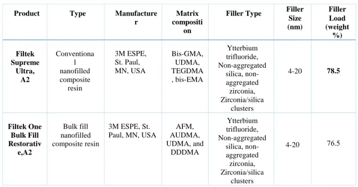
2. Materials and Methods 2.1. Materials:
Two composite resin types were used in this study (Table 1.1):
1. Filtek One Bulk Fill Restorative (FOBFR), 3M ESPE, USA 2. Filtek Supreme Ultra (FSU), 3M ESPE, USA
2.2. Specimens Distribution and Groups Description:
Specimens (N=180) were prepared and distributed into six groups (n=10) for each test
performed:
Group 1: FOBR Baseline
Group 2: FSU Baseline
Group 3: FOBR Preheated
Group 4: FSU Preheated
Group 5: FOBR Fatigued
Group 6: FSU Fatigued
34
to a temperature of (68 ± 1ºC) for 10 minutes before polymerization by utilizing a composite
warmer (HeatSync, Bioclear, Seattle, USA).
Specimens of groups 5 and 6 were subjected to fatiguing. This was achieved by cyclic loading the specimens with 50 N and 0.5 mm indentation with steatite indenter using chewing
simulator CS-4 (SD Mechatronik, Germany) for 600000 cycles at 1.4 Hz, which represent approximately 2.5 years of clinical performance. Thermocycling was simultaneously performed
using distilled water at 5ºC and 55ºC with a 30 second dwell time. Specimens were inspected for premature failure every 24 hours. Surviving specimens were tested for fracture toughness, diametral tensile strength, flexural strength and elastic modulus.
Detailed description of specimen’s preparation and testing for each test is discussed in the
following section.
Three mechanical tests were performed to determine the mechanical properties of the groups described above.
2.3. Fracture toughness
The single edge V-notched beam (SEVNB) method (ISO standard 6872) was used to measure the fracture toughness of FOBFR and FSU composite resins. Sixty beam-shaped specimens (21.0 ± 0.1mm) in length, a rectangular cross section of (4.0 ± 0.1mm) in depth and
(3.0 ± 0.1mm) in thickness were prepared for each of the composite resin materials (Figure 2.1 A). Polyvinylsiloxane impression material (PVS) was used to create a mold specific to the specimen
dimensions for easy removal after polymerization. Composite resin material was carefully injected into the mold to prevent the entrapment of air bubbles. A transparent ethylene film and glass slide
35
atmosphere during polymerization. Slight pressure was applied to the glass slide to extrude excess
material. Each specimen was carefully photo-polymerized according to the manufacturer’s recommended time of exposure using a visible light curing unit (Elipar DeepCure-S, 3M ESPE, USA) with an utilizable wavelength range 420-490 nm and mean light irradiance of 1198
mW/cm2. The wavelength and irradiance of the curing unit were calibrated and confirmed using the MARC Light Collector (BlueLight Analytics, Canada). After complete polymerization of the
specimens, they were carefully removed from the mold and examined. Specimens with obvious voids were excluded from testing. A no. 15 blade was used to remove excess composite resin from the edges of the specimen and then 600-grit silicon-carbide abrasive paper (MicroCutTM, Buehler,
Lake Bluff, Illinois, USA) was used for final smoothening. Specimens were then immersed in deionized water at 37°C for 24 hours. After removing from water, a notch depth of approximately
0.5 mm was cut into the bar specimen using a 150 um thick diamond blade. A razor blade coated with diamond polishing paste (3.5 um, Kent Supplies, USA) was positioned in the starter notch and a light force (5 N to 10 N) was applied using a gentle back and forth while maintaining a
constant horizontal motion. The depths of the V-shaped notches were measured using a calibrated microscope with magnification > 50x to three significant figures. Both sides of the specimen were
36
The width (b) and thickness (w) of each specimen was recorded prior to testing using a
digital micrometer capable of measurements to ±1µm accuracy (QuantuMike Micrometer, Mitutoyo Corporation, Japan). Testing for fracture toughness was carried out using the 4-point bending fixture (Figure 2.1 B). The 3 mm width face with the V-notch was placed down on the
testing fixture (tensile side). Specimens were loaded on an Instron Universal Testing Machine (Instron 4411, SINTECH, MTS system corporation, USA) with a crosshead speed of 0.5 mm/min
till fracture.
Figure 2.1: A, Schematic Illustration of Fracture Toughness Specimen. B, Specimen Placed on 4-Point Bending Fixture
The peak fracture load was recorded to three significant figures and the fracture toughness
37 KIC = P / bh ½ * L / h * 3a ½ / 2 (1-a)3/2 * Y.
Y=1.9887-(1.326*a)-((3.49-(0.68*a)+(1.35*(a^2)))*(a)*(1-a))/((1+a)^2)
α= average V-notch depth of group
P= fracture load
b= width of specimen
w= thickness of specimen
L = distance between support beams
2.4. Diametral Tensile Strength
The diametral tensile strength (DTS) of FOBFR and FSU was determined under specification (No. 27 of ANSI/ADA, 1993). 1 Sixty cylindrical shaped specimens (6.0 ± 0.1 mm in diameter and 3 ± 0.1 mm in height) were prepared for each composite resin material. PVS impression material was used to create a mold specific to specimen dimensions for easy removal after polymerization.
Composite was carefully injected into the mold to prevent the entrapment of air bubbles. A transparent ethylene film and glass slide were placed over the mold to confine the material and
minimize exposure to oxygen from the atmosphere during polymerization. Slight pressure was applied to the glass slide to extrude excess material. Specimens were photo-polymerized as described previously in the fracture toughness section, according to the manufacturer’s instructions
and recommended time. After complete polymerization of the specimens, they were carefully removed from the mold and examined. Specimens with obvious voids were excluded from testing.
38
for final smoothening. The diameter (d) of each specimen was calculated by taking the mean of
two measurements at right angles to each other, using a digital micrometer (Digimatic Micrometer, Mitutoyo Corporation, Japan) with an accuracy of 0.01 mm. Specimens were immersed in water at 37°C for 24 hours prior to testing.
DTS for each specimen was calculated using an Instron Universal Testing Machine (Instron 4411, SINTECH, MTS system corporation, USA). The cylindrical shaped specimens
were positioned on their side between two compression plate fixtures (Figure 2.2). Specimens were loaded at a crosshead speed of 0.5mm/min until fracture. For groups 5 and 6, specimens were fatigued as described previously prior to loading.
Figure 2.2: Schematic Illustration of Specimen Placement for Dimetral Tensile Testing
The peak load was recorded and the DTS was determined according to the formula:
DTS = 2 F/ πdt
39 d= diameter of specimen
t= thickness of specimen
2.5. Flexural Strength and Young’s Elastic Modulus:
The flexural strength of FOBFR and FSU was determined by a 3-point bending test
according to (ISO standard 4049). Sixty specimens (2.0 ± 0.1mm in thickness, 2.0 ± 0.1mm in width and 25.0 ± 0.1mm in length) were prepared for each composite resin material. PVS
impression material was used to create a mold specific to specimen dimensions for easy removal after polymerization. Composite was carefully injected into the mold to prevent the entrapment of air bubbles. A transparent ethylene film and glass slide were placed over the mold to confine the
material and minimize exposure to oxygen from the atmosphere during polymerization. Slight pressure was applied to the glass slide to extrude excess material. Specimens were
photo-polymerized as described previously, according to the manufacturer’s instructions and recommended time. After complete polymerization of the specimens, they were carefully removed from the mold and examined. Specimens with obvious voids were excluded from testing. A no. 15
blade was used to remove excess composite resin from the edges of the specimen and then 600-grit silicon-carbide abrasive paper (MicroCutTM, Buehler, Lake Bluff, Illinois) was used for final
smoothening. Specimens were then immersed in water at 37°C for 24 hours prior to testing. The width (b) and thickness (w) of each specimen was recorded using a digital micrometer capable of 0.01mm accuracy (QuantuMike Micrometer, Mitutoyo Corporation, Japan).
Testing was performed using the 3-point bending fixture (Figure 2.3). The specimens were loaded on Instron Universal Testing Machine (Instron 4411, SINTECH, MTS system corporation,