doi:10.1093/nar/gky061
The hepatitis C viral nonstructural protein 5A
stabilizes growth-regulatory human transcripts
Liang Guo
1,2,3, Suresh D. Sharma
4, Jose D. Debes
1, Daniel Beisang
1,5,
Bernd Rattenbacher
5, Irina Vlasova-St. Louis
1, Darin L. Wiesner
5, Craig E. Cameron
4,*and
Paul R. Bohjanen
1,2,3,5,*1Department of Medicine, Division of Infectious Diseases and International Medicine, Program in Infection and
Immunity, University of Minnesota, Minneapolis, MN 55455, USA,2Institute for Molecular Virology Training Program, University of Minnesota, Minneapolis, MN 55455, USA,3Graduate Program in Comparative and Molecular
Bioscience, University of Minnesota, Minneapolis, MN 55455, USA,4Department of Biochemistry & Molecular Biology, The Pennsylvania State University 201 Althouse Laboratory, University Park, PA 16802, USA and 5Department of Microbiology and Immunology, University of Minnesota, Minneapolis, MN 55455, USA
Received August 25, 2017; Revised January 17, 2018; Editorial Decision January 19, 2018; Accepted January 22, 2018
ABSTRACT
Numerous mammalian proto-oncogene and other growth-regulatory transcripts are upregulated in ma-lignancy due to abnormal mRNA stabilization. In hep-atoma cells expressing a hepatitis C virus (HCV) subgenomic replicon, we found that the viral non-structural protein 5A (NS5A), a protein known to bind to viral RNA, also bound specifically to human cellu-lar transcripts that encode regulators of cell growth and apoptosis, and this binding correlated with tran-script stabilization. An important subset of human NS5A-target transcripts contained GU-rich elements, sequences known to destabilize mRNA. We found that NS5A bound to GU-rich elementsin vitroand in cells. Mutation of the NS5A zinc finger abrogated its GU-rich element-binding and mRNA stabilizing activ-ities. Overall, we identified a molecular mechanism whereby HCV manipulates host gene expression by stabilizing host transcripts in a manner that would promote growth and prevent death of virus-infected cells, allowing the virus to establish chronic infec-tion and lead to the development of hepatocellular carcinoma.
INTRODUCTION
For viruses to survive and replicate, they often control the host cellular environment by manipulating host gene ex-pression. It is becoming increasingly clear that viral manip-ulation of host posttranscriptional regulatory mechanisms
plays critical roles in viral pathogenesis. For example, Ka-posi’s sarcoma herpes virus globally down-regulates expres-sion of host cellular transcripts by expressing the shut-off al-kaline exonuclease (SOX), which mediates transcript degra-dation (1). Herpes simplex virus selectively degrades cer-tain host transcripts through a viral endonuclease (2) but stabilizes and up-regulates a specific subset of host cellular transcripts through the viral ICP27 protein (3). By utilizing posttranscriptional mechanisms, viruses are able to selec-tively manipulate the expression of host transcripts to create a cellular environment that inhibits antiviral host defense mechanisms and allows the establishment of viral infection. Hepatitis C virus (HCV), which belongs to the family Fla-viviridae family of viruses (4), is a RNA virus with a 9.6 kb RNA genome of positive polarity that encodes struc-tural and nonstrucstruc-tural proteins required for HCV repli-cation (5). HCV infects approximately 184 million people worldwide (6) and causes hepatitis, liver cirrhosis and hep-atocellular carcinoma (7). The mechanisms leading to liver cirrhosis and cancer in the HCV-infected individual are not understood. In the current era, cure of HCV infection with new antivirals is possible, but even patients with virological cure have an increased risk of developing hepatocellular car-cinoma (8,9). Therefore, understanding the molecular pro-gression of HCV infection to hepatocellular carcinoma is a priority.
Due to its small genome, HCV must use the host protein synthesis machinery to produce viral proteins required for viral replication. One of these viral proteins, non-structural protein 5A (NS5A), is multifunctional and influences many viral and cellular processes. The entire proteprotein in-teraction network of the HCV proteome was mapped by
*To whom correspondence should be addressed. Email: [email protected]
Correspondence may also be addressed to Craig E. Cameron. Email: [email protected]
C
Present address: Bernd Rattenbacher, Institute of Medical Engineering/ Space Biology Group, Lucerne School of Engineering and Architecture, Lucerne University
of Applied Sciences and Arts, Seestrasse 41, CH 6052 Hergiswil, Switzerland.
The Author(s) 2018.
This is an Open Access article distributed under the terms of the Creative Commons Attribution Non-Commercial License
high throughput yeast two hybrid screening, and NS5A was found to interact with proteins functioning in focal adhesions,gap-junctions andhostcellularsignaling path-ways (10). NS5A alsoinfluencesc ellc yclec ontrolb y in-teractingwithp53andmodulationofp21(11–14).During viral genome replication,NS5Ainteracts with the RNA-dependentRNA-polymeraseNS5B(15,16),aninteraction thatisessentialtomaintaintheHCVsubgenomicreplicon inHuh7cells(15,17).NS5AalsobindstoG-andU-rich sequencesinHCVgenomicRNA(18).Theaminoterminal domain1 andthe adjacentunstructuredregion ofNS5A interactswithHCVgenomicRNA(16)andmodulatesits templateselection(19).Crystalstructuresofdomain1show a RNA-bindingzinc fingerw hichc oordinatesa Z n2+ion
(20,21).WehypothesizedthatNS5Afunctionstoregulate the expression of host genes at posttranscriptional levels throughitsabilitytobindtoGandUrichRNAsequences. WepreviouslydescribedaGU-richelement(GRE)that isenrichedinthe3untranslatedregion(UTR)ofrapidly degraded cellulartranscripts expressedinprimaryhuman T cells (22). GRE-containing transcripts encode numer-ous proto-oncogene proteins and other proteins involved incellgrowthregulationorapoptosis(23).Insertionofthe GREsfromJUN,JUNBorTNFRSF1BmRNAsintothe 3 UTRofa beta-globinreportertranscriptconferred in-stability ontothe otherwise stable beta-globintranscript. FurtherinvestigationshowedthattheCUGBP1and ETR-3––Like Factor 1 (CELF1) proteinfunctions as a GRE-bindingproteinandmediatesthedecayofGRE-containing transcripts(22),perhapsbyrecruitingotherenzymatically activeproteinstothetranscript.TheXenopushomologue ofCELF1,EDEN-BP,alsobindstoGREsandisinvolved indeadenylationofmRNAduringoocytematuration(24). Inmammaliancellextracts,CELF1interactswithpolyA ri-bonucleaseandmediatestranscriptdeadenylation(25).The GREandCELF1defineaposttranscriptionalmechanism forcoordinatelyregulatingtheexpressionofmultiple tran-scriptsinvolvedincellulargrowthandapoptosis,andwe hy-pothesizedthatHCVmanipulatesthismechanismthrough itsNS5Aproteintocreateacellularenvironmentthat pre-ventscelldeathandpromotesgrowthofvirus-infectedcells. Inthisreport,wedemonstratethattheHCVNS5A pro-teindoesindeedbindtohosttranscripts,includingalarge setofGRE-containingtranscriptsandmediatestheir stabi-lization.ExpressionofasubgenomicHCVrepliconinthe Huh7.5humanhepatomacelllineledtobindingbyNS5A tohostGRE-containingtranscripts,whichcorrelatedwith the stabilizationof these transcripts.NS5Aexpressed ex-ogenously in HeLa cellsbound to reporter transcripts in a GRE-dependent manner, and this bindingled to tran-script stabilization, demonstrating that NS5Ahas RNA-stabilizing activityincells. Apurifiedr ecombinantNS5A polypeptide bound to GRE sequences in a zinc finger-dependent manner.Mutationofthezinc fingerabolished the RNA-binding and the mRNA stabilizing activity of NS5A.Together,thesedatasuggestthatHCVmanipulates hostcellularmRNAdecaythroughNS5A-mediated stabi-lizationofhosttranscripts.BecauseGRE-containingNS5A targettranscriptsencodeproto-oncogenesandother impor-tantregulatorsofcellgrowthandapoptosis,HCV-induced stabilization of thesetranscripts would preventcell death
and promote growth of virus-infected cells, allowing the virus to establish a chronic infection and thereby promote the development of hepatocellular carcinoma.
MATERIALS AND METHODS
Cell culture
The human hepatoma cell line Huh 7.5 (Huh) is a deriva-tive of the Huh7 human hepatoma cell line attenuated in the RIG-I/interferon regulatory factor 3 (IRF-3) path-way (26). The Huh 7.5 SI cell line (Huh-HCV) stably expresses the HCV-Con1 replicon with an adaptive mu-tation in NS5A-coding sequence producing the S.2204.I variant (18). This cell line was generated by transfect-ing 1.6 × 10⁶ Huh 7.5 cells with 2 g of in vitro
tran-scribed replicon RNA using TransMessenger transfection system (Qiagen). After transfection, 1 × 10⁵ cells were
seeded in 100-mm diameter dishes and 12–14 h later the cells were placed under G418 selection (500g/mL) for 3 weeks and colonies were further expanded. Huh and Huh-HCV cells were propagated in Dulbecco’s modified eagle’s medium (DMEM, Gibco) supplemented with 10% FBS (Atlanta biologicals), 0.1 mM nonessential amino acids (Gibco), 1% L-Glutamine (Gibco) and 100 units/ml of penicillin/streptomycin (Gibco). For Huh-HCV cells, 500
g/ml of G418 (Calbiochem) was added to the medium. HeLa Tet-off cells (Clontech) were cultured in minimal es-sential medium alpha (Gibco) containing 10% tetracycline-free FBS (Clontech), 1% L-glutamine (Gibco) and 100
units/ml of penicillin/streptomycin (Gibco).
Plasmids
The pTracerC green fluorescence protein (GFP) expres-sion plasmid and the pcDNA3 plasmid were purchased from Invitrogen. The tet-responsive beta-globin expres-sion plasmid pTetBBB (BBB) (27) was a gift from Dr Ann-Bin Shyu (University of Texas-Houston). We pre-viously described insertion of the JUNB GRE, mutated
Plasmids for expression of recombinant NS5A polypep-tides inEscherichia coliwere created by cloning PCR am-plified sequences from NS5A into the pSUMO plasmid (LifeSensors Inc). The following PCR primers were used to amplify the NS5A-domain 1+ coding sequence from the plasmid pHCVbart.rep1b/Ava-II (29): 5-GCG GGT CTC AAG GTG GAG TCC CCT TC-3and 5-GCG CGC AAG CTT CTA TTA GGA GTC ATG CCT GGT AGT GCA TGT TGC-3. The amplified product was subcloned into the pSUMO plasmid using the BsaI and HindIII sites to generate pSUMO-NS5A-domain1+. The QuikChange Site-Directed Mutagenesis Kit (Stratagene) was used to mutate four cysteines to serines (C39S, C57S, C59S and C80S) in pSUMO-NS5A-domain1+ to create pSUMO-5A-domain1+ 4C-4S.
Purification of NS5A-domain 1+ WT and 4C-4S proteins
The NS5A polypeptides expressed from the pSUMO-based plasmids were expressed as fusion proteins with SUMO at the amino terminus. Overexpression of protein in this sys-tem was performed in the Rosetta (DE3) strain ofE.coli and purified as described previously (30).
RNA sequencing and Actinomycin D mRNA decay assays
To measure the decay of endogenous cellular transcripts in Huh or Huh-HCV cells, actinomycin D (5 g/ml, Sigma) was added to the media, and total cellular RNA was col-lected after 0, 3 and 6 h using the RNeasy kit (Qiagen). Genome-wide RNA sequencing was performed on tech-nical duplicate samples to assess mRNA expression lev-els and mRNA decay rates as described previously (31). The zero time point was used to determine mRNA ex-pression levels. Sequencing reads were mapped to the hu-man genome (hg19) using Bowtie 2.0 with default settings. Tophat (v2.0.13) and Cufflinks (v2.2.1) were used subse-quently to generate Fragments Per Kilobase of transcript per Million (FPKM) mapped reads that were quantified using custom R scripts. Transcript decay rates were de-termined following addition of actinomycin D based on a model of first order decay.
To measure the decay of beta-globin reporter transcripts in Huh and Huh-HCV cells, the cells were transfected in a 15 cm dish using 150 l Lipofectamine 2000 (Invitro-gen) and 15g of the beta-globin expression plasmids BBB, BBB-GRE or BBB-ARE as well as 8g of the pTracerC GFP expression plasmid. After transfection, each 15 cm dish of cells was split into three 10 cm dishes. Cells were treated 48 h later with 5g/ml of actinomycin D (Sigma), and total RNA was isolated after 0, 3 or 6 h using the RNeasy kit (Qiagen), following manufacturer’s recommen-dations. Residual genomic or plasmid DNA was removed by digesting 1 g of each sample with 1 unit of DNAse I (NEB) for 30 min at 37◦C. cDNA was prepared with Super-script II enzyme (Invitrogen) and oligo dT15primer from 1
g of total RNA for each time point. Controls without RT were also prepared from 1g of total RNA. Quantitative real time PCR was performed in triplicate for each sample, and relative concentrations were calculated based on stan-dard curves for each primer set. The PCR reaction primers were:
Beta-globin: forward 5-GAGGGTCTGAATCACCT GGA-3 and reverse 5-GCCAAAATGATGAGACAGC A-3.
GFP: forward 5-TGGAAACATTCTCGGACACA-3 and reverse 5-CTTTTCGTTGGGATCTTTCG-3. These primers were mixed with the template and the IQ™
SYBR® Green Supermix (Biorad) and amplified
SYBR-green amounts were measured in an iCycler (Biorad) over time. Beta-globin levels were normalized to GFP levels.
RNA-immunoprecipitation followed by RNA sequencing or RT-PCR
To identify mRNA targets of NS5A, RNA-IP was per-formed as described previously (32) on Huh or Huh-HCV cells using a rabbit polyclonal antibody for NS5A which was produced at Covance Research Products (Denver, PA) using the purified recombinant protein NS5A-His as the antigen (16,18), RNA was purified from the immunopre-cipitated material using the RNeasy kit (Qiagen) following manufacturer’s instructions. Genome-wide RNA sequenc-ing was performed as described previously (31). For each transcript, we calculated a NS5A-binding parameter called the fold change in enrichment (FCE) defined as:
FCE=(NS5A IP/input) Huh−HCV (NS5A IP/input) Huh
where the ratio of the RNA-Seq expression level for each transcript from the NS5A IP to the level from input RNA based on duplicate samples for Huh-HCV cells is divided by the same ratio for Huh cells.
To assess NS5A binding to reporter transcripts, Huh-HCV (15 cm dish) were transfected with 15g of BBB or BBB-GRE reporter plasmids and 8g of pTracerC plas-mid in 100–150 l of Lipofectamine 2000. RNA-IP was performed as described previously (32) using an anti-His antibody (Santa Cruz Biotech Inc.), anti-NS5A antibody (16), or anti-PABP antibody (Immuquest). RNA was puri-fied from the input and immunoprecipitated material using the RNeasy kit (Qiagen) following manufacturer’s recom-mendations. cDNA was prepared using Superscript II en-zyme (Invitrogen), and PCR was performed with the beta-globin and GFP PCR primers described above.
In vitroRNA–protein binding assay
The fluorescence polarization assay was performed us-ing a Beacon fluorescence polarization system (Amer-sham Biosciences) as described previously (18). Recombi-nant NS5A domain 1+ protein (0–1500 nM) and the 3 -fluorescein-labeled GRE RNA oligonucleotides or mutant RNA oligonucleotides shown in Table1were gently mixed in binding reaction buffer (20 mM HEPES, pH 7.5, 5 mM MgCl2, 10 mM 2-mercaptoethanol, 100M ZnCl2and 100
Table 1. NS5A affinity (Kd) for binding to RNA oligonucleotides
RNA Sequence Kd (nM)
1 GGCUGAGGCAGG 10±1.5
2 GGGUGGGGGUGG 20±2.7
3 UGUUUGUUUGUCCC 100±10
4 UCUUUCUUUCUCCC 700±200
5 UAUUUAUUUAUCCC 1000±400
6 AAAAAAAAAAAAAAA 1000±150
Tet-off mRNA decay assay
HeLa Tet-off cells (15 cm dish) were transfected with 15
g of the parental BBB reporter plasmid or BBB plasmids containing 3 UTR inserts along with 15g of the NS5A expression plasmid, mutated NS5A expression plasmids or mock expression plasmid, and 8g of the pTracerC GFP expression plasmid in 100–150 l of Lipofectamine 2000. After transfection each 15 cm dish was split into five 10 cm dishes and 48 hours later cells were treated with 300 ng/ml of doxycycline. After 0, 1.5, 3, 4.5 or 6 h, total RNA was extracted using the RNeasy kit (Qiagen) following manu-facturer’s recommendations, and for each sample, 10g of RNA was analyzed by northern blot using beta-globin and GFP probes as described previously (33). To ensure expres-sion of NS5A or mutated NS5A, protein was extracted from cells in duplicate plates 48 hours after transfection and ana-lyzed by western blotting as described previously (34) using an anti-NS5A antibody (16) and an anti-ERK 1/2 antibody (Cell signaling).
RESULTS
Host cellular transcripts are stabilized in HCV-expressing cells
We measured mRNA expression levels and mRNA decay rates on a genome-wide basis using Actinomycin D mRNA decay assay in the human hepatoma cell line, Huh 7.5 (Huh), and the same cell line expressing an HCV subge-nomic replicon (Huh-HCV). Actinomycin D was added to Huh and Huh-HCV cells to block transcription and total cellular RNA was collected at 0, 3, and 6 h. This RNA was analyzed by genome-wide RNA sequencing (RNA-Seq) to calculate levels of transcript expression and transcript half-life based on a model of first order decay. We found a total of 16 714 transcripts were expressed in either Huh or Huh-HCV cells, 14 894 transcripts were expressed in Huh cells, 15 542 transcripts were expressed in Huh-HCV cells, and 14 359 transcripts were expressed in both. Of these, 6012 transcripts (36%) were up-regulated by >20% and 3903 transcripts (23%) were down-regulated by >20% in Huh-HCV cells compared to Huh cells. We calculated the half-life of each transcripts in both Huh and Huh-HCV cells and found 4945 transcripts (30%) were stabilized (the log of the decay slope increased by>50%) and 5131 transcripts (31%) were destabilized (the log of the decay slope decreased by> 50%) in Huh-HCV cells compared to Huh cells. A master file containing all the primary gene expression and mRNA decay data is found in Supplementary Table S1. Overall, these data suggest that expression of the HCV subgenomic
replicon had a dramatic impact on host gene expression and mRNA decay.
NS5A binding to host cellular transcripts correlates with transcript stabilization
Since the HCV NS5A protein is an RNA-binding pro-tein known to bind to HCV genomic RNA, we hypoth-esized that NS5A might also bind to host cellular mR-NAs. To test this hypothesis, we used an NS5A anti-body to immunoprecipitate (IP) NS5A from cytoplasmic extracts prepared from Huh-HCV and Huh cells, and we used RNA-Seq to identify and quantify co-purified host cel-lular mRNA transcripts. The anti-NS5A antibody used in this study was previously shown to be specific for NS5A in Western blot and to specifically immunoprecipitate NS5A from Huh cells expressing HCV replicon (16,18). For each transcript, we calculated a NS5A-binding parameter called the fold change in enrichment (FCE) as defined in the Ma-terials and Methods. We defined NS5A targets as host tran-scripts with a FCE>3 or transcripts present in the NS5A IP from Huh-HCV cells but absent in the NS5A IP from Huh cells and identified 960 NS5A target transcripts. A pathway analysis of these NS5A target transcripts revealed that 701 transcripts were related to cancer, 294 transcripts were related to cell growth and proliferation, and 290 tran-scripts were related to cell death (Ingenuity Pathway As-sistant software). Supplementary Table S2 shows examples of NS5A target transcripts, and a complete listing of tar-get transcripts is found in Supplementary Table S3. Fig-ure1shows examples of functional pathways that contain NS5A target transcripts involved in apoptosis (Figure1A) and cell growth/proliferation (Figure1B). Thus, we found that NS5A target transcripts were highly enriched for tran-scripts encoding regulators of cell growth, cell death and cancer.
We hypothesized that cytoplasmic binding by NS5A to host cellular transcripts could alter their mRNA half-life. To evaluate this, we assessed changes in the mRNA decay rates of NS5A target transcripts in Huh-HCV cells com-pared to Huh cells. Of the 960 NS5A target transcripts, 556 (58%) were stabilized in Huh-HCV cells. In contrast, among 8731 transcripts that were not NS5A targets (FCE<or= 1), only 2116 (24%) were found to be stabilized (Figure2A, blue bars). This difference was highly significant (P<2.2× 10⁻¹⁶). In addition to enrichment in transcript stabilization, we also found that NS5A target transcripts were highly en-riched for up-regulation and stabilization (Figure2A, red bars;P<2.2×10⁻¹⁶). For a subset of NS5A target
tran-scripts, we used quantitative RT-PCR to verify that they were stabilized and upregulated in Huh-HCV cells (Sup-plementary Table S4). Overall, our data suggest NS5A tar-gets are highly enriched for transcript stabilization and up-regulation in Huh-HCV cells.
GRE-containing reporter transcripts are stabilized in Huh-HCV cells
Figure 1. GRE-containing NS5A target transcripts encode regulators of apoptosis (A) and cell growth/proliferation (B). Transcripts depicted in green
are NS5A target transcripts that contain GREs based on a FCE>3 as defined in the Materials and Methods. These pathway figures were created using
Ingenuity Pathway Assist software (Qiagen Inc).
in the 3UTR of certain human transcripts target them for rapid mRNA degradation (22). We found that 118 NS5A target transcripts contain GRE sequences in their 3UTRs, as defined previously (22,33). We derived a cumulative dis-tribution of the FCE for NS5A target transcripts that con-tain a GRE or do not concon-tain a GRE and found greater binding by NS5A to targets that contained a GRE (P=
0.003, two sample Kolmogorov-Smirnov test, Supplemen-tary Figure S1). Many other NS5A targets contain 3UTR sequences that are similar to the GRE and are rich in G or U residues. Ade novomotif search was performed using Partek software to look for conserved sequences in the 3UTRs of NS5A targets based on the RNA sequencing data. The top 12-mer motif, shown in Figure2B (top sequence), is highly G rich and resembles a consensus sequence that was previ-ously found in CELF1 target transcripts (bottom sequence) (33). Also, a motif resembling a polyU sequence was among the top 11-mer motifs identified as a consensus sequence present in NS5A target transcripts (Supplementary Figure S2). These data are consistent with previous findings that NS5A could bind to polyU or polyG sequences (18). Since G and U rich sequences, similar to known NS5A binding sites, were conserved in the 3UTRs of NS5A target tran-scripts, we hypothesized that NS5A could impact the de-cay of transcripts containing G and U rich sequences. Our finding that the consensus NS5A target sequences shown in Figure 2B (top) has similarity to previously published GRE sequences (33) suggested a possible relationship be-tween NS5A binding sites and GREs.
To determine if GU-rich sequences regulate host mRNA decay in HCV-expressing cells, we transfected Huh or Huh-HCV cells with beta-globin reporter constructs in which the
JUNB GRE (BBB-GRE) and the IL2 ARE (BBB-ARE) were inserted into the 3 UTR. TheJUNB GRE and the
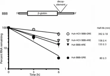
IL2ARE have been previously shown to function as me-diators of rapid mRNA decay (22,35). The cells were also co-transfected with a GFP reporter construct to control for transfection efficiency. Transcription was inhibited by the addition of actinomycin D, and total RNA was isolated af-ter 0, 3 and 6 h. Expression of the reporaf-ter transcripts was measured by quantitative RT-PCR and was normalized to the expression of the GFP transcripts. In this set of exper-iments, the BBB-GRE transcript decayed rapidly in Huh cells with a half-life of 80±3 min and was stabilized (P=
0.005) in Huh-HCV cells with a half-life of 392±18 min. In contrast, the BBB-ARE transcript decayed rapidly in Huh cells with a half-life of 133±3 min but exhibited only mi-nor and insignificant stabilization in Huh-HCV cells with a half-life of 158±4 min (Figure3). This finding that the GRE-containing reporter transcript exhibited specific sta-bilization in replicon-containing cells suggests that a mech-anism exists for selective recognition of the GRE in HCV replicon-containing cells.
Binding by the HCV NS5A protein to host cellular transcripts correlates with transcript stabilization
Figure 2. NS5A target transcripts are highly enriched for transcript
stabi-lization and up-regulation. (A) The percentage of NS5A target transcripts,
non-NS5A target transcripts and all transcripts that were stabilized (blue bars) or stabilized and up-regulated (red bars) is shown. ** represents sta-tistically significant differences (P-value<10−16, Fisher’s exact test with
R) and *represents statistically significant differences (P-value<10−11, Fisher’s extact test, R) in the percentages comparing NS5A target
tran-scripts to Non-NS5A target trantran-scripts or all trantran-scripts. (B) Top: A
mo-tif search was performed to look for conserved consensus sequences in
the 3UTRs of NS5A target transcripts. The top 12-mer motif is shown.
The position in the signal (bases) is depicted on the horizontal axis. The height of each stack of letters on the vertical axis is proportional to the residue frequency in the given position. Bottom: The motif previously found in CELF1 target transcripts that resembles the top 12-mer motif shown above.
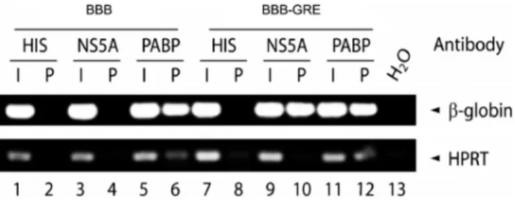
or the same reporter carrying theJUNBGRE in its 3UTR (BBB-GRE). Two days after transfection, cell lysates were immunoprecipitated with an anti-His antibody (negative control), PABP antibody (positive control) or anti-NS5A antibody, and RNA was isolated from the input ma-terial (I) and the immunoprecipitation pellet (P). Reverse transcription PCR was performed on this RNA to evalu-ate levels of beta-globin transcript and Hypoxanthine Phos-phoribosyltransferase (HPRT) transcript, which is used as a housekeeping gene control (Figure4). In cells transfected with the BBB-GRE construct, the beta-globin transcript was present in the NS5A pellet but the HPRT transcript was not (lane 10), whereas in cells transfected with the BBB construct, neither the beta-globin nor the HPRT transcripts were present in the NS5A pellet (lane 4). This result suggests that NS5A bound specifically in cells only to beta-globin reporter transcript that contained a GRE. Thus, NS5A was capable of binding to the GRE in HCV replicon-containing cells.
We performed in vitroRNA-binding assays to measure the binding affinity (Kd) of NS5A for the GRE and other sequences, including RNA sequences derived from the
con-Figure 3.GRE-containing host mRNA transcripts are stabilized in Huh
cells stably expressing an HCV subgenomic replicon (Huh-HCV). Huh or Huh-HCV cells were transfected with the BBB-GRE or BBB-ARE beta-globin reporter constructs. Actinomycin D was added to stop transcription and total cellular RNA was isolated after 0, 3 or 6 h. Specific mRNA levels were determined by quantitative real time RT-PCR. Beta-globin transcript levels at each time point were normalized to the transcript levels from a co-transfected GFP reporter. Transcript levels at the 0 time point were set to 100%, and the percent mRNA remaining was plotted as a function of time. The error bars indicate the standard error of the mean (SEM) from three experiments. Transcript half-life and SEM are shown to the right of each graph.
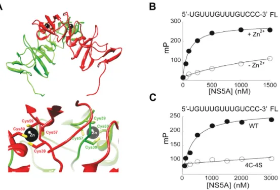
sensus sequence shown in Figure2B, top (Table1). For these assays, we used a polypeptide that contained the amino ter-minal domain (domain 1) of NS5A. The structure of do-main 1 of NS5A has been determined by X-ray crystallog-raphy (20,21). The protein crystallized as a homodimer, and the structural integrity of the dimer was dependent on the tetra-cysteine-coordinated Zn2+ ion (Figure5A). We
puri-fied a recombinant NS5A polypeptide that contained do-main 1 plus 36 additional carboxy terminal amino acids; we refer to this protein as NS5A domain 1+. We also puri-fied a NS5A derivative that was identical except the 4 cys-teines in the zinc finger were changed to serines (referred to as 4C-4S). We used a fluorescence polarization assay (18) to measure binding of these NS5A polypeptides to fluorescein-labeled RNA oligonucleotides that contained a minimal GRE sequence (RNA #3 in Table1). NS5A domain 1+ was titrated into a binding mixture containing a 3-fluorescein labeled RNA substrate. The median polarization (mP) was plotted as a function of NS5A domain 1+ concentration, and the equilibrium dissociation constant was determined by fitting the data to a hyperbola. NS5A domain 1+ bound with high affinity to the GRE and this binding depended on the presence of zinc (Figure5B). The mutated NS5A polypeptide (4C-4S), incapable of binding Zn2+because of
complete disruption of the Zn2+-binding site, failed to bind
to the GRE (Figure5C). Converting the GRE sequence into CU- or AU-rich sequences resulted in a 7- to 10-fold reduc-tion in the observed affinity of NS5A domain 1+ for these RNAs (Table1; compare RNA #4 and #5 to #3). Two RNA sequences derived from the consensus sequence in Figure
2B bound to NS5A domain 1+ with high affinities (Table
Figure 4. NS5A binds to GRE-containing transcripts in cells. Huh7-HCV cells were transfected with the BBB, or BBB-GRE reporter plasmids. Cell lysates were immunoprecipitated using specific antibodies against the His-tag (HIS), NS5A or the poly A binding protein (PABP). RNA isolated from the input (I) or the pellet fraction (P) was reverse transcribed and amplified by PCR using beta-globin and HPRT specific primers, and the
RNA was separated by electrophoresis. Water (H2O) was used as a
con-tamination control for the PCR.
specific interaction between NS5A and the GRE in cells and
in vitro.
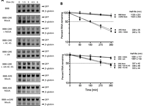
NS5A directly stabilizes GRE-containing transcripts
Based on our findings that GRE-containing transcripts were stabilized in Huh-HCV cells and that the HCV NS5A protein was found to bind to GRE sequences, we hypothe-sized that binding by NS5A to GRE-containing transcripts mediates transcript stabilization. To test our hypothesis, we transfected HeLa cells with the BBB, GRE, BBB-mGRE or BBB-ARE beta-globin reporter constructs and co-transfected them with constructs that expressed wild-type NS5A (NS5A) or the following NS5A derivatives: 4C-4S,N orN-4C-4S. A GFP expression construct was also co-transfected to control for transfection efficiency. Tran-scription from the tet-responsive promoter was blocked by the addition of doxycycline, and total RNA was extracted after 0, 1.5, 3, 4.5 or 6 h. Transcript degradation was mea-sured over time by northern blot (Figure 6) and the ex-pression of NS5A or derivatives were monitored by west-ern blot (Supplementary Figure S3). As expected, the BBB-reporter alone and the BBB-mGRE BBB-reporter were very sta-ble, whereas the BBB-GRE reporter decayed more rapidly (half-life=337±42 minutes). When the NS5A construct was co-transfected, however, the BBB-GRE transcript was stabilized (1091 ± 117 min; P = 0.0005). This stabiliz-ing effect was not observed when the four cysteines that form the NS5A zinc finger were mutated to serine (4C-4S). This indicated that a zinc finger-mediated NS5A-GRE in-teraction was required for stabilization. Published reports showed that the N-terminal helix of NS5A is required for tethering the NS5A protein to the viral replication com-partment of the endoplasmatic reticulum (36–38). Deletion of the N-terminal helix releases this protein into the cyto-plasm and decreases viral replication (39). We found that deletion of the N-terminal 36 amino acids (N) had no ef-fect on the ability of NS5A to stabilize the GRE-containing reporter transcript. Deletion of the N-terminal 36 amino acids and perturbation of the NS5A zinc finger at the same time (N-4C-4S), however, abrogated the stabilizing activ-ity of NS5A. In contrast to the GRE-reporter, the ARE re-porter transcript (BBB-ARE) displayed equal decay in the
presence or absence of NS5A with half-life of 240±15 min-utes and 228±25 minutes, respectively. This result indicates that NS5A specifically and selectively mediated the stabi-lization of GRE-containing, but not ARE-containing tran-scripts.
DISCUSSION
Introduction of a HCV subgenomic replicon into Huh cells led to the selective stabilization of host cellular transcripts through a mechanism that involved binding of NS5A di-rectly to GU-rich sequences. Not all stabilized transcripts showed increased abundance. This could be due to other mechanisms that may affect transcript abundance to main-tain homeostasis. For NS5A target transcripts we observed a significantly higher number of transcripts with increased abundance compared to non-NS5A target transcripts (Fig-ure 2A). We showed that NS5A binds directly with high affinity to GRE sequences, and may bind to other G or U-rich sequences that were conserved in the 3 UTRs of NS5A target transcripts. Our finding in HeLa cells that ex-ogenous expression of NS5A specifically stabilizes GRE-containing transcripts clearly demonstrates that NS5A pos-sesses mRNA-stabilizing activity (Figure6). This function of NS5A enables HCV to control host cellular gene expres-sion.
The results presented here support previous work sug-gesting that NS5A functions as an RNA-binding protein. NS5A interacts with HCV genomic RNA (18,19) and was shown to bind to polyU or polyG sequences (18). Our re-sults indicate that a G-rich consensus sequence was present in the 3 UTRs of NS5A target transcripts (Figure 2B). This sequence resembles the G-rich consensus sequence pre-viously found in CELF1 target transcripts (33). In addi-tion, NS5A is also reported to bind to poly(U/UC) se-quence in the 3UTR of HCV RNA and downregulates viral RNA translation (40). We found similar poly(U/UC)-rich sequences were present within the 3UTR of NS5A target transcripts (Supplementary Figure S2). It is reported that GRE sequences are very common in the genome. Although GREs were found in NS5A target transcripts, they were not enriched. Our data shows that the GRE can function as a site for NS5A, but not all GRE-containing transcripts are NS5A targets. This may be due to secondary structures, ge-nomic context, or availability for binding sites.
Our results confirm the RNA-binding activity of NS5A and suggest that the GRE sequence, UGUUUGUU-UGU (22), is a binding target for NS5A. We showed by RNA immunoprecipitation that an NS5A anti-body specifically co-immunoprecipitated GRE-containing reporter transcripts, but not reporter transcripts that lacked a GRE (Figure4). These results suggest that the GRE func-tions as a target of NS5A within cells. Ourin vitrobinding experiments showed that NS5A binding to the GRE was de-pendent upon the C4zinc finger motif in domain 1 since no
Figure 5.Recombinant NS5A binds to GRE RNA in a manner that is dependent on an intact zinc-binding site and the presence of zinc. (A) The upper panel shows a ribbon diagram of dimeric NS5A domain 1 that was prepared by using reference number 1ZH1 from the Protein Data Bank. One subunit is colored red and the other is colored green. The lower panel zooms in on the zinc-binding site of each subunit. Four conserved cysteines are required
for zinc binding. (B) The NS5A domain 1+ polypeptide was titrated into a binding reaction buffer (20 mM HEPES pH 7.5, 5 mM MgCl2, 10 mM
2-mercaptoethanol, 100 mM NaCl) in the absence (-Zn2+) or presence (+Zn2+) of 100M ZnCl
2and incubated briefly at 25◦C in a final volume of 100l.
Binding of NS5A was measured by the change in polarization (mP). The change in fluorescence polarization was plotted as a function of NS5A domain 1+
concentration and fit to a hyperbola by using KaleidaGraph (Synergy Software). (C) Experiments were performed as described in panel B in the presence
of zinc using the NS5A domain 1+ polypeptide (WT) or the derivative whose zinc-binding site was inactivated by converting the four cysteine residues to serine residues (4C-4S).
(20,21) (Figure5A). Collectively, our findings suggest that the zinc finger motif contributes to the functional form of NS5A that enables specific interaction of NS5A with GRE RNA.
We previously showed that the GRE mediates rapid mRNA decay by binding to CELF1 (22), which appears to recruit components of the cellular mRNA decay machinery such as poly A ribonuclease (25). In the presence of NS5A, the rapid decay of the GRE-containing reporter was com-pletely abolished (Figure 6), suggesting that NS5A might potentially antagonize the activity of CELF1. CELF1 and NS5A appear to have overlapping binding specificities since they both bind directly to G-rich sequences, including the GRE, but a subset of NS5A target transcripts might not be targets of CELF1. These proteins have different mech-anisms for RNA binding. NS5A binding involves zinc fin-ger domains, whereas CELF1 binding involves RNA recog-nition motifs, yet their binding target sites appear to over-lap, at least in the case of GRE sequences. Further work is needed to better define the sites for binding by these pro-teins to host transcripts.
Viruses have developed mechanisms to manipulate host gene expression at posttranscriptional levels in order to sub-vert antiviral defense mechanisms and to create an environ-ment in the virus-infected cell that will prevent cell death and allow viral replication. Kaposi’s sarcoma virus (1) and Herpes simplex virus (2) produce nucleases that mediate host mRNA decay. Herpes simplex virus also produces the
ICP27 protein which mediates the stabilization of AU-rich element-containing host transcript (3) and Epstein-Barr virus infection leads to the stabilization of mRNAs through the activation of the stress activated protein kinase p38 (41). Several studies found differential host gene expression in cell cultures infected with HCV using high-throughput ap-proaches and screens (42–46), but the mechanism of how the host gene expression is regulated by the virus is not well understood. Luna et al. proposed that microRNA-122 sequestration by HCV RNA may lead to stabilization of miR-122 targets, facilitating the oncogenesis of HCV (47). Moon et al. demonstrated another mechanism that HCV utilizes to affect host mRNA decay. They found the 5UTR of HCV genomic RNA can stall and suppress the cellu-lar 5-3exoribonuclease Xrn1, leading to global stabiliza-tion of cellular transcripts (48). The mechanism by which HCV stabilizes GRE-containing transcripts, described in this report, depends on the direct interaction between the NS5A protein and GU-rich sequences within host cellu-lar mRNA. Through this mechanism, HCV blocks the host mRNA degradation machinery and selectively stabilizes a set of GU-rich transcripts that may be necessary for estab-lishment or maintenance of chronic HCV infection.
tran-Figure 6. Exogenously expressed NS5A stabilizes GRE-containing reporter genes. (A) HeLa tet-off cells were transfected with BBB, BBB-GRE, BBB-ARE
or BBB-mGRE reporter plasmids as well as a plasmid that express NS5A, NS5A-4C-4S,N-NS5A,N-NS5A-4C-4S or a mock control plasmid. A GFP
expression plasmid was included in each to control for transfection efficiency. Transcription from the tet-responsive promoter was stopped with 300 ng
of doxycycline and RNA harvested after 0, 1.5, 3, 4.5 or 6 h was analyzed by northern blotting using GFP and beta-globin probes. (B) The experiment
shown in (A) was performed three times, and the northern blot signals were quantified by a Storm 820 phosphorimager (Amersham Biosciences). For each time point, the intensity of the beta-globin reporter was normalized to the intensity of the GFP band, and the band intensity at the 0 time point was set to 100%. The percent of mRNA remaining was plotted over time. The error bars indicate the standard error of the mean (SEM) from three experiments. The calculated transcript half-life and SEM are shown to the right of each graph.
scripts such asJUN,JUNB,JUND, andETS2, transcripts encoding regulators of apoptosis such as BCL10, BAG,
MAP3K5, andTNFRSF1, and transcripts encoding other regulators of cell growth including EIF4EBP2, EIF4G3,
SMAD7andHOXC10. After infection with HCV, NS5A-mediated stabilization and overexpression of these GRE-containing transcripts would be predicted to promote cell growth and prevent cell death in order to allow a chronic infection to be established. It is possible that new drugs that block the NS5A interaction with the GRE could be devel-oped that would prevent establishment of chronic HCV fections and inhibit the maintenance or propagation of in-fection. Indeed, drugs like ledipasvir that target NS5A but whose mechanism of action remain unclear (49) might func-tion by perturbing the ability of NS5A to bind GREs or similar G or U rich sequences and modulate host gene ex-pression.
Infection with HCV increases the risk for hepatocellu-lar carcinoma development as well as certain lymphomas (50,51). The onset of the tumors, however, occurs many years after HCV infection, suggesting that multiple events are required to transform hepatic cells to become malig-nant (52). Progression of liver fibrosis to cirrhosis in HCV-infected patients will generate a local milieu that
predis-poses to liver cancer. In this environment, changes occur to the hepatic parenchyma, with hepatocyte injury, which contributes to sequential genetic hits that culminate in ma-lignant transformation (53). However, the mechanisms in-volved in this process are largely unknown. Moreover, mul-tiple studies have showed a direct role for HCV in hep-atic carcinogenesis and transgenic mice expressing the HCV polyprotein can develop liver cancer in the absence of in-flammation, hepatic cirrhosis or immune recognition of the transgene (13,54,55). The stabilization and overexpression of GRE-containing transcripts including proto-oncogene transcripts and transcripts encoding regulators of apoptosis may contribute to the development of cancer by promoting cell growth and preventing the death of genetically damaged cells. Later events that finally lead to uncontrolled growth of these cells might destroy the fail-safe mechanisms that are initially able to cope with the increased stability of these im-portant messages.
DATAAVAILABILITY
Geneexpressionomnibus(GEO)accessionnumberGSE1 02910.
SUPPLEMENTARYDATA
SupplementaryDataareavailableatNARonline.
ACKNOWLEDGEMENTS
Wethank DrIbrahimMoustafaforpreparingFigure5A andDrAnn-BinShyuforprovidingplasmids.The Univer-sity of MinnesotaSupercomputing Instituteprovidedthe accesstoIngenuityPathwayAssistant(QiagenInc).None oftheauthorshasaconflictofinterest.
FUNDING
NationalInstitutesofHealth(NIH)[AI057484,AI072068 toP.R.B.,AI053531toC.E.C.,T32AI83196toL.G.];Swiss National Science Foundation (to B.R.); Lymphoma Re-searchFoundationandstart-upfundsfromtheDepartment of Medicine at the University of Minnesota (to I.V.-S.). Funding foropenaccesscharge:Universityof Minnesota InstitutionalFunds.
Conflictofintereststatement.Nonedeclared.
REFERENCES
1. Glaunsinger,B., Chavez,L. and Ganem,D. (2005) The exonuclease and host shutoff functions of the SOX protein of Kaposi’s
sarcoma-associated herpesvirus are genetically separable.J. Virol.,79,
7396–7401.
2. Elgadi,M.M., Hayes,C.E. and Smiley,J.R. (1999) The herpes simplex virus vhs protein induces endoribonucleolytic cleavage of target
RNAs in cell extracts.J. Virol.,73, 7153–7164.
3. Corcoran,J.A., Hsu,W.L. and Smiley,J.R. (2006) Herpes simplex virus ICP27 is required for virus-induced stabilization of the
ARE-containing IEX-1 mRNA encoded by the human IER3 gene.J.
Virol.,80, 9720–9729.
4. Lindenbach,B.D. and Rice,C.M. (2001)Flaviviridae: The Viruses and
their Replication. 4th edn. Lippincott-Raven, Philadelphia. 5. Grakoui,A., McCourt,D.W., Wychowski,C., Feinstone,S.M. and
Rice,C.M. (1993) Characterization of the hepatitis C virus-encoded serine proteinase: determination of proteinase-dependent polyprotein cleavage sites.J. Virol.,67, 2832–2843.
6. Thrift,A.P., El-Serag,H.B. and Kanwal,F. (2016) Global
epidemiology and burden of HCV infection and HCV-related disease.
Nat. Rev. Gastroenterol. Hepatol.,14, 122–132.
7. Poynard,T., Yuen,M.F., Ratziu,V. and Lai,C.L. (2003) Viral hepatitis C.Lancet,362, 2095–2100.
8. Debes,J.D., Janssen,H.L and Boonstra,A. (2017) Hepatitis C
treatment and liver cancer recurrence: cause for concern?Lancet
Gastroenterol. Hepatol.,2, 78–80.
9. Reig,M., Marino,Z., Perello,C., Inarrairaegui,M., Ribeiro,A.,
Lens,S., Diaz,A., Vilana,R., Darnell,A., Varela,M.et al.(2016)
Unexpected high rate of early tumor recurrence in patients with
HCV-related HCC undergoing interferon-free therapy.J. Hepatol.,
65, 719–726.
10. de Chassey,B., Navratil,V., Tafforeau,L., Hiet,M.S., Aublin-Gex,A., Agaugue,S., Meiffren,G., Pradezynski,F., Faria,B.F., Chantier,T.
et al.(2008) Hepatitis C virus infection protein network.Mol. Syst. Biol.,4, 230.
11. Majumder,M., Ghosh,A.K., Steele,R., Ray,R. and Ray,R.B. (2001) Hepatitis C virus NS5A physically associates with p53 and regulates
p21/waf1 gene expression in a p53-dependent manner.J. Virol.,75,
1401–1407.
12. Ghosh,A.K., Steele,R., Meyer,K., Ray,R. and Ray,R.B. (1999) Hepatitis C virus NS5A protein modulates cell cycle regulatory genes
and promotes cell growth.J. Gen. Virol.,80, 1179–1183.
13. Lan,K.H., Sheu,M.L., Hwang,S.J., Yen,S.H., Chen,S.Y., Wu,J.C.,
Wang,Y.J., Kato,N., Omata,M., Chang,F.Y.et al.(2002) HCV NS5A
interacts with p53 and inhibits p53-mediated apoptosis.Oncogene,
21, 4801–4811.
14. Arima,N., Kao,C.Y., Licht,T., Padmanabhan,R., Sasaguri,Y. and Padmanabhan,R. (2001) Modulation of cell growth by the hepatitis C
virus nonstructural protein NS5A.J. Biol. Chem.,276, 12675–12684.
15. Shirota,Y., Luo,H., Qin,W., Kaneko,S., Yamashita,T., Kobayashi,K. and Murakami,S. (2002) Hepatitis C virus (HCV) NS5A binds RNA-dependent RNA polymerase (RdRP) NS5B and modulates
RNA-dependent RNA polymerase activity.J. Biol. Chem.,277,
11149–11155.
16. Huang,L., Sineva,E.V., Hargittai,M.R., Sharma,S.D., Suthar,M., Raney,K.D. and Cameron,C.E. (2004) Purification and
characterization of hepatitis C virus non-structural protein 5A
expressed in Escherichia coli.Protein. Expr. Purif.,37, 144–153.
17. Shimakami,T., Hijikata,M., Luo,H., Ma,Y.Y., Kaneko,S., Shimotohno,K. and Murakami,S. (2004) Effect of interaction between hepatitis C virus NS5A and NS5B on hepatitis C virus RNA replication with the hepatitis C virus replicon.J. Virol.,78,
2738–2748.
18. Huang,L., Hwang,J., Sharma,S.D., Hargittai,M.R., Chen,Y., Arnold,J.J., Raney,K.D. and Cameron,C.E. (2005) Hepatitis C virus
nonstructural protein 5A (NS5A) is an RNA-binding protein.J. Biol.
Chem.,280, 36417–36428.
19. Ivanov,A.V., Tunitskaya,V.L., Ivanova,O.N., Mitkevich,V.A., Prassolov,V.S., Makarov,A.A., Kukhanova,M.K. and
Kochetkov,S.N. (2009) Hepatitis C virus NS5A protein modulates
template selection by the RNA polymerase in in vitro system.FEBS
Lett.,583, 277–280.
20. Tellinghuisen,T.L., Marcotrigiano,J. and Rice,C.M. (2005) Structure of the zinc-binding domain of an essential component of the hepatitis
C virus replicase.Nature,435, 374–379.
21. Love,R.A., Brodsky,O., Hickey,M.J., Wells,P.A. and Cronin,C.N. (2009) Crystal structure of a novel dimeric form of NS5A domain I
protein from hepatitis C virus.J. Virol.,83, 4395–4403.
22. Vlasova,I.A., Tahoe,N.M., Fan,D., Larsson,O., Rattenbacher,B., Sternjohn,J.R., Vasdewani,J., Karypis,G., Reilly,C.S., Bitterman,P.B.
et al.(2008) Conserved GU-rich elements mediate mRNA decay by
binding to CUG-binding protein 1.Mol. Cell,29, 263–270.
23. Vlasova,I.A. and Bohjanen,P.R. (2008) Posttranscriptional regulation
of gene networks by GU-rich elements and CELF proteins.RNA
Biol.,5, 201–207.
24. Graindorge,A., Le Tonqueze,O., Thuret,R., Pollet,N., Osborne,H.B.
and Audic,Y. (2008) Identification of CUG-BP1/EDEN-BP target
mRNAs in Xenopus tropicalis.Nucleic. Acids. Res.,36, 1861–1870.
25. Moraes,K.C., Wilusz,C.J. and Wilusz,J. (2006) CUG-BP binds to
RNA substrates and recruits PARN deadenylase.RNA,12,
1084–1091.
26. Zhong,J., Gastaminza,P., Cheng,G., Kapadia,S., Kato,T., Burton,D.R., Wieland,S.F., Uprichard,S.L., Wakita,T. and
Chisari,F.V. (2005) Robust hepatitis C virus infection in vitro.Proc.
Natl. Acad. Sci. U.S.A.,102, 9294–9299.
27. Peng,S.S., Chen,C.Y. and Shyu,A.B. (1996) Functional
characterization of a non-AUUUA AU-rich element from the c-jun proto-oncogene mRNA: evidence for a novel class of AU-rich
elements.Mol. Cell Biol.,16, 1490–1499.
28. Ogilvie,R.L., Abelson,M., Hau,H.H., Vlasova,I., Blackshear,P.J. and Bohjanen,P.R. (2005) Tristetraprolin down-regulates IL-2 gene
expression through AU-rich element-mediated mRNA decay.J.
Immunol.,174, 953–961.
29. Blight,K.J., Kolykhalov,A.A. and Rice,C.M. (2000) Efficient
initiation of HCV RNA replication in cell culture.Science,290,
1972–1974.
30. Hwang,J., Huang,L., Cordek,D.G., Vaughan,R., Reynolds,S.L., Kihara,G., Raney,K.D., Kao,C.C. and Cameron,C.E. (2010) Hepatitis C virus nonstructural protein 5A: biochemical
characterization of a novel structural class of RNA-binding proteins.
31. Beisang,D., Reilly,C. and Bohjanen,P.R. (2014) Alternative
polyadenylation regulates CELF1/CUGBP1 target transcripts
following T cell activation.Gene,550, 93–100.
32. Tenenbaum,S.A., Lager,P.J., Carson,C.C. and Keene,J.D. (2002) Ribonomics: identifying mRNA subsets in mRNP complexes using
antibodies to RNA-binding proteins and genomic arrays.Methods,
26, 191–198.
33. Rattenbacher,B., Beisang,D., Wiesner,D.L., Jeschke,J.C., von Hohenberg,M., St Louis-Vlasova,I.A. and Bohjanen,P.R. (2010) Analysis of CUGBP1 targets identifies GU-repeat sequences that
mediate rapid mRNA decay.Mol. Cell Biol.,30, 3970–3980.
34. Kiser,K.F., Colombi,M. and Moroni,C. (2006) Isolation and characterization of dominant and recessive IL-3-independent
hematopoietic transformants.Oncogene,25, 6595–6603.
35. Chen,C.Y., Gherzi,R., Ong,S.E., Chan,E.L., Raijmakers,R., Pruijn,G.J., Stoecklin,G., Moroni,C., Mann,M. and Karin,M. (2001) AU binding proteins recruit the exosome to degrade ARE-containing
mRNAs.Cell,107, 451–464.
36. Penin,F., Brass,V., Appel,N., Ramboarina,S., Montserret,R., Ficheux,D., Blum,H.E., Bartenschlager,R. and Moradpour,D. (2004) Structure and function of the membrane anchor domain of hepatitis
C virus nonstructural protein 5A.J. Biol. Chem.,279, 40835–40843.
37. Brass,V., Bieck,E., Montserret,R., Wolk,B., Hellings,J.A., Blum,H.E., Penin,F. and Moradpour,D. (2002) An amino-terminal amphipathic alpha-helix mediates membrane association of the hepatitis C virus
nonstructural protein 5A.J. Biol. Chem.,277, 8130–8139.
38. Sapay,N., Montserret,R., Chipot,C., Brass,V., Moradpour,D., Deleage,G. and Penin,F. (2006) NMR structure and molecular dynamics of the in-plane membrane anchor of nonstructural protein
5A from bovine viral diarrhea virus.Biochemistry,45, 2221–2233.
39. Sauter,D., Himmelsbach,K., Kriegs,M., Carvajal Yepes,M. and Hildt,E. (2009) Localization determines function: N-terminally truncated NS5A fragments accumulate in the nucleus and impair
HCV replication.J. Hepatol.,50, 861–871.
40. Hoffman,B., Li,Z. and Liu,Q. (2015) Downregulation of viral RNA translation by hepatitis C virus non-structural protein NS5A requires
the poly(U/UC) sequence in the 3’ UTR.J. Gen. Virol.,96,
2114–2121.
41. Vockerodt,M., Pinkert,D., Smola-Hess,S., Michels,A.,
Ransohoff,R.M., Tesch,H. and Kube,D. (2005) The Epstein-Barr virus oncoprotein latent membrane protein 1 induces expression of
the chemokine IP-10: importance of mRNA half-life regulation.Int.
J. Cancer,114, 598–605.
42. Hojka-Osinska,A., Budzko,L., Zmienko,A., Rybarczyk,A., Maillard,P., Budkowska,A., Figlerowicz,M. and Jackowiak,P. (2016) RNA-Seq-based analysis of differential gene expression associated
with hepatitis C virus infection in a cell culture.Acta. Biochim. Pol.,
63, 789–798.
43. Woodhouse,S.D., Narayan,R., Latham,S., Lee,S., Antrobus,R., Gangadharan,B., Luo,S., Schroth,G.P., Klenerman,P. and
Zitzmann,N. (2010) Transcriptome sequencing, microarray, and proteomic analyses reveal cellular and metabolic impact of hepatitis
C virus infection in vitro.Hepatology,52, 443–453.
44. Papic,N., Maxwell,C.I., Delker,D.A., Liu,S., Heale,B.S. and Hagedorn,C.H. (2012) RNA-sequencing analysis of 5’ capped RNAs identifies many new differentially expressed genes in acute hepatitis C virus infection.Viruses,4, 581–612.
45. Walters,K.A., Syder,A.J., Lederer,S.L., Diamond,D.L., Paeper,B., Rice,C.M. and Katze,M.G. (2009) Genomic analysis reveals a potential role for cell cycle perturbation in HCV-mediated apoptosis
of cultured hepatocytes.PLoS Pathog.,5, e1000269.
46. Blackham,S., Baillie,A., Al-Hababi,F., Remlinger,K., You,S., Hamatake,R. and McGarvey,M.J. (2010) Gene expression profiling indicates the roles of host oxidative stress, apoptosis, lipid metabolism, and intracellular transport genes in the replication of hepatitis C virus.J. Virol.,84, 5404–5414.
47. Luna,J.M., Scheel,T.K., Danino,T., Shaw,K.S., Mele,A., Fak,J.J.,
Nishiuchi,E., Takacs,C.N., Catanese,M.T., de Jong,Y.P.et al.(2015)
Hepatitis C virus RNA functionally sequesters miR-122.Cell,160,
1099–1110.
48. Moon,S.L., Blackinton,J.G., Anderson,J.R., Dozier,M.K., Dodd,B.J., Keene,J.D., Wilusz,C.J., Bradrick,S.S. and Wilusz,J. (2015) XRN1 stalling in the 5’ UTR of Hepatitis C virus and Bovine Viral Diarrhea
virus is associated with dysregulated host mRNA stability.PLoS
Pathog.,11, e1004708.
49. Kwon,H.J., Xing,W., Chan,K., Niedziela-Majka,A., Brendza,K.M.,
Kirschberg,T., Kato,D., Link,J.O., Cheng,G., Liu,X.et al.(2015)
Direct binding of ledipasvir to HCV NS5A: mechanism of resistance
to an HCV antiviral agent.PLoS One,10, e0122844.
50. Craxi,A., Laffi,G. and Zignego,A.L. (2008) Hepatitis C virus (HCV)
infection: a systemic disease.Mol. Aspects. Med.,29, 85–95.
51. Hassan,M., Selimovic,D., Ghozlan,H. and Abdel-kader,O. (2009) Hepatitis C virus core protein triggers hepatic angiogenesis by a
mechanism including multiple pathways.Hepatology,49, 1469–1482.
52. Hanahan,D. and Weinberg,R.A. (2000) The hallmarks of cancer.
Cell,100, 57–70.
53. Debes,J.D., de Knegt,R.J. and Boonstra,A. (2017) The path to cancer, and back: Immune modulation during hepatitis C virus infection, progression to fibrosis and cancer, and unexpected roles of new
antivirals.Transplantation.101, 910–915.
54. Moriya,K., Fujie,H., Shintani,Y., Yotsuyanagi,H., Tsutsumi,T., Ishibashi,K., Matsuura,Y., Kimura,S., Miyamura,T. and Koike,K. (1998) The core protein of hepatitis C virus induces hepatocellular
carcinoma in transgenic mice.Nat. Med.,4, 1065–1067.
55. Fukutomi,T., Zhou,Y., Kawai,S., Eguchi,H., Wands,J.R. and Li,J. (2005) Hepatitis C virus core protein stimulates hepatocyte growth:
correlation with upregulation of wnt-1 expression.Hepatology,41,